• Skip to primary navigation
  • Skip to main content
  • Skip to primary sidebar
  • Skip to footer

Bowdoin Science Journal

  • Home
  • About
    • Our Mission
    • Our Staff
  • Sections
    • Biology
    • Chemistry and Biochemistry
    • Math and Physics
    • Computer Science and Technology
    • Environmental Science and EOS
    • Honors Projects
    • Psychology and Neuroscience
  • Contact Us
  • Fun Links
  • Subscribe

Luke Taylor '24

What Causes Aging? An Epigenetics Study From The Sinclair Lab May Have an Answer

April 2, 2023 by Luke Taylor '24

Dr. David Sinclair, A.O., Ph.D. Photo from the Sinclair Lab, Harvard Medical School (2023).

Aging, also known as “senescence,” is an inevitable process in all living things. Organisms small and large eventually break down, accumulating enough wear and tear in their cells that ultimately causes the body to stop functioning as a whole. While medicine and lifestyle improvements stave off aging, identifying its fundamental causes has been more challenging. In January 2023, scientists in Dr. David Sinclair’s lab at Harvard Medical School published a paper with experimental evidence supporting what Sinclair calls the “Information Theory of Aging,” where damage to the epigenome can cause aging.

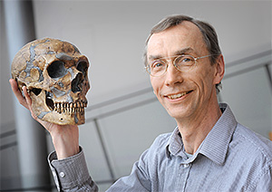
After receiving his Ph.D. in molecular genetics from the University of New South Wales, Sinclair completed his postdoctorate at MIT where he co-discovered the role of sirtuin enzymes in limiting age-related cellular damage in yeast. In addition to teaching genetics and translational medicine at Harvard Medical School since 1999, Sinclair authored the popular book Lifespan: Why We Age – and Why We don’t Have To (2019). His breakthroughs in the science of aging have earned him a great deal of attention from the public eye, resulting in appearances on several popular media outlets, including CBS’s “60 Minutes” and TIME magazine (The Sinclair Lab, 2023).

Sinclair’s newest discovery, published as “Loss of epigenetic information as a cause of mammalian ageing” in January 2023, focused on the role of epigenetics in aging. The title specifies that epigenetic information loss, rather than genetic information loss, is a cause of aging. Genetics refers to the raw molecular information sequences stored in cells as deoxyribonucleic acid (DNA), which is physically condensed inside the nucleus into pairs of chromosomes. The material inside a chromosome is known as “chromatin.” The central dogma of molecular biology states that information in DNA sequences is read by the cell in the form of messenger RNA (mRNA) through transcription, and then ribosomes in the cell read this mRNA to make proteins through translation. DNA sequences that correspond to the production of a specific molecule are genes. The prefix epi- means “on top of,” so “epigenetics” refers to mechanisms that function “above” the molecules of DNA themselves, including reading DNA sequences, regulating gene transcription, and repairing mutated DNA. Like the DNA sequence, epigenetic changes are inheritable from parents to offspring.

The differences between genetics and epigenetics influence cellular reaction to damage. Damage to genes causes mutations, which are changes in the sequence of the DNA of that gene. Cells have mechanisms of repairing mutated DNA, but failure of these mechanisms can lead to cell death, or worse, cancer. Some DNA mutations, like those where a nucleotide is deleted, are irreversible.

By contrast, epigenetic mechanisms are more easily reversed. One epigenetic mechanism is DNA methylation, where a methyl group (-CH3) is added to a cytosine nucleotide by the DNA methyltransferase enzyme. This extra functional group blocks transcription factors from binding to promoter regions nearby the methylated cytosine, in effect “silencing” the gene as it cannot be transcribed into mRNA. DNA methylation is important for differentiating cells into specific cell types by enabling cells to only express the most pertinent genes while still containing the entire genome (Moore et al., 2012). DNA methylation is reversible with the help of TET dioxygenase enzymes (Wu and Zhang, 2014). Geneticists have found DNA methylation to be a way to assess molecular aging in cells as a sort of “epigenetic clock”. By analyzing methylation patterns of the genome (the “methylome”), scientists can find the biological age as well as the rate of aging in an organism’s cells (Hannum et al., 2012).

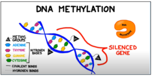
Figure 1: Methyl groups attached to cytosine bases in a gene block the enzyme RNA polymerase from binding to the promoter region of a gene, preventing transcription. Adapted from BOGOBiology (2017)

To investigate how changes to the epigenome affect aging in mice, Sinclair used a mouse system with induced changes to the epigenome (ICE). The genetically modified mice had a higher frequency of double-strand breaks (DSBs) in the DNA, which cause changes in the epigenome as cells are required to use their mechanisms of DNA repair more often . Sinclair’s method reduced the frequency of mutations by breaking the DNA strands in a way that left more whole unpaired nucleotides in the severed strand, making it more difficult for the cell to repair the DNA strand with a different sequence than before. While ICE mice had no significant difference in mutation frequencies versus control mice, DSBs in specific locations of the genome were observed as expected (Yang et al., 2023). Thus, any apparent changes in aging in the ICE mice of this study could be ascribed to the epigenetic DSB changes rather than mutations.

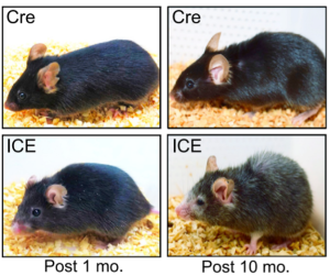
Figure 2: ICE treated mice after 10 months appear to have the physical hallmarks of aging, such as hair loss and reduced body mass, early compared to CRE control mice (Yang et. al. 2023).

The haggard appearance of the ICE 10 month old mice confirmed the early aging effects of the epigenetic changes (Figure 2). At the molecular level, analysis of DNA methylation at genome sites associated with age showed that ICE cells were approximately 1.5 times “older” than the control cells. With this artificially increased age came physiological consequences. ICE mice showed diminished short-term memory retention compared to Cre-control mice, as assessed by fear conditioning tests, and the ICE mice performed half as well in a Barnes Maze test as control, indicating decreased long-term memory. Additionally, ICE mice had decreased muscle mass and grip strength after 16 months. The authors attributed the cause of this accelerated aging to increased “faithful DNA repair” from the induced DSB breaks in the ICE mice, meaning that there were not significant mutations in the repair of DSBs.(Yang et al., 2023). During DSB repair, chromatin modifying factor proteins activate and move within a cell in a process known as “relocalization,” with repeated activation of this process known to cause epigenetic changes that silence genes normally expressed in young mice. Sinclair’s lab hypothesized that the relocalization of chromatin modifiers that occurs from repeated DSB repair associated with induced epigenetic changes lead to a gradual loss of cellular function associated with aging (Yang et al., 2023).

It may seem ironic that DNA strand break repair, a process meant to keep cells functioning when critical genes are damaged, is part of what ultimately causes the death of organisms. The fact that the mechanism implemented is epigenetic rather than genetic suggests that the effects of ageing may be reversible, like epigenetic mechanisms. In fact, Sinclair has shown that it is possible to partially undo the damaging effects of aging: after inducing OSK expression, which is a set of proteins known as “Yamanaka factors,” ICE mice exhibited some signs of rejuvenation in their eyes, kidneys, and muscles. Yamanaka factors like OSK are important in the fields of aging and regenerative medicine because they are keys to the synthesis of induced pluripotent stem cells, where somatic cells can become stem cells and potentially re-differentiate into other cell types (Takahashi and Yamanaka, 2006). The OSK treatment decreased the expression of age-associated markers in the kidney and muscle cells of the mice (Yang et al., 2023).

Sinclair’s experiments have shown the epigenome to be a key front in the investigation of aging, as the reversibility of changes to the epigenome can allow it to be a more accessible interface for scientists to interact with. The plasticity of the epigenome as demonstrated from the ability of Yamanaka factors to reverse the molecular indicators of aging mice show that there may be hope for science to bring this phenomenon to human epigenomes. Indeed, the news of reversing aging in mice made rounds in the media when Sinclair’s paper was published earlier this winter, and for good reason.

Works Cited

Al Aboud, N. M., Tupper, C., & Jialal, I. (2022). Genetics, Epigenetic Mechanism. In StatPearls. StatPearls Publishing. http://www.ncbi.nlm.nih.gov/books/NBK532999/

Bannister, A. J., & Kouzarides, T. (2011). Regulation of chromatin by histone modifications. Cell Research, 21(3), Article 3. https://doi.org/10.1038/cr.2011.22

BOGObiology (Director). (2017, October 11). Epigenetics: Nature vs. Nurture. https://www.youtube.com/watch?v=Q8BMP6HDIco

CDC. (2022, August 15). What is Epigenetics? | CDC. Centers for Disease Control and Prevention. https://www.cdc.gov/genomics/disease/epigenetics.htm

David Sinclair | The Sinclair Lab. (n.d.-a). Retrieved March 5, 2023, from https://sinclair.hms.harvard.edu/people/david-sinclair

Fernandez, A., O’Leary, C., O’Byrne, K. J., Burgess, J., Richard, D. J., & Suraweera, A. (2021). Epigenetic Mechanisms in DNA Double Strand Break Repair: A Clinical Review. Frontiers in Molecular Biosciences, 8, 685440. https://doi.org/10.3389/fmolb.2021.685440

Gilbert, S. F. (2000). Methylation Pattern and the Control of Transcription. Developmental Biology. 6th Edition. https://www.ncbi.nlm.nih.gov/books/NBK10038/

Hannum, G., Guinney, J., Zhao, L., Zhang, L., Hughes, G., Sadda, S., Klotzle, B., Bibikova, M., Fan, J.-B., Gao, Y., Deconde, R., Chen, M., Rajapakse, I., Friend, S., Ideker, T., & Zhang, K. (2013). Genome-wide Methylation Profiles Reveal Quantitative Views of Human Aging Rates. Molecular Cell, 49(2), 359–367. https://doi.org/10.1016/j.molcel.2012.10.016

Jin, B., Li, Y., & Robertson, K. D. (2011). DNA Methylation. Genes & Cancer, 2(6), 607–617. https://doi.org/10.1177/1947601910393957

Kulis, M., & Esteller, M. (2010). 2—DNA Methylation and Cancer. In Z. Herceg & T. Ushijima (Eds.), Advances in Genetics (Vol. 70, pp. 27–56). Academic Press. https://doi.org/10.1016/B978-0-12-380866-0.60002-2

Molecules discovered that extend life in yeast, human cells. (n.d.). EurekAlert! Retrieved March 5, 2023, from https://www.eurekalert.org/news-releases/664233.

Moore, L. D., Le, T., & Fan, G. (2013). DNA Methylation and Its Basic Function. Neuropsychopharmacology, 38(1), 23–38. https://doi.org/10.1038/npp.2012.112

Offord, Catherine. Two research teams reverse signs of aging in mice. (n.d.). Retrieved March 14, 2023, from https://www.science.org/content/article/two-research-teams-reverse-signs-aging-mice.

Takahashi, K., & Yamanaka, S. (2006). Induction of Pluripotent Stem Cells from Mouse Embryonic and Adult Fibroblast Cultures by Defined Factors. Cell, 126(4), 663–676. https://doi.org/10.1016/j.cell.2006.07.024

What is Epigenetics? The Answer to the Nature vs. Nurture Debate. (n.d.). Center on the Developing Child at Harvard University. Retrieved March 5, 2023, from https://developingchild.harvard.edu/resources/what-is-epigenetics-and-how-does-it-relate-to-child-development/

Wu, H., & Sun, Y. E. (2009). Reversing DNA Methylation: New Insights from Neuronal Activity–Induced Gadd45b in Adult Neurogenesis. Science Signaling, 2(64), pe17–pe17. https://doi.org/10.1126/scisignal.264pe17

Wu, H., & Zhang, Y. (2014a). Reversing DNA Methylation: Mechanisms, Genomics, and Biological Functions. Cell, 156(0), 45–68. https://doi.org/10.1016/j.cell.2013.12.019

Yang, J.-H., Hayano, M., Griffin, P. T., Amorim, J. A., Bonkowski, M. S., Apostolides, J. K., Salfati, E. L., Blanchette, M., Munding, E. M., Bhakta, M., Chew, Y. C., Guo, W., Yang, X., Maybury-Lewis, S., Tian, X., Ross, J. M., Coppotelli, G., Meer, M. V., Rogers-Hammond, R., … Sinclair, D. A. (2023). Loss of epigenetic information as a cause of mammalian aging. Cell, 186(2), 305-326.e27. https://doi.org/10.1016/j.cell.2022.12.027


Filed Under: Biology, Science

Examining the work of 2022 Nobel Prize in Physiology or Medicine Laureate Svante Pääbo

November 6, 2022 by Luke Taylor '24


Svante Pääbo. Max Planck Institute for Evolutionary Anthropology. Retrieved November 6, 2022. https://www.eva.mpg.de/genetics/staff/paabo/#c28042


            Have you ever wondered how humans lived on Earth before the first major civilizations formed? It turns out that we were not the only hominid species on Earth in those times: other relatives of humans, such as Neanderthals, lived and intermingled with humans. The genetic relationship between humans and these hominids is a subject of great interest to the scientific community, since the traits of our species’s distant relatives can explain genetic phenomena in modern humans. On October 3rd, 2022, the Nobel Prize Committee announced they would be awarding Swedish geneticist Svante Pääbo the prize in Physiology or Medicine “for his discoveries concerning the genomes of extinct hominins and human evolution” (NobelPrize.org, 2022). One of Dr. Pääbo’s most significant achievements leading to his prize was sequencing the Neanderthal genome. A closer look at Dr. Paabo’s work in the field of genetics elucidates how his work led to the foundation of the field of paleogenetics.

            Originally a student of Egyptology, Dr. Pääbo received a Ph.D. in molecular immunology from the University of Uppsala in 1986, but his interest in the former remained: his first Nature publication was about cloning ancient DNA from ancient Egyptian mummies (Gruber Foundation, 2022; Pääbo, 1985). Despite the possibility of degradation or contamination from the mummification process itself and from the millenia that passed since, he found that surface-level tissue samples in a one-year old boy yielded DNA that could be cloned using DNA recombination techniques (Pääbo, 1985). Since publishing this, Dr. Pääbo has made a career of refining techniques that allow the sequencing of genomes from many other types of ancient humans or hominins.
            Dr. Pääbo’s discoveries have advanced the field of paleogenomics, the study of genomes belonging to extinct species. Of primary concern is recovering ancient DNA (aDNA) from specimens in ideal physical conditions, as in dry and high-salinity environments, since in those environments long DNA molecules will not degrade as fast (Lan, 2019). Then, with techniques such as polymerase chain reaction (PCR) and Sanger sequencing, the aDNA molecules can be cloned and amplified to allow scientists to study copies of genes without needing more of the original sample (Lan, 2019). Storing individual genes from these recovered genomes in bacteria allows scientists to form “libraries” of specimen genomes. The contents of these genomic libraries can then be analyzed to fully sequence the genome of the specimen. After sequencing the genome of one species, scientists can then compare the genome to a related species to identify the differences (Genome.gov, 2020).
            In 2010, Dr. Pääbo and colleagues published “A Draft Sequence of the Neandertal Genome,” where they collected and analyzed aDNA from three individual Neanderthal specimens in Europe using the techniques described above (Green, 2010). Using data from this draft genome of the Neanderthal, they identified key genetic differences between modern humans and ancestral species. In particular, there were mutations in certain genes associated with disorders in modern humans, such as in RUNX2. Mutations of the RUNX2 gene can lead to cleidocranial dysplasia, a disorder that causes protruded frontal bones on the cranium and bell-shaped rib cages. These symptoms resemble the known skeletal morphologies of Neanderthals, which gives researchers a clue as to how humans and Neanderthals diverged genetically from each other (Green, 2010).
            The discoveries that Dr. Pääbo made in the field of paleogenomics have brought to light how molecular differences between Neanderthals and humans translate to their defining features as species. With the techniques that he developed, scientists can now examine the genomes of specimens thousands of years old without fear of contamination. Future developments in the field of paleogenomics could be expanding upon the links between Neanderthal DNA in human genomes and risk factors for diseases like COVID-19, which Dr. Pääbo himself has contributed to (Zeberg & Pääbo, 2021). Svante Pääbo’s work will help scientists uncover further links between our distant ancestors and modern humans for decades to come.


Works Cited:

Callaway, E., & Ledford, H. (2022). Geneticist who unmasked lives of ancient humans wins medicine Nobel. Nature, 610(7930), 16–17. https://doi.org/10.1038/d41586-022-03086-9


DNA Sequencing Fact Sheet. (n.d.). Genome.Gov. Retrieved November 6, 2022, from https://www.genome.gov/about-genomics/fact-sheets/DNA-Sequencing-Fact-Sheet


Green, R. E., Krause, J., Briggs, A. W., Maricic, T., Stenzel, U., Kircher, M., Patterson, N., Li, H., Zhai, W., Fritz, M. H.-Y., Hansen, N. F., Durand, E. Y., Malaspinas, A.-S., Jensen, J. D., Marques-Bonet, T., Alkan, C., Prüfer, K., Meyer, M., Burbano, H. A., … Pääbo, S. (2010b). A Draft Sequence of the Neandertal Genome. Science, 328(5979), 710–722. https://doi.org/10.1126/science.1188021


Lan, T., & Lindqvist, C. (2019). Paleogenomics: Genome-Scale Analysis of Ancient DNA and Population and Evolutionary Genomic Inferences. In O. P. Rajora (Ed.), Population Genomics: Concepts, Approaches and Applications (pp. 323–360). Springer International Publishing. https://doi.org/10.1007/13836_2017_7


Pääbo, S. (1985). Molecular cloning of Ancient Egyptian mummy DNA. Nature, 314(6012), Article 6012. https://doi.org/10.1038/314644a0

The Nobel Prize in Physiology or Medicine 2022. (n.d.-a). NobelPrize.Org. Retrieved October 16, 2022, from https://www.nobelprize.org/prizes/medicine/2022/advanced-information/


The Nobel Prize in Physiology or Medicine 2022. (n.d.-b). NobelPrize.Org. Retrieved October 16, 2022, from https://www.nobelprize.org/prizes/medicine/2022/press-release/


The Nobel Prize in Physiology or Medicine 2022. (n.d.-c). NobelPrize.Org. Retrieved October 16, 2022, from https://www.nobelprize.org/prizes/medicine/2022/paabo/facts/

Svante Pääbo | Gruber Foundation. (n.d.). Retrieved October 16, 2022, from https://gruber.yale.edu/genetics/svante-p-bo


Svante Pääbo—Max Planck Institute for Evolutionary Anthropology. (n.d.). Retrieved October 22, 2022, from https://www.eva.mpg.de/genetics/staff/paabo/#c28042


Warren, M. (2018). Mum’s a Neanderthal, Dad’s a Denisovan: First discovery of an ancient-human hybrid. Nature, 560(7719), 417–418. https://doi.org/10.1038/d41586-018-06004-0
Zeberg, H., & Pääbo, S. (2021). A genomic region associated with protection against severe COVID-19 is inherited from Neandertals. Proceedings of the National Academy of Sciences, 118(9), e2026309118. https://doi.org/10.1073/pnas.2026309118

Zeberg, H., & Pääbo, S. (2021). A genomic region associated with protection against severe COVID-19 is inherited from Neandertals. Proceedings of the National Academy of Sciences, 118(9), e2026309118. https://doi.org/10.1073/pnas.2026309118

Filed Under: Biology, Science

The Power of Plant Cells: An Interview with Luis Vidali, PhD

December 5, 2021 by Luke Taylor '24

Walking around campus, we are surrounded by plants of various sizes — pines, grass, bushes, mosses. Despite the variety of size and characteristics, all these plants share a similar structure: their cytoskeleton. The cytoskeleton is the protein fibers found within the liquid cytoplasm of plant cells that maintain and modify their physical structure. It performs the same function in plants as the bone skeleton does in animals. But how does it function? How does a tiny seed develop into a large, sturdy tree?  

On October 11th I met with Dr. Luis Vidali, a scientist researching mechanisms in plant cell growth and reproduction, with a focus on studying the cytoskeleton of the moss species Physcomitrium patens. Born in Mexico before moving to the US to continue his studies after college, Dr. Vidali received his doctorate at the University of Massachusetts, Amherst, and is currently Associate Professor of Biology and Biotechnology at the Worcester Polytechnic Institute in Worcester, MA. 


Interview Transcript*: 

*At the time of the interview verbatim quotes could not be recorded. This transcript is based on notes taken during the interview and the transcript was submitted to Professor Vidali prior to publication to make sure his words were accurately represented. 


Luke Taylor: If you were to explain the implications of your research to the general public in a few words or sentences, what would you say? 

Dr. Luis Vidali: I study how plant cells grow, especially how plant cells take up more space as they grow. Studying the growth of plant cells is important because plants are integral to our everyday lives- from providing food, fibers, and fuels. Plants are responsible for all of these and all plants are made of microscopic cells with defined shapes. If you want to understand how plants grow, you need to understand plant cell growth.  


LT: Why is moss such a good model? 

LV: The model I use is Physcomitrium patens: spreading earth moss. We want plant models that grow fast and have a short reproductive cycle, to expedite the pace of researching the cells. Additionally, plants have a reproduction cycle that consists of alternation of generations, where the gametophyte is haploid (has one copy of each chromosome in its cells) and the sporophyte is diploid (has double the number of chromosomes in its cells). The generation we use is primarily the gametophyte, so it having fewer chromosomes in its cell for each generation allows us to identify mutations in its genome and find a demonstrated phenotype much faster than with diploid cells. The moss cells will eventually become identical to each other, allowing for easy control of experimentation without self-breeding techniques.  


LT: How do you circumvent the alternation of generations cycle with moss cells if you primarily work with the gametophyte? 

LV: The diploid sporophyte of the moss we work with makes brown capsules. We are not interested in these capsules; we are interested in the more dominant gametophyte. The reason we can circumvent the alternation of generations is because the spores develop protonemata, which are the filaments of cells growing from the moss gametophyte. We grind the moss every week to prevent the sporophyte from developing and propagating spores. The tools we use to perform this grinding are blenders not too unlike the ones in a kitchen blender, but could use a two-shaft, two-probe homogenizer as well.  


LT: You state that the cytoskeleton is one of the most conserved structures in plants, animals, and fungi. From what you have researched in plant cytoskeletal structure and function, which functions in plant cytoskeletons do you think may be conserved (paralleled) by fungi and animals, and which structures and functions do you believe diverged? 

LV: Conserved structures in all eukaryotic cells include the separation of chromosomes by the microtubules in the mitotic spindle, and the polarized transport of vesicles  mediated by actin. In evolution, cell division divergence includes the use of the phragmoplast exclusively in some green algae and plants. The phragmoplast is a complex including microtubules and actin which mediates the production of the cell plate during cytokinesis of the plant cell. In contrast, fungi and animal cells use actin and myosin to make the contractile ring, which squeezes the two daughter cells apart. 


LT: Myosin is one of the proteins of study in your lab. From my understanding, myosin is associated with animal muscle cells. How does myosin in plant cells relate to myosin use in animal cells? 

LV: Plant cells only have two classes of myosin proteins, whereas animal cells have several more classes, the most abundant one is myosin II,  (Which explains why animal muscle contraction may be the first thing to come to mind when one hears of myosin. Myosin class II in animal cells make contractile filaments with actin, whereas myosin class I mediate vesicle transportation with actin. These myosins in animal cells are related to stress fibers and their contractile nature. Plant cells lack these myosins: they only have myosin class VIII and XI. Myosin class XI is functionally homologous with myosin class V. Myosin V was present within the last common ancestor between plants and animals and mediates the transport of vesicles. The presence of myosin XI in plant cells shows the conserved nature of vesicle transportation in eukaryotic cells. In fungi, class V, I, and II myosins are present. And class II has a function like the one in the contractile ring seen in animal cells. This is an example of the phylogenetic closeness of the fungi kingdom to the animal kingdom in comparison to the plant kingdom to the animal kingdom. Myosin VIII in plant cells specifically mediates vesicle transportation of the phragmoplasts and plasmodesmata, a function specific to plant cells.   


LT: You have a collaboration with the department of physics at WPI. What does this entail in terms of your research and methods? Do you find the interdisciplinary nature of your research to be more enlightening about phytological research? How do you apply the principles of physics in your research?  

LV: In my lab we use biophysical and mathematical techniques to model the diffusion of vesicles and molecules in the cell. To do this, we first need to measure the diffusion coefficient of the particles. The diffusion coefficient provides information of how fast particles are moving in space and has units of µm2/s. Because the motion of particles is difficult to measure directly, we instead use the diffusion coefficient to estimate how fast particles may cover a given area in space. By using the plane of coverage as a function of time, we know it takes longer for the molecule or vesicle with a larger diffusion coefficient to cover a larger space.  In our experiments, we use reaction-diffusion calculations to measure how long vesicles bind to myosin, and we see that the vesicles bind to the myosin for very brief periods of time. These mathematical and physical models of diffusion allow us to understand the systems better and model changes in the rate of vesicle transport and secretion. 

We also use physics and mathematics to study the mechanical properties of plant cell walls. For our purposes, we model plant cell walls as a thin shell of a complex polysaccharide matrix, which behaves like a balloon. Having osmotic pressure of the plant cell will apply turgor pressure to the wall of the cells, causing it to expand and assume a more rigid form. The level we study the cell wall at is at the material rather than molecular level, generally speaking. We use an elastic model for the material properties of the cell wall, including considering tensions, stressors, and strains from the turgor pressure on the wall from osmosis. The purpose of our model is to make predictions about how the plant cell behaves, and as we continue to test these predictions, we update our model’s parameters accordingly.  


Related papers by Dr. Vidali and colleagues:  

Bibeau, J.P., Furt, F., Mousavi, S.I., Kingsley, J.L., Levine,M.F., Tüzel, E. and Vidali, L. (2020) In vivo interactions between myosin XI, vesicles and filamentous actin are fast and transient in Physcomitrella patens. J. Cell Sci. (2020) 133, jcs234682 doi: 10.1242/jcs.234682  

Chelladurai, D., Galotto, G., Petitto, J., Vidali, L., and Wu, M. (2020). Inferring lateral tension distribution in wall structures of single cells. Eur Phys J Plus 135, 662. https://doi.org/10.1140/epjp/s13360-020-00670-8. 

Galotto, G., Abreu, I., Sherman, C.A., Liu, B., Gonzalez-Guerrero, M., and Vidali, L. (2020) Chitin triggers calcium-mediated immune response in the plant model Physcomitrella patens. Molecular Plant-Microbe Interactions. doi: 10.1094/MPMI-03-20-0064-R 

Kingsley, J.L., Bibeau, J.P., Mousavi, S.I., Unsal, C., Chen, Z., Huang, X., Vidali, L., and Tüzel, E. (2018) Characterization of cell boundary and confocal effects improves quantitative FRAP analysis. Biophysical Journal. 114:1153-1164. doi:10.1016/j.bpj.2018.01.01. 

Filed Under: Biology, Math and Physics, Science Tagged With: Cell Biology, Cytoskeleton, Luis Vidali, Moss, Plants

Primary Sidebar

CATEGORY CLOUD

Biology Chemistry and Biochemistry Computer Science and Tech Environmental Science and EOS Honors Projects Math and Physics Psychology and Neuroscience Science

RECENT POSTS

  • Biological ChatGPT: Rewriting Life With Evo 2 May 4, 2025
  • Unsupervised Thematic Clustering for Genre Classification in Literary Texts May 4, 2025
  • Motor Brain-Computer Interface Reanimates Paralyzed Hand May 4, 2025

FOLLOW US

  • Facebook
  • Twitter

Footer

TAGS

AI AI ethics Alzheimer's Disease antibiotics artificial intelligence bacteria Bathymetry Beavers Biology brain Cancer Biology Cell Biology Chemistry and Biochemistry Chlorofluorocarbons climate change Computer Science and Tech CRISPR Cytoskeleton Depression dreams epigenetics Ethics Genes honors Luis Vidali Marine Biology Marine Mammals Marine noise Medicine memory Montreal Protocol Moss neurobiology neuroscience Nutrients Ozone hole Plants Psychology and Neuroscience REM seabirds sleep student superintelligence Technology therapy

Copyright © 2025 · students.bowdoin.edu