CRISPR-Cas9 has become a standard method for editing the nuclear genome, but researchers have long debated whether the mitochondrial genome could be edited too. At last, we have an answer: yes.
When you hear the word “genome,” what comes to mind? The collection of DNA present in the nucleus? While technically correct, our old friend, the “powerhouse of the cell,” would like to object: it too has a genome! In humans, the mitochondrial genome is a circularized strand of DNA that is around 16,500 base pairs long, and within a single cell, it can have hundreds to thousands of copies [1]. At first glance, mitochondrial DNA (mtDNA) doesn’t seem that impressive, especially compared to the 3.4 billion base pairs of the nuclear genome. However, it has significant relevance to human health: mtDNA mutations are associated with numerous diseases from the cardiomyopathy of Barth syndrome to the neurodegeneration of Leigh syndrome [2]. Thus, efforts have been made to “fix” these defects, particularly through CRISPR-Cas9 gene editing.
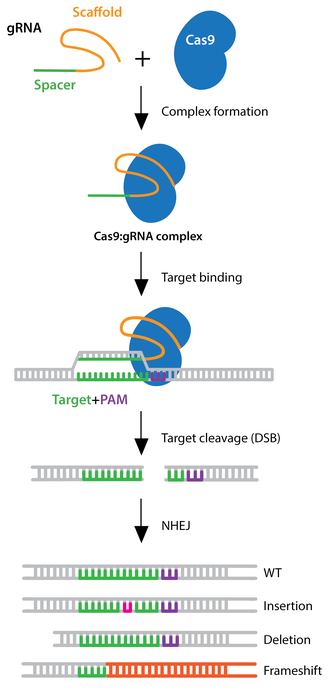
The CRISPR-Cas9 system consists of an enzyme (Cas9) and a guide RNA (sgRNA) that can locate a specific region of the genome and induce a double-stranded break in the DNA. Using DNA repair mechanisms, this can be exploited to delete or insert a new sequence. For instance, homology-directed repair (HDR) uses a DNA template, either natural or supplied by researchers, to rebuild the broken strand with that template’s sequence. This has been especially powerful for editing nuclear genomes, but it has been debated whether this method could be used for mtDNA as well [3].
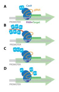
This is where Bi et al. enter the scene with their recent study [4]. Their goal was simple: to demonstrate that the mitochondrial genome can, in fact, be “CRISPR-ized.” The first step was to design a CRISPR-Cas9 system capable of entering the mitochondrion. To accomplish this, they took an existing system for nuclear editing and inserted “localization signals” that tell the cell where the protein needs to go. One of these was the mitochondrial targeting signal (MTS) derived from a nuclear-encoded gene, COX8A, that allows the protein to travel to the mitochondrion. Next, an sgRNA was designed that targets the ND4 gene of the mitochondrial genome. Combining these, Bi et al. created a system that could be tested in human cells. Indeed, with the use of fluorescent markers and microscopy, they demonstrated that both the Cas9 protein and sgRNA could enter the mitochondrion (Figure 1).
Next, the researchers needed to determine whether this system could edit mtDNA once inside the organelle. In addressing this, they added the CRISPR-Cas9 system alongside a template strand (called an ssODN) that contained an inserted sequence, GAATTC, that they could screen for (Figure 1). Once the cells were treated, they confirmed the insertion through two methods: PCR and direct sequencing. The PCR method used a primer that could only bind to the inserted sequence, so if the mtDNA was amplified, that indicated a successful “knockin.” The sequencing method, “PCR-free 3rd generation sequencing,” provided additional confirmation without the risk of false-positives from PCR artifacts (Figure 1).

Finally, as a proof of concept, Bi et al. altered the sgRNA and template strand to insert a pathogenic mtDNA mutation (changing ACCTTGC to GCAAGGT on the ND1 gene). This is associated with mitochondrial myopathy (a disease that causes muscular problems), and if successfully inserted, it could provide researchers with a new cell that models this mutant. Indeed, PCR and sequencing confirmed its knockin, demonstrating the power of editing mtDNA.
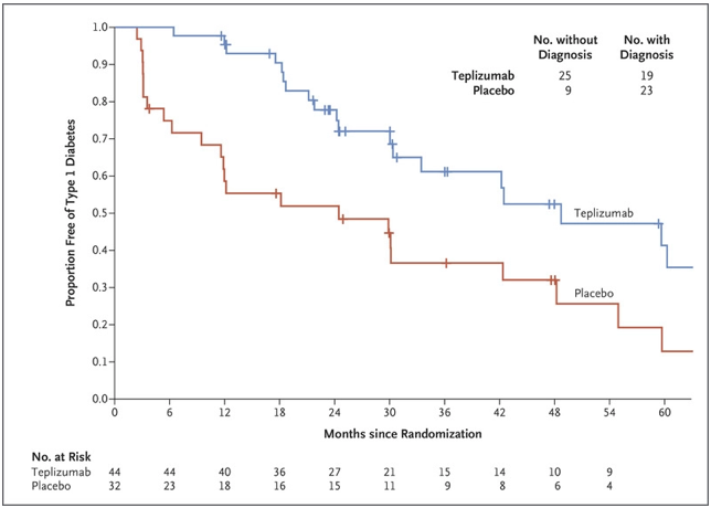
The authors of this paper directly confirmed that mtDNA could be edited via CRISPR-Cas9 for the first time. This is in contrast to previous studies which have shown mitochondrial localization of the Cas9-sgRNA complex, but with no clear indication of knockin because of PCR-based artifacts or indirect screening [5]. However, the present study is not without its own limitations, particularly in the method’s feasibility at a clinical level. First and foremost, an individual cell has many more copies of mtDNA than nuclear DNA, so editing every strand is more difficult. Indeed, the “knockin efficiency,” or proportion of DNA with the GAATTC insertion, was only 0.03-0.05% (compared to the 20-30% seen in some nuclear genome editing studies) [6]. Furthermore, Bi et al. reported an increase of reactive oxygen species (ROS) formation upon the addition of CRISPR-Cas9, a class of reactive molecules that can degrade DNA and induce cell death.
Work remains to be done, whether it is through improving the efficacy of the CRISPR-CAS9 system or confirming the method’s safety amidst ROS production. Nonetheless, Bi et al. give us a powerful starting point for treating mitochondrial dysfunction in ways never previously thought possible. In the coming years, CRISPR-izing mtDNA may even become a standard technique, just as we saw with nuclear DNA in the last decade.
References:
- Falkenberg M, Larsson NG, Gustafsson CM. (2007). DNA replication and transcription in mammalian mitochondria. Annu Rev Biochem, 76: 679-99. https://doi.org/10.1146/annurev.biochem.76.060305.152028.
- Stenton SL, Prokisch H. (2020). Genetics of mitochondrial diseases: Identifying mutations to help diagnosis. EBioMedicine, 56: 102784. https://doi.org/10.1016/j.ebiom.2020.102784.
- Gammage PA, Moraes CT, Minczuk M. (2018). Mitochondrial Genome Engineering: The Revolution May Not Be CRISPR-Ized. Trends Genet, 34(2): 101-110. https://doi.org/10.1016/j.tig.2017.11.001.
- Bi R et al. (2022). Direct evidence of CRISPR-Cas9-mediated mitochondrial genome editing. Innovation (Camb), 3(6): 100329. https://doi.org/10.1016/j.xinn.2022.100329.
- Antón Z et al. (2020). Mitochondrial import, health and mtDNA copy number variability seen when using type II and type V CRISPR effectors. J Cell Sci, 133(18): jcs248468. https://doi.org/10.1242/jcs.248468.
- Zhang JP et al. (2017). Efficient precise knockin with a double cut HDR donor after CRISPR/Cas9-mediated double-stranded DNA cleavage. Genome Biol, 18(1): 35. https://doi.org/10.1186/s13059-017-1164-8.


 There are millions of women taking steroids every day. But how is this possible? Are they just getting really buff? It feels like we always hear stories about how performance-enhancing drugs, namely steroids, are giving world-class athletes the boost they need to beat out their competition. But women across the globe are taking steroids every day as well, in the form of hormonal birth control. Despite their widespread use, side effects of hormonal contraceptives are largely unstudied, or have been until recently. In the last ten years, several studies have come out about the effect of taking a daily dose of steroids on women’s brains and mental health, which until now has been a severely neglected area where lack of knowledge affects millions of people worldwide.
  Thes
Thes

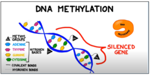 Figure 1: Methyl groups attached to cytosine bases in a gene block the enzyme RNA polymerase from binding to the promoter region of a gene, preventing transcription. Adapted from BOGOBiology (2017)