With a long history dating back nearly 700 years, Tarot cards have maintained a presence in society as a tool that is considered to predict the future and understand one’s inner issues, desires, and motivations. There are many conflicting theories regarding the origin of Tarot cards, with the predominant notion pointing to 14th century northern Italy (Tarot Heritage). Researchers claim that the major arcana of Tarot is based on the Egyptian hieroglyphic book of Thoth (the Egyptian god of wisdom), which is also known as the book of Tarot (Willis 1988). But why do people still use Tarot cards, and what do we get out of using them? The phenomenon surrounding the use and interpretation of Tarot cards can be broken down into two juxtaposing explanations: paranormal and nonparanormal. The paranormal explanation claims that Tarot cards reveal hidden motives, portray opportunities, and offer a reflection of a person’s inner processes, allowing the cards to provide clarity regarding a person’s questions or conflicts. Meanwhile, the nonparanormal explanation claims that the entire phenomenon of Tarot cards can be explained by examining two simple psychological effects: The Barnum effect and “cold reading” (Ivtzan 2007). Additionally, several modern therapeutic approaches have employed the use of Tarot cards as a tool for self-reflection, with Tarot card readings offering clients a sense of order and control in their own lives (Hofer 2009). There are several different reasons for why people use Tarot cards, and the associated applications of the cards can help to improve a person’s mental health when the cards are utilized in a therapeutic context (Hofer 2009).
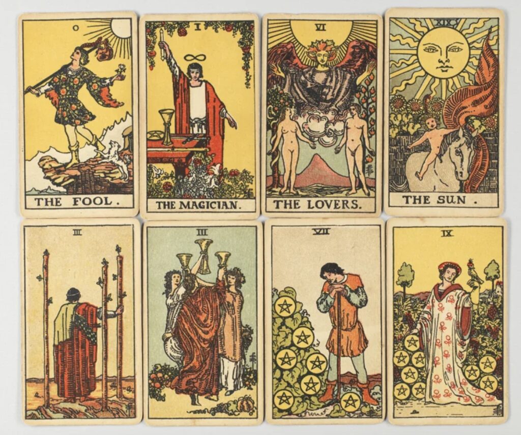
Many standard Tarot decks follow the same 78-card structure, which is divided into the minor arcana (56 cards), and the major arcana (22 cards). The cards in the major arcana represent the main themes of human life, such as love, death, spirituality, acceptance, etc. The cards in the minor arcana represent subtle mysteries of life, and are considered to be lesser compared to the major arcana (Ivtzan 2007). Additionally, there are several different techniques for choosing the cards in a reading, with the most popular option being for the reader to ask the client to shuffle the cards while focusing on a question, spread the deck, and choose the cards that they feel the most drawn to (Ivtzan 2007). The use of Tarot cards has continued to flourish, even in western societies, and the popularity of Tarot cards is not an indication of reliability or validity, but rather a look into how using the cards can influence our thought processes and mental state.

Figure 1: The major arcana of Tarot (Medium).
The paranormal explanation surrounding the phenomenon of Tarot cards is the approach that is acclaimed by occultists who believe that the cards reveal information about the quality of a moment for an individual (Ivtzan 2007). They do not believe that the cards predict the future as if it is fixed, but rather reveal information and potential circumstances about changeable events. By creating more awareness about the meaning of a specific moment for a client, this can help to provide the client with important insights, as well as a drive to take control of their own life and make changes that will be beneficial to them in the long run. Comparatively, the nonparanormal explanation examines the use of Tarot cards through the lens of psychological effects, with the Barnum effect being the most emphasized. The Barnum effect is the tendency to believe that vague predictions or general personality descriptions, such as those offered by Tarot or astrology, have specific applications to one’s unique circumstances (American Psychological Association). A Tarot reader may make general, trivial statements that could apply to anyone, and a client, eager to seek guiding information about their life, will accept these statements as truth. The major arcana of Tarot deals with themes that concern every individual’s life, so it is not difficult to come up with general statements about these themes that any person could be susceptible to (Ivtzan 2007). The other psychological effect that the nonparanormal explanation examines is “cold reading,” which is a set of deceptive psychological techniques that give a client the impression that a reader has paranormal abilities. The Barnum effect falls under the umbrella of “cold reading,” and the techniques behind “cold reading” involve the use of sharp observational skills and a good memory when examining a client. Cues such as a client’s clothing, physical characteristics, and manner of speech can reveal a lot of valuable information to a reader, that a reader can then use to inform the statements that they make to a client regarding the topic of their reading (Ivtzan 2007).
Although there are underlying psychological influences behind the use of Tarot cards, Tarot card readings can still have beneficial effects on a person’s mental health when used in a therapeutic context. A 2009 study investigated how regular users of Tarot cards employed the cards as a tool for self-reflection (rather than for divination). The study involved conducting interviews with several co-researchers who used Tarot cards regularly and in a self-reflective manner, and the interviews from the study were transcribed, with the common themes and qualities that existed between the interviews being extracted (Hofer 2009). Overall, the results of the study found that the co-researchers used Tarot cards as a way to gain insight into their current life situations. The cards were found to be used the most often during difficult times where they could offer a source of comfort. This source of comfort involved providing confirmation that everything was okay and that life had a sense of order.
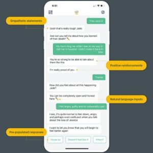
On top of this, Tarot cards were also used as a tool for positive reinforcement, where cards were drawn both intentionally and randomly to provide insights about what the co-researchers were seeking in their own lives. With a goal in mind, some of the co-researchers drew a card and then kept it with them until what they were working on or towards had been resolved. They claim that Tarot does not reveal new information to them, but that the use of Tarot cards can help to provide a new perspective on an issue that can influence a plan for a possible course of action (Hofer 2009).
By examining how therapeutic techniques involving Tarot have been successful for co-researchers who have consistently employed these techniques in their own lives, this study outlines how Tarot has the potential to be used as an effective therapeutic tool. Despite the foundational psychological effects behind the mainstream use of Tarot, Tarot cards can still have beneficial impacts on a person’s mental health and inner psychological processes. Further research surrounding the beneficial impacts of Tarot in a therapeutic setting would involve examining a greater number of participants from a wider variety of backgrounds, so that this research could be generalized to a larger audience. Regardless of the reasoning behind why a person may use Tarot cards, there is no doubt that Tarot cards have maintained a strong presence in society, and these cards have the potential to do more than just “predict the future.”
Literature Cited
- APA Dictionary of Psychology. “APA Dictionary of Psychology.” Apa.org, 2014, dictionary.apa.org/barnum-effect.
- “History.” Tarot Heritage, 24 July 2011, tarot-heritage.com/history-4/. Accessed 13 Apr. 2024.
- Hofer, Gigi Michelle. “Tarot cards: an investigation of their benefit as a tool for self reflection.” University of Victoria PhD diss (2009).
- Ivtzan, Itai. “Tarot cards: a literature review and evaluation of psychic versus psychological explanations.” Journal of Parapsychology 71 (2007).
- Macsparrow, Mark. “Many Major Arcana Cards in a Reading Means Many Changes Ahead.” Medium, 12 May 2021, tarotreadings.medium.com/many-major-arcana-cards-in-a-reading-means-many-changes-ahead-516becf2faf5. Accessed 13 Apr. 2024.
- Willis, T. Magick and the tarot. Wellingborough, UK: Aquarian (1988).