• Skip to primary navigation
  • Skip to main content
  • Skip to primary sidebar
  • Skip to footer

Bowdoin Science Journal

  • Home
  • About
    • Our Mission
    • Our Staff
  • Sections
    • Biology
    • Chemistry and Biochemistry
    • Math and Physics
    • Computer Science and Technology
    • Environmental Science and EOS
    • Honors Projects
    • Psychology and Neuroscience
  • Contact Us
  • Fun Links
  • Subscribe

Genes

Smoke Signals: The Unexpected Long Term Effects of Smoking on the Immune System

April 30, 2024 by Divya Bhargava

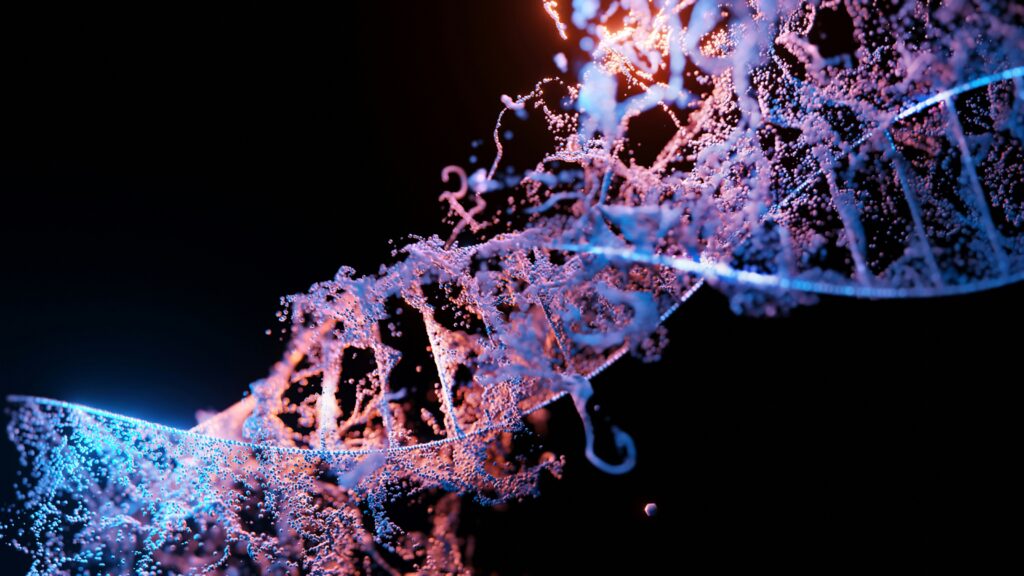
Image Source: “Smoking has a Lasting Impact on the Immune System, 2024”

When we get sick, our bodies’ immune systems work to fight off infections by invading pathogens, or organisms like bacteria and viruses that cause disease. However, many factors such as lack of sleep and poor nutrition weaken our immune system, meaning that we are less able to stay healthy. It has been known that smoking is another one of these factors that weaken our immune systems, but a recent study from a group at the Institut Pasteur in France looking at the effects of a variety of factors on the immune system showed that the extent to which smoking plays a role is much higher than many would think. But to understand the results of this study, it is important to first understand the mechanisms the immune system uses to fight infection. 

The immune system has many different moving components, including two distinct branches. The first is the faster, more general innate immune system which has a similar response to all infections. The second is the adaptive immune system which is slower, memory-based, and is involved in pathogen specific response. Although the innate immune system involves general molecules that interact with all cells and the adaptive immune system has specialized molecules that interact with pathogens based on memory of past infection, they share one important class of signaling molecules. These molecules are called cytokines and their role is to coordinate both of these types of immune response. Cytokines are small molecules that are released by immune cells to communicate with other parts of the body and each other. This signaling results in deployment of a response by other immune cells against invading pathogens. However, levels of cytokine production exist in a very fine balance. In order to get the desired immune response, you need the exact right level of cytokines present. If levels are too high or too low, they could cause abnormalities including overactive immune response and inflammation or impaired immune responses. 

To investigate the effects of a variety of different factors on the immune system and cytokine responses of healthy individuals, a project called the Milieu Intérieur put together a cohort of 1000 healthy participants and has been studying variability in the immune system between these individuals (“The Milieu Intérieur Project”). In an investigation of this data, the group from Institut Pasteur, Saint André et al, analyzed 136 variables measured in the Milieu Intérieur Project that could be causing differences in cytokine secretion and immune response (Luo and Stent 2024). These variables included everything from demographics, to diet, to health habits like smoking, to social and environmental characteristics (Saint-André et al. 2024).

After they performed their initial statistical analysis, Saint André et al measured production of 13 disease relevant cytokines as a quantitative measure of immune response in populations with different demographics, health habits, and other characteristics. In the lab, they exposed blood samples from their sample population to 12 different molecules meant to serve as stimulants for the immune system (these molecules included things like viral and bacterial proteins). After this exposure, the authors tested cytokine production in both innate and adaptive immune cells, and once they had that data, they took their results one step further. The group also used epigenetics, or the study of changes in gene expression rather than the DNA code that makes up the genome to investigate possible reasons for variability in immune responses associated with factors tested. Their epigenetic evaluation consisted of analyzing the extent to which one epigenetic process, DNA methylation, occurred at specific regulators of signaling and metabolism (Saint-André et al. 2024) to assess changes associated with smoking. 

As previously stated, one of the authors’ main findings from the initial statistical analysis was that smoking had a large effect on cytokine response. In fact it had the same effect as age, sex, and genetics, three things many would consider much more directly impactful to the immune system than smoking. In their in vitro simulations, they found that smoking had a temporary effect on the ability of the innate immune system to function properly. This result is a relatively intuitive one. If you do something that is considered bad for you, it makes sense that you would get sick more easily. 

However, more surprisingly, they also found that smoking leaves a lasting effect on memory based adaptive immune responses even after cessation of smoking, meaning that even after people quit smoking, their immune systems still are impacted. They found that in samples from individuals who smoked there were higher levels of cytokine expression, especially of an inflammatory cytokine called CXCL5 that is secreted in response to bacterial infection. Secretion of this cytokine is associated with the presence of an inflammatory protein called CEACAM6 in the blood. Consistent upregulation of levels of this protein has been found to have links with multiple cancers such as colon cancer (Wu et al. 2024). In Saint André et al’s epigenetic investigation of this association, they found that DNA methylation, which results in a downregulation of gene expression and in this case an increase in cytokine production, is linked to smoking’s lasting effect on the immune system (Greenberg and Bourc’his 2019). DNA methylation was decreased at many of the sites they tested which are involved in regulation of signaling genes and metabolism. Decreased DNA methylation was likely impacting levels of cytokines in response to detection of pathogens. In these populations, smoking caused lasting changes in gene expression which resulted in long term changes in addition to the expected short term effects on the immune system. 

This study demonstrates that smoking can have lasting negative impacts on your health which are not limited to just lung damage. It is also associated with pro-inflammatory cancer pathways and epigenetic markers that cause increased cytokine production. This overproduction of cytokines can confuse cells and also cause increased inflammation. Over time the extra inflammation can damage tissues and lead to developments of other conditions, like the cancers previously mentioned and complications associated with overproduction of cytokines (“What are Cytokines”). These recent findings emphasize that it is important to consider the possible implications of smoking and all things that we expose ourselves to, and to keep in mind that new data is still being discovered.

Works Cited

The Milieu Intérieur Project Institut Pasteur. Luo,Y. and Stent,S. (2024) Smoking’s lasting effect on the immune system. Nature, 626,  724–725.

Saint-André,V., Charbit,B., Biton,A., Rouilly,V., Possémé,C., Bertrand,A., Rotival,M., Bergstedt,J., Patin,E., Albert,M.L., et al. (2024) Smoking changes adaptive immunity with persistent effects. Nature, 626, 827–835.

Wu,G., Wang,D., Xiong,F., Wang,Q., Liu,W., Chen,J. and Chen,Y. (2024) The emerging roles of CEACAM6 in human cancer (Review). International Journal of Oncology, 64, 1–15.

Greenberg,M.V.C. and Bourc’his,D. (2019) The diverse roles of DNA methylation in mammalian development and disease. Nat Rev Mol Cell Biol, 20, 590–607.

What are Cytokines? Types and Function Cleveland Clinic.

Smoking has a lasting impact on the immune system, a new study finds (2024) Euronews.

Filed Under: Biology Tagged With: Biology, Genes, Medicine

The Solution to Alzheimer’s May Lie in Depression

April 21, 2024 by Nicholas Enbar-Salo '27

Despite being discovered by Alois Alzheimer almost 120 years ago, Alzheimer’s Disease (AD) still remains incurable (Hippius & Neundörfer, 2003). AD causes the brain to break down over time, which is also known as neurodegeneration. AD begins by deterioration of the hippocampus, which is the part of the brain responsible for memory and emotion. It then slowly spreads to other parts of the brain, eventually breaking apart the brain stem, which is responsible for involuntary movements such as breathing and swallowing (Lee et al., 2015). Given that around 39 million people have Alzheimer’s worldwide and that this disease has a 100% fatality rate, scientists across the world have tried to find a cure to this ravaging disease (World Health Organization, 2023). While there is yet to be a cure, recent developments by Stephanie Langella could help with mitigating one of the earliest signs of Alzheimer’s: depressive symptoms.

In this study, Langella and her team studied Presenilin-1 (PSEN1) gene mutations, a major cause of early-onset Alzheimer’s Disease. The PSEN1 gene provides instructions for making the presenilin-1 protein. This protein is an essential part of a protein complex known as gamma-secretase. This complex cleaves toxic proteins such as the amyloid precursor protein (APP) to create nontoxic proteins. When the PSEN1 gene mutates, gamma-secretase struggles to form and break down these toxic proteins, causing APP molecules to join together to create amyloid-beta (also known as amyloid-ꞵ or A-ꞵ), the protein responsible for the neurodegeneration seen in Alzheimer’s Disease (Bagaria et al., 2022). 

A figure of gamma-secretase, APP processing, and generation of Amyloid-β (Aβ). Cleavages of C99 by gamma secretase (ε/ζ/γ) release sAPPβ, a type of APP which is beneficial to cells. When the PSEN1 gene mutates, gamma secretase (γ) produces AICD, a harmful type of APP, into a cell’s liquid (cytosol) and amyloid-β 37-43 into cell organelles. Aβ42 is the form of amyloid-β responsible for neurodegeneration in AD patients. (Steiner et al., 2018).

In particular, they studied its relationship to the neurodegeneration of the hippocampus and depressive symptoms (Langella et al., 2023). They began by creating two groups:  the first group consisted of carriers of the PSEN1 mutation but that had not yet been diagnosed with AD, and the second group consisted of the family members of the respective PSEN1 carriers that did not have the mutation and were not diagnosed with Alzheimer’s. Then, two structural MRIs – a method of neuroimaging which models the brain structures of a patient– with a one-year gap in between the two images were taken of the participants’ hippocampuses to measure the change in the volume of the hippocampus over a year. Participants also took the Geriatric Depression Scale, a 15-item survey that measures depressive symptoms, such as the subjects’ feelings of hopelessness and rating their interest in hobbies, to measure depressive symptoms over one year. 

Once the study was concluded, Langella found that there was no significant difference in the severity of the depressive symptoms between those carrying the PSEN1 mutation and those that did not. However, within the group carrying the PSEN1 mutation, those with smaller hippocampal volumes experienced more depressive symptoms. This association remained even after accounting for the age differences in the participants. This same association was not present in the non-PSEN1 carriers (Langella et al., 2023). Since the volume of the hippocampus did not have any relationship with depressive symptoms with non-PSEN1 carriers, there is likely some relationship between Alzheimer’s and depressive symptoms caused by hippocampal neurodegeneration. 

A).  Structural MRI of the hippocampus from the back of the head (shown in yellow)

C). Top-down structural MRI of the hippocampus (shown in yellow)  (Sato et al., 2021)

There are several important implications of this research. To start, if there is indeed a relationship between the severity of depressive symptoms and the size of the hippocampus in someone with AD, there is a chance that trying to mitigate these depressive symptoms through therapy and antidepressant medication could slow down the deterioration of the hippocampus. By keeping the hippocampus intact for a longer time, people with AD could have better emotional control and memory later in life, which would greatly improve their quality-of-life (Langella et al., 2023). Also, since AD first deteriorates the hippocampus, it is possible that the onset of depressive symptoms in people with the PSEN1 mutation could be used as an indicator to doctors on the severity of the neurodegeneration. For instance, if someone with the PSEN1 gene mutation suddenly begins displaying depressive symptoms, it is possible that AD has just recently started decaying the hippocampus. Doctors can then try to intervene and slow the decay of the hippocampus through administering antidepressants and therapy, but also through encouraging lifestyle changes such as increased exercise. This way, those with Alzheimer’s can live a longer time before their hippocampus fully degrades, letting them keep their memories for a longer time. 

Since this is one of the first studies relating depression and hippocampal decay in people with PSEN1 mutations, there is no theorized mechanism behind why this relationship exists in people with the PSEN1 mutation but not in those without. However, Langella et al. did find a particularly strong association between hippocampal decay in those with the PSEN1 mutation and displaying apathy, one of the measured depressive symptoms in the study (2023). More research should be done on the potential role of certain depressive symptoms on hippocampal decay, along with more research on the neural underpinnings relating the PSEN1 mutation, depression symptoms, and hippocampal decay. There is some evidence linking the formation of amyloid-ꞵ to depression in late-life major depression, but further research into the mechanism underlying this relationship is required (Pomara et al., 2022). 

However, there is a pressing issue with this study; it had a fairly small sample size, with the PSEN1 carrier group having 27 participants and the non-PSEN1 group having 26. Since AD is a disease that affects everyone slightly differently, having such a small sample size makes the results unreliable and hard to generalize to everyone with AD. Regardless of the issues in the study, developments such as the ones created by this study serve to improve the quality of life and life expectancy of people with AD, which promises to improve the lives of almost 39 million people and their families. With every passing discovery into Alzheimer’s, scientists are also getting more information on the mechanisms behind the disease, which could eventually lead humanity to curing the disease altogether. 


Citations

Bagaria, J., Bagyinszky, E., & An, S. S. A. (2022). Genetics, Functions, and Clinical Impact of Presenilin-1 (PSEN1) Gene. International journal of molecular sciences, 23(18), 10970. https://doi.org/10.3390/ijms231810970


Hippius, H., & Neundörfer, G. (2003). The discovery of Alzheimer’s disease. Dialogues in clinical neuroscience, 5(1), 101–108. https://doi.org/10.31887/DCNS.2003.5.1/hhippius


Langella S,  Lopera F,  Baena A, et al.  Depressive symptoms and hippocampal volume in autosomal dominant Alzheimer’s disease. Alzheimer’s Dement.  14 Oct. 2023, 986–994. https://doi.org/10.1002/alz.13501


Lee, J. H., Ryan, J., Andreescu, C., Aizenstein, H., & Lim, H. K. (2015). Brainstem morphological changes in Alzheimer’s disease. Neuroreport, 26(7), 411–415. https://doi.org/10.1097/WNR.0000000000000362


Pomara, N., Bruno, D., Plaska, C.R. et al. Plasma Amyloid-β dynamics in late-life major depression: a longitudinal study. Transl Psychiatry 12, 301 (2022). https://doi.org/10.1038/s41398-022-02077-8


Sato, Jinya, et al. “Lower Hippocampal Volume in Patients with Schizophrenia and Bipolar  Disorder: A Quantitative MRI Study.” Journal of Personalized Medicine, vol. 11, no. 2, 13 Feb. 2021, p. 121, https://doi.org/10.3390/jpm11020121.


Steiner, H., Fukumori, A., Tagami, S., & Okochi, M. (2018, October 28). Making the final cut: Pathogenic amyloid-β peptide generation by γ-secretase. The Journal of Cellular Pathology. https://www.cell-stress.com/researcharticles/making-the-final-cut-pathogenic-amyloid-%ce%b2-peptide-generation-by-%ce%b3-secretase


World Health Organization. “Dementia.” Dementia, 2023, www.who.int/news-room/fact-sheets/detail/dementia.

Filed Under: Psychology and Neuroscience, Science Tagged With: Alzheimer's Disease, Depression, Genes

Your DNA Remembers: Correlating Epigenetics and Early Childhood Trauma through DNA Methylation

April 21, 2024 by Lia Scharnau

What’s your earliest memory? Can you remember everything that happened between the ages of three to five? No? Me neither. Despite the gaps in your memory, your body—specifically your DNA—knows what happened to you. It shouldn’t be surprising that DNA has such a good memory. After all, it stores genetic information tracing back thousands of years. This of course prompts several questions, namely the following; how does DNA store my memories and how does that affect me?

Let’s start from the beginning. Epigenetics encompasses all heritable changes in gene activity that do not stem from a change in DNA sequences (Moore et al, 2013). DNA methylation introduces a particularly salient new epigenetic mechanism for gene regulation and cell differentiation. The key to DNA methylation lies within the special enzymes that modify the cytosine DNA base by adding a methyl group (Moore et al, 2013; Suelves, 2016). Interestingly, cell differentiation is partly driven by differing levels of DNA methylation. An overall increase in DNA methylation occurs during the differentiation process, while a decrease in it at cell-specific loci helps define cellular identity (Suelves et al, 2016). Additionally, the progressive decrease in overall DNA methylation can contribute to physiological aging and the development of cancer (Suelves et al, 2016). Overall, these altered cytosines play key roles in human development and health issues (Moore et al, 2013).  

This brings us to the second question: how does my DNA’s photographic memory affect me? Well, it turns out that choices such as diet, drinking, exercise, illness, and environmental conditions can all have an impact on genomic stability and gene-specific DNA methylation (Lim et al, 2012). So while you might not remember what happened as a kid, your DNA may already be internalizing those experiences. The Avon Longitudinal Study of Parents and Children patiently waited for years so they could specifically study how childhood experiences affect epigenetic markers in adolescence. A cohort of 13,988 children with due dates between April 1991 and 1992 were monitored for exposure to childhood adversity from birth to the age of eleven. Using changes in DNA methylation at the age of fifteen, researchers investigated whether the timing of adversity has epigenetic consequences across childhood and adolescence (Lussier et al, 2023). 

Each mother reported whether their child faced any of the following seven types of childhood adversity; caregiver physical or emotional abuse, sexual or physical abuse, maternal psychopathology, one-adult households, family instability, and neighborhood disadvantage as well as the timing that the adversity was present. Out of the 13,988 children, 609-665 showed signs of both adversity and a decrease in DNA methylation at 15 years old (Lussier et al, 2023). 

Within this sample from the original cohort, further DNA analysis identified twenty-two loci that showed significant associations between exposure to adversity and altered DNA methylation at the age of 15 (Lussier et al, 2023). Of the loci identified to be associated with decreased DNA methylation, the highest percent of loci were correlated with growing up in one-adult households (Lussier et al, 2023). None of the identified loci indicated that adversity may alter DNA methylation at birth or the age of seven, but instead only emerged in adolescence (Lussier et al, 2023).  

Researchers concluded that the ages between three and five years old are when children are vulnerable to adversity and the consequences of this adversity may biologically embed itself and later manifest itself in adolescence (Lussier et al, 2023). Additionally, the adversity-associated decrease in DNA methylation is correlated to have effects on the central nervous system (Lussier et al, 2023). Of course, it’s great timing that the altered DNA methylation becomes an issue during puberty. Especially since it’s associated with poor self-esteem and increased depressive symptoms. 

What’s the upside? There has to be a silver lining or else this article is just a doomsday proclamation. The ability to finally trace health complications back to epigenetic mechanisms due to latent childhood trauma provides an important piece of the puzzle of understanding complex diseases. However, the puzzle is far from solved. The cohort in this study was predominantly of European descent and further research into how the complexities of race factor into DNA methylation and childhood adversity is the next step in this journey.

Literature Cited:

Lim , U., & Song, M. (2012). Dietary and Lifestyle Factors of DNA Methylation. In Cancer Epigenetics (Vol. 863, pp. 359–376). Humana Press.

 Lussier, A. A., Zhu, Y., Smith B. J., Cerutti J., Fisher J., Melton P. E., Wood N. M., Cohen-Woods S., Huang R., Mitchell C., et al. (2023). Association between the timing of childhood adversity and epigenetic patterns across childhood and adolescence: findings from the Avon Longitudinal Study of Parents and Children (ALSPAC) prospective cohort. Lancet Child Adolesc Health. 7(8), 532-43. https://doi.org/10.1016/S2352-4642(23)00127-X

Moore, L. D., Le, T., & Fan, G. (2013). DNA methylation and its basic function. Neuropsychopharmacology, 38(1), 23–38. https://doi.org/10.1038/npp.2012.112

Suelves, M., Carrió, E., Núñez-Álvarez, Y., & Peinado, M. A. (2016). DNA methylation dynamics in cellular commitment and differentiation. Briefings in Functional Genomics, elw017. https://doi.org/10.1093/bfgp/elw017

Filed Under: Biology Tagged With: epigenetics, Genes, Trauma

Unpacking the Ethical Implications of Human Germline Editing

December 3, 2023 by Kennedy Kirkland

On December 30th, 2019, biophysicist Dr. He Jiankui was sentenced to three years in prison for forging ethical review documents and misleading doctors into unknowingly implanting gene-edited embryos into two women in China (Normile 2019). One of these women had a set of twins and the other had a single child. These children are now the first genetically modified humans in history to be resistant to HIV. They can pass this modification to the next generation, and their whereabouts are still unknown (Greely 2019). Jiankui used CRISPR/Cas9, a gene editing tool, to modify their germlines and edit the CCR5 gene, a contributor to broad immune responses (his focus was its importance to HIV viruses).

Based on the National Natural Science Foundation of China, there has been a total of 3.7 billion yuan (roughly $576 million) government spending on embryonic stem cell research from 1997 to 2019 (Lou 2021). Germline gene editing research is allowed, but establishing a pregnancy with genetically modified embryos has been outlawed by multiple regulations (Xinqing 2014). Similarly to China, the National Institutes of Health within the US estimates that human embryonic stem cell research has received $1.48 billion in government funding since 2009. There are no current laws or regulations that ban germline gene editing conducted through private funding within the US, but it would have to be approved by the FDA for marketing and clinical studies. No proposals have been submitted (​​Genetic Literacy Project 2019). Dr. Jiankui has since been disgraced by the scientific community around the world for his actions. Some scientists believe that germline gene editing is not only an unethical practice but a potentially dangerous one that could lead to a new era of eugenics with irreversible harm (Genetic Literacy Project 2019). Little is known about the intricate details of Dr. Jiankui’s experiment, but what has been revealed poses several hard-hitting ethical questions. Should we manipulate the next generation of humans? What are the ethical dilemmas in the advancement of this technology? To create an informed opinion on the matter, it is helpful to first understand the intricacies of Dr. Jiankui’s experiment, and how exactly he made the first genetically modified children.

Dr. Jiankui conducted human germline genome editing, which refers to the technique of modifying not only the genetic information of a subject but also what can be passed down to the next generation (Normile 2019). Germ cells can create a new generation (ex. sperm and eggs). So germ cells, and cells that produce germ cells, are known as the germline. These are different from somatic cells, which are body cells that continuously divide throughout a person’s lifetime and play a minimal role in gene inheritance (Greely 2019). Certain alleles or alterations to CCR5 within the human genome can provide resistance to HIV. Thus, Jiankui used CRISPR to alter CCR5 in HIV-susceptible patients to make them resistant to the disease.

CRISPR, which stands for clustered regularly interspaced short palindromic repeats, is a technique originally found in bacteria as a defense system to render inserted viral DNA ineffective. It also allows for the modification of DNA (Rapini 2023). The enzyme Cas-9 can cut pieces of DNA and is guided to its location based on an attached strand of RNA (called the guide RNA strand). By cutting a targeted sequence of DNA, it can render protein-coding genes inactive or disfunctional. After Cas-9 has cut the target DNA, researchers can choose to place a modified DNA sequence in the vacant space (Fig 1).

Figure 1. The editing technique of CRISPR involves the guide RNA strand, guide RNA sequence, Cas-9 enzyme, and target DNA (Roach 2015).

While little is known about Dr. Jiankui’s specific technique in using the CRISPR/Cas-9 system, we know that he modified the gene CCR5 (Greely 2019). CCR5 is a gene on chromosome 3 that encodes for a protein called C-C chemokine receptor type 5 (also shortened to CCR5) (Normile 2019). CCR5, along with another receptor called CD4, is utilized by the HIV virus to bind to bacteria-detecting/destruction cells called macrophages (white blood cells) and infect them. The HIV virus protein envelope binds to the primary receptor CD4 on the macrophage with the gp120 protein, which are exterior “protective” protein around the HIV cell. If the CCR5 coreceptor is present as well, it can successfully enter the macrophage, release viral RNA and enzymes, and infect the macrophage. This infection alters the function of the macrophage, causing it to assemble and release viruses. Eventually, the gene that codes for the gp120 receptor is altered by a mutation and can now bind to a different co-receptor called CXCR4 which is found on the CD4 plus T-cells. (Prakash 2019). The same process of virus construction occurs within T-cells, but as the viruses leave the T-cell it ruptures the plasma membrane. This kills the T-cells, causing a weakened immune response and eventually resulting in the onset of AIDS.

Figure 2. The mechanism by which the HIV viral envelope infects the macrophage creates mutated viral cells with different gp120 receptors and destroys T-cells. (Prakash 2019) 

It is believed that Dr. Jiankui used the CRISPR/Cas-9 system to delete 32 base pairs of the CCR5 gene, therefore making it produce non-functional copies of the CCR5 protein. This was done in the hope that HIV would be unable to infect the white blood cells of the babies born from the embryos (Normile 2019). If Jiankui only modified somatic cells using this technique, the children may or may not have gained protection against HIV and could not pass it down to the next generation.

In 2015 (before He’s experiment was released) Jennifer Doudna, one of two researchers who published their findings on CRISPR, convened with the U.C Berkeley’s Institute for Genome Innovation, which was composed of Nobel laureates and esteemed professors of bioethics. They concluded that conducting germline editing would be irresponsible until matters such as balancing the potential risks and benefits have been concluded and that there is a broad societal consensus about the appropriate use of proposed applications of this technology (Cohan, 2018). Jennifer Doudna and Emmanuelle Charpentier created this technology to find ways to edit out faulty genes and cure diseases at the source—within our genetic code (Doudna 2019). However, after the release of He’s experiment, Doudna publicly stated her evolving concerns relating to this application of her discovery. During her national press tour in 2019, she shared a frightening dream she had in which a colleague of her’s asked about CRISPR but later turned out to be Hitler (Than 2019). This likely stems from the horrifying history of Nazis and the eugenics movement, which stemmed from the idea of improving the genetic quality of humans and sterilizing and or eliminating those with unfavorable traits. She also voiced that her opinion on germline genome editing has been evolving from an outright ban to warranting it in certain circumstances. However, her underlying opinion remains that it would have to be under transparent and safe circumstances and concern a medical need that was unmet by any technology. In addition to Doudna, countless other scientists have come out in disagreement with He Jianku’s work including researchers from Stanford University, Harvard Medical School, and the NIH (Cohen 2018). Specifically, a journalist from Science magazine states that representatives from eight countries who attended the International Summit on Human Genome Editing in Hong Kong came to a consensus that his actions were irresponsible, violated international norms, lacked transparency, and did not have sufficient medical justification (Cohen 2018).

Jennifer Doudna stated that her goal was to edit out faulty genes, but that definition leaves a lot to interpretation. How can we as a society come together to define the parameters of a faulty gene? From treating HIV to Down Syndrome, these conditions hold more than just health implications, but social ones as well. If the scientific community deems it accessible to prevent Down syndrome using human germline genome editing, are they stating that humans that currently possess this condition are “wrong” and have “faulty genes” in need of correction? Whoever holds the power to manipulate this technology essentially has the power to decide what the next generation of humans will be capable of and what genes are deemed “faulty” within our society. Jianku’s reckless use of technology was majorly deemed wrong for its lack of transparency, safety, and following set regulations upheld by currency bioethical standards. However, in situations like parents wanting their children to be healthy, it is completely understandable that human germline editing sounds like an intriguing way to protect their children. For these reasons, it is imperative to familiarize ourselves with the development of this technology, and its continued push for regulations across the world to maintain ethical standards. Further, it is also important to ask ourselves hard-hitting questions such as: should one person, scientist, or government have the power to determine what the next generation will possess, look like, or become? We as members of society must continue to stay informed in a world with the capacity to genetically modify human beings.

References:

  1. Bhanu Prakash. “How the HIV Infection Cycle Works.” Proceum Pvt. Ltd. January 2019. https://www.youtube.com/watch?v=GyofqO1TRjU
  2. Brianna Rapini, Sarina Peterson, “Genetic Engineering.” September 2023. https://www.youtube.com/watch?v=CDw4WPng2iE
  3. Dennis Normile. “Chinese scientist who produced genetically altered babies sentenced to 3 years in jail.” Science. December 2019.https://www.science.org/content/article/chinese- scientist-who-produced-genetically-altered-babies-sentenced-3-years-jail
  4. Deng Luo, Zihui Xu, Zhongjing Wang, Wenzhuo Ran. “China’s Stem Cell Research and Knowledge Levels of Medical Practitioners and Students.” Stem Cells International. 2021. https://doi.org/10.1155/2021/6667743.
  5. Doudna J. 2019. Faculty Research Page. Department of Molecular & Cell Biology. http://mcb.berkeley.edu/faculty/BMB/doudnaj.html.
  6. Dylan Roach, Tanya Lewis. “CRISPR, the gene-editing tech that’s making headlines, explained in one graphic.” Insider. December 2015. https://www.businessinsider.com /crispr-gene-editing-explained-2015-12
  7. Genetic Literacy Project. 2019 July 23. United States: Germline / Embryonic. Global Gene Editing Regulation Tracker. https://crispr-gene-editing-regs-tracker.geneticliteracy project.org/united-states-embryonic-germline-gene-editing/.
  8. Henry T Greely. “CRISPR’d babies: human germline genome editing in the ‘He Jiankui affair.’” Journal of Law and the Biosciences. Volume 6, Issue 1. October 2019, Pages 111–183, https://doi.org/10.1093/jlb/lsz010
  9. Indra Mani. “CRISPR-Cas9 for treating hereditary diseases.” Progress in molecular biology and translational science. Volume 181. February 2017. https://pubmed.ncbi.nlm.nih.gov/34127193/
  10. John Cohen. “After last week’s shock, scientists scramble to prevent more gene-edited babies.” Science. December 2018.https://www.science.org/content/article/after-last- weeks-shock-scientists-scramble-prevet-more-gene-edited-babies
    Join David Ignatius. “Walter Isaacson & Jennifer Doudna join Washington Post Live to discuss CRISPR.” Washington Post. March 2021. https://www.youtube.com/ watch?v=cKHuuALENZk
  11. Than K. 2019 Nov 12. AI and gene-editing pioneers to discuss ethics. Stanford News. [accessed 2023 Dec 3]. https://news.stanford.edu/2019/11/12/ai-gene-editing-pioneers- discuss-ethics/#:~:text=Doudna%20decried%20the%20act%20but.
  12. Yi Zheng et al. “Structure of CC Chemokine Receptor 5 with a Potent Chemokine Antagonist Reveals Mechanisms of Chemokine Recognition and Molecular Mimicry by HIV.” Immunity. Volume 46, Issue 6. June 2017. https://www.cell.com/immunity /pdf/S1074-7613(17)30218-2.pdf
  13. ‌Zhang Xinqing, Zhang Wenxia, Zhao Yandong. “The Chinese Ethical Review System and its Compliance Mechanisms.” TRUST. September 2014. https://trust-project.eu/ wp-content/uploads/2016/03/Chinese-Ethics-Review-System.pdf

Filed Under: Biology, Chemistry and Biochemistry, Science Tagged With: Bioethics, Genes, HIV

Primary Sidebar

CATEGORY CLOUD

Biology Chemistry and Biochemistry Computer Science and Tech Environmental Science and EOS Honors Projects Math and Physics Psychology and Neuroscience Science

RECENT POSTS

  • Biological ChatGPT: Rewriting Life With Evo 2 May 4, 2025
  • Unsupervised Thematic Clustering for Genre Classification in Literary Texts May 4, 2025
  • Motor Brain-Computer Interface Reanimates Paralyzed Hand May 4, 2025

FOLLOW US

  • Facebook
  • Twitter

Footer

TAGS

AI AI ethics Alzheimer's Disease antibiotics artificial intelligence bacteria Bathymetry Beavers Biology brain Cancer Biology Cell Biology Chemistry and Biochemistry Chlorofluorocarbons climate change Computer Science and Tech CRISPR Cytoskeleton Depression dreams epigenetics Ethics Genes honors Luis Vidali Marine Biology Marine Mammals Marine noise Medicine memory Montreal Protocol Moss neurobiology neuroscience Nutrients Ozone hole Plants Psychology and Neuroscience REM seabirds sleep student superintelligence Technology therapy

Copyright © 2025 · students.bowdoin.edu