• Skip to primary navigation
  • Skip to main content
  • Skip to primary sidebar
  • Skip to footer

Bowdoin Science Journal

  • Home
  • About
    • Our Mission
    • Our Staff
  • Sections
    • Biology
    • Chemistry and Biochemistry
    • Math and Physics
    • Computer Science and Technology
    • Environmental Science and EOS
    • Honors Projects
    • Psychology and Neuroscience
  • Contact Us
  • Fun Links
  • Subscribe

Biology

Microplastic burden in marine benthic invertebrates depends on feeding strategies

May 8, 2024 by Cindy Dai '27

Microplastic pollution is a global issue effectively impacting all aquatic systems from the poles to tropical reefs. Current emission patterns project to around 35 – 98 metric tons of annual microplastic emission by 2030. Yet, this may only be an underestimation, as our current understanding of microplastic concentrations based on traditional sampling practices overlooks smaller debris (Lindeque et al. 2020, Borrelle et al. 2020). With this scale of rapid increase in concentrations, the implications of microplastic accumulation in marine systems have become an increasing concern. In response to this global concern, Adam Porter and his team looked towards the ocean’s floor to better understand how microplastics interact with dynamic ecosystems.

Microplastics emitted into the marine environment can adversely impact a wide range of processes from cellular metabolism to digestive functions, fertility, locomotion, and growth (Foley et al. 2018; Bour et al. 2018). Furthermore, bioaccumulation, or trophic transfer when contaminated prey is consumed by predators, magnifies microplastic burdens in organisms higher in the food chain. These above properties, in conjunction to the rapidly increasing environmental concentrations, highlight the pressing need to quantify how much microplastics marine organisms are ingesting.

Historically, our understanding of individual microplastic burdens has often assumed that levels of environmental contamination directly map onto their uptake by marine organisms. However, studies have found that this isn’t always the case. Other factors, such as feeding strategies and community composition, also impact a species’ uptake rate (Pagter et al. 2021; Bour at al. 2018). 

To bridge the mismatch of environmental concentration and individual burden, Porter et al. reviewed 412 studies on marine invertebrates from around the globe to investigate how different species traits could influence microplastic uptake. First, they gathered data from each study and assigned a geographic sector to each sampling site. Next, they evaluated each observation for a variety of variables, including feeding mode, position within the sediment, and wet weight (mass) of the individual. Then, Porter’s team used statistical tests to examine the potential influence each parameter had on plastic uptake with statistical analyses tests and visualized their findings. 

Geographically, the Pacific Northwest, Yellow Sea and Japan Trench, had the highest mean individual microplastic burden. In terms of animal class, the highest mean burden occurred in the Malacostraca class. Malacostraca encompasses common commercial species such as crabs and lobsters, which could have commercial implications on industries like lobster fishing and aquaculture. 

Of all the outlined parameters, feeding strategies had the greatest impact on microplastic uptake. Omnivores were shown to have the highest rate of uptake, followed by predators, herbivores, grazers, suspension feeders, deposit feeders, and lastly scavengers. These findings support the bioaccumulation theory, one of several hypotheses concerning microplastic uptake patterns (Wang 2014). According to the bioaccumulation theory, microplastics enter the food web through primary consumers like suspension feeders, grazers, and filter feeders. The plastic they retain in their systems will then be ingested by higher trophic levels like secondary and tertiary consumers that are omnivores,predators, and scavengers. Accordingly, the microplastic burdens would be highest in predators and omnivores, which matches the study’s findings.

In addition to the quantity of microplastics retained, feeding patterns were also found to influence the size and type of microplastics consumed were also different across groups. The most reported shape was fibers. The mean sizes of these fragments ranged from 0.2 micrometers to 17 centimeters, and herbivores in general retained the largest particles, but the precise mechanisms driving these patterns remain unclear.  

These findings precisely highlight our gap in knowledge of microplastic distribution amongst marine communities. As Porter et al. highlights, a holistic consideration of subtle processes related to feeding patterns is essential in fine tuning our understanding of how our world is changing. Thus, although the study describes general trends on a global scale, future research focusing on regional subtleties is important. Subsequently, applying these findings as policy is crucial, as many marine organisms are frequently consumed commercial species. Being major consumers of seafood, the microplastic accumulation in marine animals can directly impact humans. This is particularly concerning in context of our status as the apex predator, and therefore the final stop in the chain of bioaccumulation. As the microplastic burden in marine organisms is rising at an alarming pace, the need for action is more urgent than ever.


Works Cited

Borrelle, S. B., Ringma, J., Law, K. L., Monnahan, C. C., Lebreton, L., McGivern, A., Murphy, E., Jambeck, J., Leonard, G. H., Hilleary, M. A., Eriksen, M., Possingham, H. P., De Frond, H., Gerber, L. R., Polidoro, B., Tahir, A., Bernard, M., Mallos, N., Barnes, M., & Rochman, C. M. (2020). Predicted growth in plastic waste exceeds efforts to mitigate plastic pollution. Science, 369(6510), 1515–1518. https://doi.org/10.1126/science.aba3656 

Bour, Agathe, Carlo Giacomo Avio, Stefania Gorbi, Francesco Regoli, and Ketil Hylland. “Presence of Microplastics in Benthic and Epibenthic Organisms: Influence of Habitat, Feeding Mode and Trophic Level.” Environmental Pollution (Barking, Essex: 1987) 243, no. Pt B (December 2018): 1217–25. https://doi.org/10.1016/j.envpol.2018.09.115.   

Foley, Carolyn J., Zachary S. Feiner, Timothy D. Malinich, and Tomas O. Höök. “A Meta-Analysis of the Effects of Exposure to Microplastics on Fish and Aquatic Invertebrates.” The Science of the Total Environment 631–632 (August 1, 2018): 550–59. https://doi.org/10.1016/j.scitotenv.2018.03.046.   

Lindeque, P. K., Cole, M., Coppock, R. L., Lewis, C. N., Miller, R. Z., Watts, A. J. R., Wilson- McNeal, A., Wright, S. L., & Galloway, T. S. (2020). Arewe underestimating microplastic abundance in the marine environment? A comparison of microplastic capture with nets of different mesh-size. Environmental Pollution, 265, 114721. https://doi.org/10.1016/j.envpol.2020.114721

Pagter, Elena, Róisín Nash, João Frias, and Fiona Kavanagh. “Assessing Microplastic Distribution within Infaunal Benthic Communities in a Coastal Embayment.” Science of The Total Environment 791 (October 15, 2021): 148278. https://doi.org/10.1016/j.scitotenv.2021.148278. 

​​Porter, A., Godbold, J. A., Lewis, C. N., Savage, G., Solan, M., & Galloway, T. S. (2023). Microplastic burden in marine benthic invertebrates depends on species traits and feeding ecology within biogeographical provinces. Nature Communications, 14(1), 8023. https://doi.org/10.1038/s41467-023-43788-w 

Wang, W. -X. “Chapter 4 – Bioaccumulation and Biomonitoring.” In Marine Ecotoxicology, edited by Julián Blasco, Peter M. Chapman, Olivia Campana, and Miriam Hampel, 99–119. Academic Press, 2016. https://doi.org/10.1016/B978-0-12-803371-5.00004-7.

Filed Under: Biology, Environmental Science and EOS, Science

Engineered Nanoparticles Enable Selective Gene Therapy in Brain Tumors

May 8, 2024 by Sophie Nigrovic '24

Inborn protective mechanisms present challenges for therapies targeting cancers of the brain. Engineered nanoparticles permit the selective delivery of CRISPR-Cas9 to glioblastoma tumors.

Glioblastoma accounts for almost half of all cancerous tumors originating in the brain.1 Even with maximum safe treatment, the median survival period for patients is less than 1.5 years.2 However, survival varies widely by age, from a 0.9% 5-year survival rate for patients over 75 years old to an 18.2% 5-year survival rate for patient 0-19 years old.3 Nevertheless, the overall 5-year survival rate remains low at 5%2 and novel therapies are urgently needed for glioblastoma treatment. Zou et al. present a highly specific CRISPR-Cas9-based gene therapy for glioblastoma.4

Gene therapies offer an attractive treatment for cancers. The goal of gene therapy is to mutate or remove deleterious DNA sequences such that they are unable to be transcribed and translated into functioning proteins. In the most prevalent technique, CRISPR-Cas9, single guide RNA (sgRNA) identifies and binds to the target DNA sequence. It then recruits Cas9 proteins to excise parts of the target DNA sequence. Mutations are generated as the cell tries to repair the damaged DNA.5

While a powerful tool, researchers have struggled to effectively deliver CRISPR-Cas9 to their cellular target. Delivery is particularly complex in brain tumors such as glioblastoma. Traditionally medications are trafficked through the body and delivered to their target through the bloodstream. However, the brain is a more complex and protected system. Primarily, selective delivery of therapeutics to tumor cells is necessary to protect neuronal function. Moreover, the brain is separated from the blood stream by a thin layer of cells termed the blood-brain barrier (BBB). The BBB allows for selective permeation of compounds into the brain, shielding neurons from toxins while permitting the passage of essential nutrients.6 Previous studies have sought to transport CRISPR-Cas9 therapies across the BBB using viruses as delivery capsules7 or circumvent the BBB altogether via intercranial injection of therapeutics.8 Yet these methods carry risk, either an immune response to the viral vector or complications from the invasive injection.

Zou et al. sought to resolve the issues of specific cell targeting and BBB permeability in CRISPR-Cas9 delivery by encapsulating the gene editing complex within a nanoparticle. The researchers chose to target Polo-like kinase 1 (PLK1) using CRISPR-Cas9 gene therapy. PLK1 is an attractive for selective gene therapy due to its higher overexpression by glioblastoma cells and by the proliferative glioblastoma subtype in particular.9 Moreover, inhibition of PLK1 has been shown to reduce tumor growth and induce cell death.9

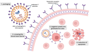
The researchers encapsulated Cas9 and sgPLK1 in a neutrally charged nanoparticle for delivery. The small size of nanoparticles, which are measured in nanometers, allow for easy transport through the bloodstream and uptake by cells. Taking advantage of the high expression of lipoprotein receptor-related protein-1 (LRP-1) on both BBB endothelial cells and glioblastoma tumor cells, they decorated the nanocapsule surface with LRP-1 ligand angiopep-2 peptide to facilitate selective uptake by BBB and glioblastoma cells (Figure 1). Zou and her colleagues bound the nanoparticle together with disulfide bonds as an added layer of selectivity for glioblastoma cell delivery. In the high glutathione environment of a glioblastoma cell, the nanoparticle dissolves, releasing its contents. However, glutathione concentrations are lower in BBB endothelial cells and healthy neurons, reducing the dissolution of the nanoparticles and leaving healthy DNA alone.


Figure 1. Nanoparticles enable permeation of the blood brain barrier (BBB) and selective delivery of the CRISPR/Cas9 system to glioblastoma (GBM) cells. Angiopep-2 peptides, which decorate the nanoparticle’s surface, bind with lipoprotein receptor-related protein-1 (LRP-1) overexpressed on BBB and GBM cells. Following uptake into GBM cells, the nanoparticles dissolve in the high glutathione environment, releasing the Cas9 nuclease and single guide RNA (sgRNA) targeting Polo-like kinase 1 (PLK-1) genes. Zou et al. demonstrated the selective mutation of PLK-1 induced apoptosis in GBM cells with minimal off-target effects.


Through Cas9/sgPLK1 delivery by nanoparticle, Zou et al. demonstrated a 53% reduction in expression of the targeted gene in vitro. PLK1 gene editing was cell selective, with negligible genetic mutation in the healthy surrounding brain tissue. While nanoparticles with and without disulfide cross-linking were capable of gene editing, the disulfide cross-linked nanoparticle induced almost 400% more mutations. Glioblastoma cells treated with disulfide cross-linked nanoparticles were also over 3 times more likely to undergo apoptosis cell death. Mice grafted with patient glioblastoma tumors treated with Cas9/sgPLK1 nanocapsules experienced an almost 3-fold extension in life expectancy, suggesting this treatment as a viable anti-glioblastoma therapy.5

Yet in order to be effectively applied as a cancer therapy, the efficiency of this nanoparticle delivery system must be increased. In mouse glioblastoma models, Zou et al. only achieved a maximum accumulation of 12% and effected a 38% knockdown of PLK1.5 Despite their low magnitude, these values are far greater than similar gene therapy treatments currently studied, suggesting nanoparticles present an innovation in the delivery of CRISPR-Cas9 to difficult to access tumors.

In addition to the improved efficacy over existing systems, the work of Zou et al. opens the door for less invasive administration of treatment. In contrast to previous gene therapies administered directly to the tumor through intercranial injection, the BBB penetration and tumor-specific accumulation of the nanoparticles may permit systemic administration. Zou et al. injected the nanoparticles intravenously, but treatment may even be given as a pill taken orally, eliminating any surgical intervention. Moreover, due to their modular design, the engineered nanoparticles may be adapted as targeted treatments for other tumors. Exchanging the angiopep-2 peptides for another ligand would facilitate uptake by cells expressing the corresponding receptor. The load carried within the nanoparticle could be altered to contain a different sgRNA targeting a new gene or another therapeutic entirely as a complementary treatment. Nanoparticle delivery systems like that studied by Zou et al. contains many layers of selectivity, offering hope for effective delivery of treatment to previously inaccessible tumors.


Works Cited:

  1. Wirsching, H.-G. & Weller, M. Glioblastoma. in Malignant Brain Tumors : State-of-the-Art Treatment (eds. Moliterno Gunel, J., Piepmeier, J. M. & Baehring, J. M.) 265–288 (Springer International Publishing, Cham, 2017). doi:10.1007/978-3-319-49864-5_18.
  2. Delgado-López, P. D. & Corrales-García, E. M. Survival in glioblastoma: a review on the impact of treatment modalities. Clin Transl Oncol 18, 1062–1071 (2016).
  3. Ostrom, Q. T. et al. CBTRUS Statistical Report: Primary Brain and Central Nervous System Tumors Diagnosed in the United States in 2007–2011. Neuro Oncol 16, iv1–iv63 (2014).
  4. Lino, C. A., Harper, J. C., Carney, J. P. & Timlin, J. A. Delivering CRISPR: a review of the challenges and approaches. Drug Delivery 25, 1234–1257 (2018).
  5. Zou, Y. et al. Blood-brain barrier–penetrating single CRISPR-Cas9 nanocapsules for effective and safe glioblastoma gene therapy. Sci Adv 8, eabm8011.
  6. Dotiwala, A. K., McCausland, C. & Samra, N. S. Anatomy, Head and Neck: Blood Brain Barrier. in StatPearls (StatPearls Publishing, Treasure Island (FL), 2024).
  7. Song, R. et al. Selection of rAAV vectors that cross the human blood-brain barrier and target the central nervous system using a transwell model. Molecular Therapy Methods & Clinical Development 27, 73–88 (2022).
  8. Lee, B. et al. Nanoparticle delivery of CRISPR into the brain rescues a mouse model of fragile X syndrome from exaggerated repetitive behaviours. Nat Biomed Eng 2, 497–507 (2018).
  9. Lee, C. et al. Polo-Like Kinase 1 Inhibition Kills Glioblastoma Multiforme Brain Tumor Cells in Part Through Loss of SOX2 and Delays Tumor Progression in Mice. Stem Cells 30, 1064–1075 (2012).

Filed Under: Biology, Chemistry and Biochemistry, Psychology and Neuroscience, Science Tagged With: blood-brain barrier, CRISPR, glioblastoma, nanoparticles

Invasive Species: Ecological Shapeshifters?

May 2, 2024 by Lex Renkert '27

Watershed reeds of midcoast Maine provide a deeper look into the field of epigenetics

Forests, grasslands, and marshes are ecological battlegrounds. In the fight to hold territory, maintain access to resources, and reproduce, many organisms compete directly to occupy the same niche– the role played by a specific organism in an ecosystem. An organism’s ability to carry out these roles is dictated by its “fitness” or capacity to survive and contribute its genes to the next generation. Naturally, relative reproductive success is incredibly environmentally dependent. Most organisms are tailor-made to thrive within their native habitats via natural selection. However, this biological narrative is challenged by the proliferation of invasive species in competition with their native counterparts. In their 2016 study, Spens and Douhovnikoff argue that epigenetics may be key to understanding ecological invasiveness and that the common reed (Phragmites australis) is “an ideal model species” (Spens & Douhovnikoff, 2016) for studying this rapidly expanding subfield of genetics.

Among other things, greater phenotypic plasticity, or “the ability of individual genotypes to produce different phenotypes when exposed to different environmental conditions” (Fusco & Minelli, 2010), increases an organism’s potential to adjust to its surroundings and occupy a vast variety of niches. This becomes possible through epigenetics.  Epigenetic modifications alter gene expression without changing the underlying DNA sequence (Weinhold, 2006). Methylation, the process by which methyl groups are added to DNA, is the key turning genes “on” and “off” (Menezo et al., 2020). The addition of methyl groups prevents DNA-transcribing proteins from accessing the DNA strand, stopping the gene’s expression as a protein. This has the potential to create significant differences in structural and even cellular function among individuals that are otherwise genetically identical.

Clonal plants provide a unique opportunity to study environmental pressures on epigenetics, as these individuals can act as their own genetic control. Reeds are an excellent example of this: as facultatively clonal plants, they can utilize both sexual and asexual reproduction. Exploiting this integral feature, and the existence of multiple subspecies of reed in midcoast Maine, researchers studied the genomes of both native and invasive reeds in two separate locations, Libby and Webhannet. They addressed two questions: Do introduced subspecies exhibit greater epigenetic variation (indicating that epigenetics plays a role in the success of an invasive species)? And will the variation between subspecies genotypes be lesser than the variation within a single genotype’s epigenetic markers (suggesting that epigenetic variation can be used to adapt to an incredibly variable environment)?

Researchers sought answers by studying clusters of reeds called ramets. Since all the reeds within a ramet were genetically identical, they could selectively measure epigenetic variation. These clones were grown within heterogeneous microhabitats that contain varying combinations of nutrients and conditions. Extracted DNA fragments were compared based on the level of methylation among subspecies, genotype, and ramet.

In both sites, the invasive reed demonstrated greater epigenetic diversity than the native reed (Figure 1). Up to 71% of epigenetic variation at the Webhannet site is attributed to differences among genotypes. These results suggest that clones adjust to the demands of their environments via epigenetics, rather than genotypic adaptation. Flexibility of this kind allows for rapid specialization in response to the hyper-individualized environmental conditions of each ramet. Additionally, each site developed an epigenetic “signature” with both subspecies exhibiting distinct, location specific, morphological characteristics. The significant differences in epigenetic markers between sites hint at the potential for large scale shifts due to epigenetics, should genotype not be a factor in these differences. The distinct characteristics displayed by each species demonstrate the vast alterations necessary to survive in an environment with subtle differences.

Figure 1. Epigenetic markers clustered by species (native, introduced) and location (Libby, Webhannet). Differences within a single genotype were greater than variation between genotypes, particularly for the introduced species. Figure adapted from Spens and Douhovnikoff

While this study was small scale, it supports the position that epigenotype variation provides a strong competitive advantage in the natural world. It also suggests that further study would provide more valuable information about the relevance of epigenetics in ecology. In our rapidly changing environment, due to climate change and other human influences, these native genotypes are in danger of being displaced from their niches. Despite a species’ history with its habitat, subtle alterations can have vast impact on individuals that demonstrate low plasticity or tolerance for change. Introduced organisms who demonstrate more flexible epigenotypes have the potential to outcompete their neighbors, eroding local ecosystems beyond repair. This reality drives ecological research in the direction of epigenetics, not only for the sake of discovery, but also in hopes of protecting species who cannot adapt as quickly as we disrupt.


Works Cited

Fusco, G., & Minelli, A. (2010). Phenotypic plasticity in development and evolution: Facts and concepts. Philosophical Transactions of the Royal Society B: Biological Sciences, 365(1540), 547–556. https://doi.org/10.1098/rstb.2009.0267

Menezo, Y., Clement, P., Clement, A., & Elder, K. (2020). Methylation: An Ineluctable Biochemical and Physiological Process Essential to the Transmission of Life. International Journal of Molecular Sciences, 21(23), 9311. https://doi.org/10.3390/ijms21239311

Spens, A., & Douhovnikoff, V. (2016). Epigenetic variation within Phragmites australis among lineages, genotypes, and ramets. Springer International Publishing. https://link.springer.com/article/10.1007/s10530-016-1223

Weinhold, B. (2006). Epigenetics: The Science of Change. Environmental Health Perspectives, 114(3), A160–A167.

Filed Under: Biology, Environmental Science and EOS Tagged With: climate change, epigenetics, invasive, reed

Smoke Signals: The Unexpected Long Term Effects of Smoking on the Immune System

April 30, 2024 by Divya Bhargava

Image Source: “Smoking has a Lasting Impact on the Immune System, 2024”

When we get sick, our bodies’ immune systems work to fight off infections by invading pathogens, or organisms like bacteria and viruses that cause disease. However, many factors such as lack of sleep and poor nutrition weaken our immune system, meaning that we are less able to stay healthy. It has been known that smoking is another one of these factors that weaken our immune systems, but a recent study from a group at the Institut Pasteur in France looking at the effects of a variety of factors on the immune system showed that the extent to which smoking plays a role is much higher than many would think. But to understand the results of this study, it is important to first understand the mechanisms the immune system uses to fight infection. 

The immune system has many different moving components, including two distinct branches. The first is the faster, more general innate immune system which has a similar response to all infections. The second is the adaptive immune system which is slower, memory-based, and is involved in pathogen specific response. Although the innate immune system involves general molecules that interact with all cells and the adaptive immune system has specialized molecules that interact with pathogens based on memory of past infection, they share one important class of signaling molecules. These molecules are called cytokines and their role is to coordinate both of these types of immune response. Cytokines are small molecules that are released by immune cells to communicate with other parts of the body and each other. This signaling results in deployment of a response by other immune cells against invading pathogens. However, levels of cytokine production exist in a very fine balance. In order to get the desired immune response, you need the exact right level of cytokines present. If levels are too high or too low, they could cause abnormalities including overactive immune response and inflammation or impaired immune responses. 

To investigate the effects of a variety of different factors on the immune system and cytokine responses of healthy individuals, a project called the Milieu Intérieur put together a cohort of 1000 healthy participants and has been studying variability in the immune system between these individuals (“The Milieu Intérieur Project”). In an investigation of this data, the group from Institut Pasteur, Saint André et al, analyzed 136 variables measured in the Milieu Intérieur Project that could be causing differences in cytokine secretion and immune response (Luo and Stent 2024). These variables included everything from demographics, to diet, to health habits like smoking, to social and environmental characteristics (Saint-André et al. 2024).

After they performed their initial statistical analysis, Saint André et al measured production of 13 disease relevant cytokines as a quantitative measure of immune response in populations with different demographics, health habits, and other characteristics. In the lab, they exposed blood samples from their sample population to 12 different molecules meant to serve as stimulants for the immune system (these molecules included things like viral and bacterial proteins). After this exposure, the authors tested cytokine production in both innate and adaptive immune cells, and once they had that data, they took their results one step further. The group also used epigenetics, or the study of changes in gene expression rather than the DNA code that makes up the genome to investigate possible reasons for variability in immune responses associated with factors tested. Their epigenetic evaluation consisted of analyzing the extent to which one epigenetic process, DNA methylation, occurred at specific regulators of signaling and metabolism (Saint-André et al. 2024) to assess changes associated with smoking. 

As previously stated, one of the authors’ main findings from the initial statistical analysis was that smoking had a large effect on cytokine response. In fact it had the same effect as age, sex, and genetics, three things many would consider much more directly impactful to the immune system than smoking. In their in vitro simulations, they found that smoking had a temporary effect on the ability of the innate immune system to function properly. This result is a relatively intuitive one. If you do something that is considered bad for you, it makes sense that you would get sick more easily. 

However, more surprisingly, they also found that smoking leaves a lasting effect on memory based adaptive immune responses even after cessation of smoking, meaning that even after people quit smoking, their immune systems still are impacted. They found that in samples from individuals who smoked there were higher levels of cytokine expression, especially of an inflammatory cytokine called CXCL5 that is secreted in response to bacterial infection. Secretion of this cytokine is associated with the presence of an inflammatory protein called CEACAM6 in the blood. Consistent upregulation of levels of this protein has been found to have links with multiple cancers such as colon cancer (Wu et al. 2024). In Saint André et al’s epigenetic investigation of this association, they found that DNA methylation, which results in a downregulation of gene expression and in this case an increase in cytokine production, is linked to smoking’s lasting effect on the immune system (Greenberg and Bourc’his 2019). DNA methylation was decreased at many of the sites they tested which are involved in regulation of signaling genes and metabolism. Decreased DNA methylation was likely impacting levels of cytokines in response to detection of pathogens. In these populations, smoking caused lasting changes in gene expression which resulted in long term changes in addition to the expected short term effects on the immune system. 

This study demonstrates that smoking can have lasting negative impacts on your health which are not limited to just lung damage. It is also associated with pro-inflammatory cancer pathways and epigenetic markers that cause increased cytokine production. This overproduction of cytokines can confuse cells and also cause increased inflammation. Over time the extra inflammation can damage tissues and lead to developments of other conditions, like the cancers previously mentioned and complications associated with overproduction of cytokines (“What are Cytokines”). These recent findings emphasize that it is important to consider the possible implications of smoking and all things that we expose ourselves to, and to keep in mind that new data is still being discovered.

Works Cited

The Milieu Intérieur Project Institut Pasteur. Luo,Y. and Stent,S. (2024) Smoking’s lasting effect on the immune system. Nature, 626,  724–725.

Saint-André,V., Charbit,B., Biton,A., Rouilly,V., Possémé,C., Bertrand,A., Rotival,M., Bergstedt,J., Patin,E., Albert,M.L., et al. (2024) Smoking changes adaptive immunity with persistent effects. Nature, 626, 827–835.

Wu,G., Wang,D., Xiong,F., Wang,Q., Liu,W., Chen,J. and Chen,Y. (2024) The emerging roles of CEACAM6 in human cancer (Review). International Journal of Oncology, 64, 1–15.

Greenberg,M.V.C. and Bourc’his,D. (2019) The diverse roles of DNA methylation in mammalian development and disease. Nat Rev Mol Cell Biol, 20, 590–607.

What are Cytokines? Types and Function Cleveland Clinic.

Smoking has a lasting impact on the immune system, a new study finds (2024) Euronews.

Filed Under: Biology Tagged With: Biology, Genes, Medicine

Novel Gene Drive Demonstrates Promise in Reducing Transmission of Malaria

April 24, 2024 by Isabelle Fitzgibbon '27

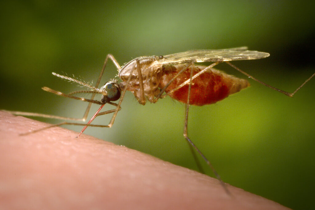
Recent progress has been made in malaria elimination using insecticides, bed nets, and clinical management (CDC). However, efficient mosquitos carrying deadly species of malaria parasites paired with favorable climate in regions where the disease is prevalent, weak infrastructure, and high intervention costs create difficult barriers to malaria elimination (CDC). Similarly, new threats are arising from drug- and insecticide-resistant parasites that pose a risk of expansion to densely populated areas with no prior exposure to the parasites and an unprepared public health system (Garrood et al., 2022). Amidst malaria control efforts, many scientists are investigating a promising new method of inhibiting mosquitos’ ability to transmit malaria: gene drives.

Gene drives, or “selfish” genes, are transmitted at a frequency greater than 50%, above what is predicted by Mendelian genetics. These genes occur naturally in populations; however, we can also engineer gene drives using CRISPR/Cas9 technology to insert a desired gene into a population (Bier, 2022, Hillary et al., 2020). The CRISPR/Cas9 system targets a certain DNA sequence and cuts it, after which the DNA will repair itself either randomly (end-joining, or EJ) or following a provided template (homology-directed repair, or HDR) (Adolfi et al., 2020, Hillary et al., 2020). In the case of gene drives, researchers create a drive, or set of genes, containing the desired genes to be inserted as well as genes encoding necessary CRISPR/Cas9 elements. The presence of CRISPR/Cas9 elements ensures that offspring can make the CRISPR/Cas9 proteins necessary to insert the desired gene into their genome. The desired inserted genes can serve a variety of purposes; in the case of malaria, scientists are inserting antimalarial effector genes into gene drives (Gantz et al, 2015, Isaacs et al., 2012).

Prior research demonstrates use of a gene drive, nRec, to insert antimalarial effector genes that inhibit parasite transmission into a mosquito population (Gantz et al, 2015, Isaacs et al., 2012). The drive targets the essential kynurenine hydroxylase (kh) gene, causing a loss-of-function mutation that reduces female survival and reproduction (Gantz et al., 2015). This type of drive, although effective in preventing malaria transmission, ultimately proves unsustainable given that the loss-of-function results in poor fitness and selection against the drive. A recent study proposes a solution to this problem via a gene drive rescue system that restores the function of the kh gene (Adolfi et al., 2020). The restoration of kh gene function ensures sustainable propagation of the drive and alleviates the fitness cost of the drive.

In a study led by Adriana Adolfi at University of California, Irvine, researchers aimed to target the same essential kh gene while also providing a rescue sequence that maintains the function of the gene (Adolfi et al. 2015). Although their drive does not include the antimalarial effector genes used in prior drives, they do not anticipate any challenges to incorporating the genes. Their approach addresses two problems: it minimizes the impact of drive-resistant alleles and reduces the fitness cost of the drive. By targeting the essential kh gene, drive-resistant mosquitoes are selected out because of the fitness cost that they take on while those that are not resistant do not suffer a fitness cost. The EJ repair that occurs in drive-resistant mosquitoes results in a loss-of-function mutation to the kh gene that makes the mosquitoes less fit for survival than their nonresistant counterparts who repair the Cas9 cleavage with HDR. HDR restores the original sequence of the kh gene by repairing the DNA according to a provided template, thus preventing the loss-of-function mutation, the fitness cost, and ultimately a population crash. In contrast, if the researchers had chosen to target a non-essential gene, this undesirable EJ event would not confer a fitness cost and would then be maintained while preventing transmission of the drive. Ultimately, the drive acts by replacing the previously targeted portion of the kh gene with the original kh sequence to alleviate prior fitness costs of the drive and minimize survival of drive-resistant individuals.

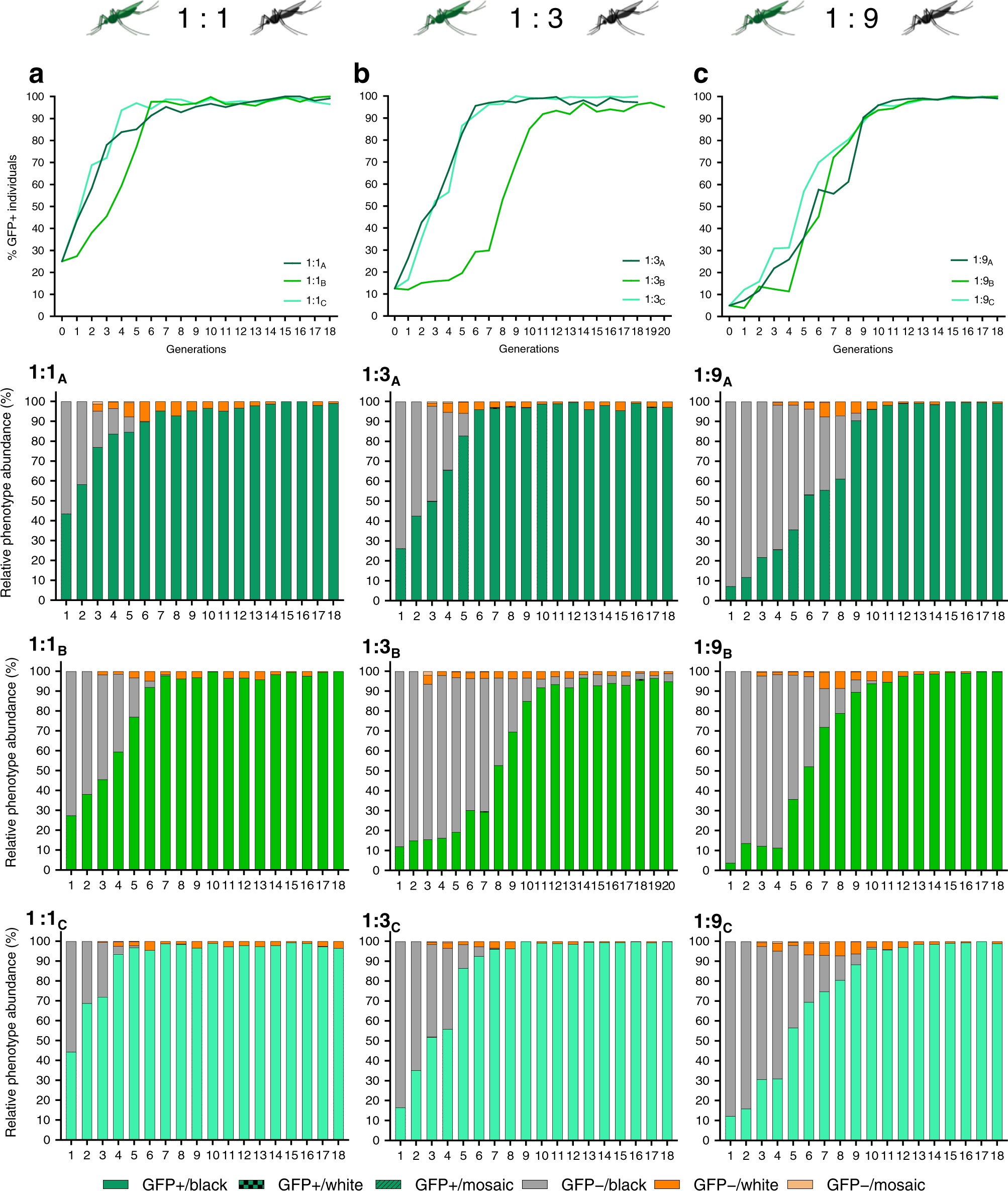
Reckh mosquitos, those that acquired the drive, demonstrated fitness equal to wild-type (WT) mosquitos, while mosquitos with a loss-of-function mutation in the kh gene demonstrated decreased fitness and were eliminated from the population. The drive was released into caged mosquito populations with varied ratios of Reckh males to WT males (1:1, 1:3, and 1:9). In all cages except for one 1:3 cage, the drive was present in more than 95% of mosquitoes by the eleventh generation. Notably, the population did not crash, and the drive was not selected out of the population. In prior research with the nRec drive, few cages reached greater than 95% introduction of the drive before the population crashed or the drive was selected out. The consistent spread of the Reckh drive demonstrates its ability to combat previous challenges of drive sustainability. 


Fig. 3

Figure 1: The drive reached >95% introduction by the eleventh generation in populations seeded with ratios of Reckh:WT males of 1:1, 1:3, and 1:9 while drive-resistant individuals were eliminated from the population. Green represents individuals with the drive, gray represents those without the drive, and orange represents drive-resistant individuals. (Adapted from Adolfi et al., 2020)

Furthermore, analysis of mosquitos with non-functional kh– alleles revealed that they were selected out of the population at a rate higher than expected. The researchers propose that lethal/sterile mosaicism factors into elimination of individuals with non-functional kh– alleles. During reproduction, offspring inherit one kh allele from their mother and one kh allele from their father. In the proposed model, CRISPR/Cas9 elements inherited from a mother with a non-functional kh– allele target and cut the WT kh+ paternal allele. This results in EJ mutations in the WT paternal allele that render the offspring less fit and accelerates its elimination from the population. The recoded kh gene sequence is not present in the mothers because they have a non-functional kh– allele, thus the mosquitoes cannot use HDR to repair the cut paternal allele and must instead use EJ. This lethal/sterile mosaicism ensures that non-functional kh– alleles act dominantly to eliminate drive-resistant individuals from the population.

The proposed gene drive shows promise towards sustainability of antimalarial gene drives in caged trials. To further explore the potential of this gene drive, the researchers propose further experimentation with the antimalarial effector genes inserted into their drive. Based on prior research, they do not anticipate any fitness disadvantage to the insertion of the antimalarial genes nor any challenges due to the large size of the drive. Although much research is required before this gene drive can be implemented in wild populations, it introduces a sustainable and effective method of malaria control.


References

  1. Adolfi, A., Gantz, V. M., Jasinskiene, N., Lee, H.-F., Hwang, K., Terradas, G., Bulger, E. A., Ramaiah, A., Bennett, J. B., Emerson, J. J., Marshall, J. M., Bier, E., & James, A. A. (2020). Efficient population modification gene-drive rescue system in the malaria mosquito Anopheles stephensi. Nature Communications, 11(1), 5553. https://doi.org/10.1038/s41467-020-19426-0
  2. Bier, E. (2022). Gene drives gaining speed. Nature Reviews Genetics, 23(1), 5–22. https://doi.org/10.1038/s41576-021-00386-0
  3. Gantz, V. M., Jasinskiene, N., Tatarenkova, O., Fazekas, A., Macias, V. M., Bier, E., & James, A. (2015). Highly efficient Cas9-mediated gene drive for population modification of the malaria vector mosquito Anopheles stephensi. Proceedings of the National Academy of Sciences of the United States of America, 112(49), E6736–E6743. https://doi.org/10.1073/pnas.1521077112
  4. Garrood, W. T., Cuber, P., Willis, K., Bernardini, F., Page, N. M., & Haghighat-Khah, R. E. (2022). Driving down malaria transmission with engineered gene drives. Frontiers in Genetics, 13, 891218. https://doi.org/10.3389/fgene.2022.891218
  5. Hillary, V. E., Ceasar, S. A., & Ignacimuthu, S. (2020). Chapter 18 – Genome engineering in insects: Focus on the CRISPR/Cas9 system. In V. Singh & P. K. Dhar (Eds.), Genome Engineering via CRISPR-Cas9 System (pp. 219–249). Academic Press. https://doi.org/10.1016/B978-0-12-818140-9.00018-0
  6. How Can Malaria Cases and Deaths Be Reduced? (2019, January 28). Centers for Disease Control and Prevention. https://www.cdc.gov/malaria/malaria_worldwide/reduction/index.html
  7. Isaacs, A. T., Jasinskiene, N., Tretiakov, M., Thiery, I., Zettor, A., Bourgouin, C., & James, A. A. (2012). Transgenic Anopheles stephensi coexpressing single-chain antibodies resist Plasmodium falciparum development. Proceedings of the National Academy of Sciences of the United States of America, 109(28), E1922–E1930. https://doi.org/10.1073/pnas.1207738109

Filed Under: Biology Tagged With: gene drive, malaria

S. glomerata show resistance to the negative effects of ocean acidification on marine microbes

April 21, 2024 by Layla Silva '27

As more CO2 enters the ocean, the water’s pH and temperature change in processes called ocean warming and acidification. Both processes pose a risk to marine microbes, as they are unaccustomed to their new, more acidic environment. Several marine species depend on the microbes that dwell in the ocean, and if the change in pH negatively impacts the oceanic microbiome, there would be negative implications for a large number of organisms.

Microbes are essential to the development of many species in the world’s oceans. They are able to activate genes, sculpt the bodies of multicellular organisms, and provide vital life information to juvenile species (Yong 2016). But these abilities may be disrupted if the ocean’s change in pH negatively affects the microbiomes both in the water and living within ocean creatures.

Dr. Elliot Scanes and his colleagues at the University of Technology Sydney evaluated the effects of ocean acidification on the Sydney rock oysters’ (S. glomerata) ability to transfer its microbiome down to its offspring during reproduction. Oysters reproduce via broadcast spawning, a process in which sedentary organisms release all of their eggs and sperm into the surrounding water in hopes that a portion of the gonads will be fertilized (Bondar, 2018). Because these broadcasted embryos are now exposing their microbiomes to warmer, more acidic environments than the microbiomes of previous generations have been accustomed to, the microbes living within these embryos are not well adapted to the new conditions. This poorly equipped microbiome is causing fewer and fewer embryos to develop properly. An oyster’s microbiome is a necessary part of its body, and without it, a juvenile oyster may not be able to develop and function as effectively (Scanes et al. 2023).

Scanes set out to examine whether exposure to ocean warming and acidification during both broadcast spawning and early reproduction would alter an oyster’s microbiome strength.

The lab team acclimated these oysters to the lab tanks and then harvested their eggs and sperm, later fertilizing them (Figure 1.) (Scanes et al. 2023). Half of the oyster embryos were raised in tanks with a normal pH, and the other half were raised in tanks with decreased pH to mimic ocean acidification. The team conditioned both sets of S. glomerata for reproduction, then used eggs and sperm from each set to breed the next generation of oysters. The next generation was divided into four groups: first, the oyster embryos collected in tanks with a normal pH were split into two groups, with one group being raised in another tank with a normal pH and the other being raised in a tank with a low pH that mimics ocean acidification. Then, the oyster embryos collected in tanks with a low pH were also split into two groups, with one group being raised in another tank with a low pH

Figure 1. Scanes et al. depicts their experimental design. The PCO2 that appears in several of the diagram labels means partial pressure of carbon dioxide, which is a term used to describe how much carbon dioxide exists within a system (Messina 2022). Ambient PCO2 means normal pH. Elevated PCO2 means acidic water.

and the other being raised in a tank with a normal pH.

The embryos produced from these four sets of oysters informed Scanes et al. of the physiological differences that occur between oyster microbiomes that are exposed to ocean acidification at different steps in the reproductive process. The team found significant alteration of the microbiome in the parent oysters exposed to ocean acidification and concluded that when oyster parents were exposed, more oyster embryo microbiomes were prepared for the new conditions, and so the more protected oyster embryos survived (Scanes et al. 2023). This information is of much consequence because it provides a baseline for studying other microbe–sea creature relationships in the future. The marine microbiome plays a critical role in the development and wellbeing of animals like the Hawaiian bobtail squid and the Hydroides elegans, otherwise known as the “squiggly worm,” who depend on them for gene activation and information on safe places to live, respectively (Yong 2016). Now that there is evidence that the changing conditions of ocean water harms microbes, and therefore harms the creatures that depend on them, as well as evidence that exposure to these conditions protects the microbes in future generations, scientists are better informed about how to protect marine species moving forward.

Literature Cited

Bondar C. Wild Moms. 2018.

Messina Z et al. Partial Pressure of Carbon Dioxide. National Library of Medicine. 2022.

Scanes E et al. Transgenerational transfer of the microbiome is altered by ocean acidification in oyster larvae. Aquaculture. 2023.

Yong E. Body Builders. I Contain Multitudes: The Microbes Within Us and a Grander View of Life. 2016. 49-59.

Filed Under: Biology, Environmental Science and EOS, Science

SMART Conservation Software aids wildlife management teams in conservation efforts

April 21, 2024 by Layla Silva '27

Those who work in the field of wildlife management aim to protect the biodiversity of ecosystems, which is critical in maintaining the health of the environment. But wildlife management workers around the world frequently experience serious challenges such as poaching, logging, illegal farming, forest fires, and insufficient resources. For example, poachers use snare loops (wire traps that tighten around the necks of animals) to catch protected species. In 2014, tiger poachers in the Sundarbans Reserved Forest of Bangladesh placed thousands of these snare loops across the entire reserve, in locations too far from guard posts to be monitored full time (Abdul Aziz et al., 2017). In most conservation groups, there are not enough funds, employees, or volunteers to efficiently manage wildlife and simultaneously prevent poachers from killing protected animals. Thus, wildlife management teams are calling for improved tools that will better protect endangered animals from further harm.

Figure 1. Snare loop around a lion’s neck. Loops can tighten around any part of the body, holding the animal in place until poachers arrive or weakening it until it dies of its injuries.

Companies such as SMART, Re:Wild, and the World Wildlife Fund developed SMART Conservation Software in 2011 to better support wildlife conservation groups. SMART is short for Spatial Monitoring and Reporting Tool, and it is a digital platform capable of collecting and evaluating data on wildlife management sites. Workers within the same management system can input data as they come across new information, allowing the platform to record what they find in real time like where animals are mating, as well as where and when poacher traps are found (https://smartconservationtools.org/). Using these inputs, SMART plots a management team’s efforts, impacts, and shortcomings over time, highlighting areas that need improvement. Once those improvements are made, management groups are better able to conserve biodiversity, enforce the law, encourage and oversee tourism, and use natural resources properly.

Figure 2. Wildlife management employees use SMART device to log important conservation information.

Companies such as SMART, Re:Wild, and the World Wildlife Fund developed SMART Conservation Software in 2011 to better support wildlife conservation groups. SMART is short for Spatial Monitoring and Reporting Tool, and it is a digital platform capable of collecting and evaluating data on wildlife management sites. Workers within the same management system can input data as they come across new information, allowing the platform to record what they find in real time like where animals are mating, as well as where and when poacher traps are found (https://smartconservationtools.org/). Using these inputs, SMART plots a management team’s efforts, impacts, and shortcomings over time, highlighting areas that need improvement. Once those improvements are made, management groups are better able to conserve biodiversity, enforce the law, encourage and oversee tourism, and use natural resources properly.

SMART is used by conservation organizations around the world, one example being the Chirripó National Park in the Talamanca Mountain Range of Costa Rica. For years, the Chirripó management team had been struggling to precisely locate and record the illegal activities taking place on protected land, making it impossible to remove offenders or convince authorities that their ongoing complaints were valid (Madrigal). But SMART software can be downloaded on personal devices, so when the park introduced SMART to their employees and surrounding members of the community, citizens who were not involved in full-time park conservation were still able to contribute (Madrigal). This added many more eyes, ears, and hands to the conservation effort, and within one year, Chirripó was able to report the exact dates and locations of 44 cases of illegal activity across the park to law enforcement (Madrigal). Once law enforcement gained access to this concrete information, they were able to operate efficiently, driving down the crime rate. More importantly to Chirripó National Park, the added coverage helped protected species such as the Baird’s tapir, the spider monkey, the puma, the agouti, and the jaguar (Madrigal). Chirripó’s experience with SMART demonstrates how useful this technology is for organizing and communicating the issues conservationists face on a daily basis.

Figure 3. SMART conservation software helps Chirripó National Park to protect animals like the Baird’s tapir pictured above.

Like most technology, SMART software is exciting, innovative, and solves modern day problems – but it also comes with some challenges. The Zimbabwe Parks and Wildlife Management Authority (ZPWMA), an organization that works to protect lions, elephants, leopards, and buffalo across all of Zimbabwe, points out that implementing SMART conservation technology can present capacity and resource issues for conservation management employees (Kavhu et al, 2021). Many workers were unfamiliar and uncomfortable with the technology, there were not enough electronic devices such as computers to collect all field data, and many of the patrol routes were without internet access (Kavhu et al, 2021). While it is possible that technological innovation is not a priority for Zimbabwe, it is also important to remember that Zimbabwe’s history is one marred by British colonialism, and the country only gained its independence in the late 1970’s (Ingham et al., 2023). These setbacks help to explain why Zimbabwe has been unable to progress as in the world of electronic technology, even if the progress is desired. These issues of technological access can be applied to other countries that do not yet have a strong electronic infrastructure, meaning that SMART works best in more electronically informed countries and falls short in countries that have not expanded their electronic bandwidth.

Figure 4. Parks in Zimbabwe aim to protect their buffalo populations.

There are some solutions to these technological problems. For example, building a strong implementation plan, motivating the discouraged workers, following the example of other institutions that have implemented SMART technology, and, most importantly, raising funds to buy more computers would make the use of SMART technology easier in Zimbabwe parks (Kavhu et al, 2021). Adding more volunteers to the conservation effort is also a great solution. If ZPWMA advertised volunteer opportunities in their communities using layperson terms, supporters of the conservation effort would be more likely to help manage the wildlife in Zimbabwe’s parks. Of course, volunteers would need to be trained so that they are able to properly identify notable occurrences in the parks, but their contributions have the potential to greatly strengthen the conservation effort.

SMART Conservation Software is off to a great start in helping to better manage parks around the world. Though SMART does find its faults in countries unaccustomed to the devices needed for software implementation, this problem will only grow smaller as the world continues to progress in the realm of personal electronic devices (given that countries like Zimbabwe want to prioritize electronic familiarity moving forward). Its ability to collect, organize, and present data across long distances and multiple devices allows wildlife management teams to care for protected species much more efficiently, making SMART a tool that revolutionizes the realm of conservation.

Works Cited

Abdul Aziz, M. et al. Investigating patterns of tiger and prey poaching in the Bangladesh Sundarbans: Implications for improved management. ScienceDirect, vol. 9, 2017, pp. 70-81.

Barrantes Madrigal, Jimmy. “Community-based SMART patrolling in one of the Great Five Forests of Mesoamerica: the Talamanca Highlands.” SMART, https://smartconservationtools.org/en-us/SMART-Community/Your-stories/Case-Study?CaseStudyID=27.

Ingham, Kenneth, et al. “History of Zimbabwe”. Encyclopedia Britannica, 12 Dec. 2023, https://www.britannica.com/topic/history-of-Zimbabwe. 

Jones, J.J.. “Snared Lioness in Kruger National Park.” Wildestofficial.com, 20 September 2019, https://wildestofficial.com/news/snare-poaching-increasing-in-kruger-national-park/.

Kavhu, Blessing, et al. Spatial Monitoring and Reporting Tool (SMART) in Mid‐Zambezi Valley, Zimbabwe: Implementation challenges and practices. ProQuest, vol. 3, 2021.

San Diego Zoo. Baby Baird’s Tapir. animals.SandiegoZoo.com, https://animals.sandiegozoo.org/animals/tapir.

Slade, James. Conservationists operating SMART device. Smartconservationtools.com, https://smartconservationtools.org/en-us/SMART-in-Practice/How-we-use-SMART. 

SMART. “About Us.” SMART, https://smartconservationtools.org/en-us/About/About-us. The Great Projects. Buffalo in Zimbabwe. thegreatprojects.com, https://www.thegreatprojects.com/volunteer-in-zimbabwe.

Filed Under: Biology, Environmental Science and EOS, Science

New Cancer Vaccine Harnesses the Human Immune System

April 21, 2024 by Olivia Miller

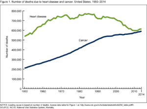
Right now, cancer is the second most common cause of death in the world, and it’s set to become the leading cause within the next several decades (Drexler) (Figure 1). Unlike many other diseases that have plagued humans throughout our history, even revolutionary advancements in medicine have not been able to fully prevent or cure it. To understand why, it’s important to recognize that cancer comes not from the outside, but from within ourselves, when our own cells begin to divide uncontrollably and spread throughout our bodies. Because cancer arises in our bodies, we can’t treat it using antibiotics or vaccines that target a particular pathogen. However, a 2023 study has proposed a new vaccine treatment that fights pancreatic cancer (which has a five-year survival rate of only 12%) using our own immune system.

Figure 1. Cancer deaths are expected to surpass deaths due to heart disease in the United States. Source: CDC

To understand this discovery, it’s important to learn a little about how the immune system works. One important component of our body’s defense system are our T cells, which fight invaders by identifying and killing body cells infected with pathogens such as viruses. A T cell only knows what to attack after it’s activated by a foreign particle called an antigen. Antigens are often tiny pieces of invading viruses or bacteria, like “keys” which unlock a T cell immune response. Now, what does all this have to do with cancer? The idea behind this proposed treatment is to use neoantigens (a type of antigen) to help the body recognize cancer cells. Neoantigens are mutant proteins which only form on tumor cells, so helping T cells recognize them could help the body learn to attack cancerous cells.

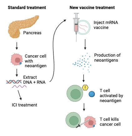
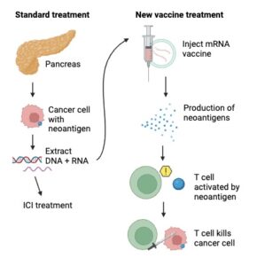
Figure 2. In combination with ICI treatment, these novel mRNA vaccines prompt the immune system to attack cancer cells by manufacturing the neoantigens on their surface. Adapted from Huff & Zaidi (2023) and created with BioRender.com.

This study’s major discovery was a new way to prompt the immune system to recognize neoantigens. The researchers took genetic material (such as DNA and RNA) from the surgically removed tumors of sixteen people with pancreatic cancer. Then they gave each patient a kind of treatment called an immune-checkpoint inhibitor (ICI). Immune checkpoints are proteins on the surfaces of healthy cells which usually tell our T cells not to attack them. The problem is that sometimes, immune checkpoints on cancerous cells prevent T cells from attacking them too. ICIs can treat some kinds of cancers by blocking these interactions between T cells and cancerous cells, allowing T cells to kill them, but they often do not work well on pancreatic cancer. To solve this problem, the researchers used the genetic information collected from the tumors to create a personalized vaccine containing mRNA (a type of RNA) that codes for the exact neoantigen formed on an individual’s cancer cells. After receiving this vaccine, a person’s cells would be able to use the mRNA to produce this neoantigen in abundance. Then, their T cells could theoretically recognize it, become activated, and know to go after the cancer cells with the neoantigen on their surfaces (Figure 2). To maximize the chances of triggering this T cell response, the vaccines included mRNA coding for as many as twenty neoantigens.

Now for the big question: did it work? Well, cancer unfortunately came back an average of 13.4 months after treatment for half of the study’s participants. The other eight, however, all had T cells which recognized the artificial neoantigens in their bodies after vaccine treatment and also remained cancer-free after ~18 months (Huff & Zaidi, 2023). This vaccine is clearly not perfect, and future research is still needed to understand why half the patients’ immune systems did not respond to the treatment. However, it is still an incredible breakthrough that could shift the direction of cancer treatment. A clinical trial, the second phase of this study, is already well underway and includes over 250 patients (Stallard, 2024). Given the small sample size of this first study, the new clinical trial will help clarify how effective this vaccine treatment really is. Immunotherapy is emerging as a leading factor in the fight against cancer, and this study gives reason to believe that new treatment possibilities could be on the horizon for cancer patients, even those with aggressive tumors.


References

Drexler, M. (n.d.). The Cancer Miracle Isn’t a Cure. It’s Prevention. Harvard Public Health. https://www.hsph.harvard.edu/magazine/magazine_article/the-cancer-miracle-isnt-a-cure-its-prevention/. 

Heron, M. & Anderson, R.N. (2016). Changes in the leading cause of death: Recent patterns in heart disease and cancer mortality. CDC: NCHS data brief, 254. https://www.cdc.gov/nchs/products/databriefs/db254.htm.

Huff, A.L. & Zaidi, N. (2023, May 10). Vaccine boosts T cells that target pancreatic tumours. Nature. https://www.nature.com/articles/d41586-023-01526-8.

Rojas, L.A., Sethna, Z., Soares, K.C. et al. (2023). Personalized RNA neoantigen vaccines stimulate T cells in pancreatic cancer. Nature 618, 144–150. https://doi.org/10.1038/s41586-023-06063-y. 

Stallard, J. (2024, April 7). Investigational mRNA Vaccine Induced Persistent Immune Response in Phase 1 Trial of Patients With Pancreatic Cancer. Memorial Sloan Kettering Cancer Center. https://www.mskcc.org/news/can-mrna-vaccines-fight-pancreatic-cancer-msk-clinical-researchers-are-trying-find-out.

Filed Under: Biology, Science

Chernobyl Wolves Living with Cancer

April 21, 2024 by Natalie Hayden

A peculiar phenomenon is occurring in Chernobyl, Ukraine. It has been a ghost town since the nuclear accident in 1986, and radiation levels are too high for humans to live safely. When the accident occurred, it released over 100,000 lbs of radioactive material into the atmosphere which was dispersed across Europe and the USSR. Over 100,000 people were evacuated from over 200 villages. An exclusion zone with a 30 km radius was created to protect humans from the radiation that still remains (NPR, 2024). The level of radiation varies within the CEZ because wind and rain spread the radiation when the accident occurred. Wolves are in a unique position as the apex predators– they encounter radiation exposure at every level of the food chain. From a contaminated deer eating contaminated grass, wolves are one of the most radiation-exposed animals in the CEZ. The wolves in the CEZ are exposed to much higher levels of radiation than are considered safe for humans. For humans, the annual limit is 1 mGy, but for wolves, they are exposed to 2.6-35.7 mGy on a daily basis (MIT News, 1994). Despite this high level of radiation, the wildlife is thriving, filled with elk, deer, boar, and wolves. While radiation is typically thought of as a risk for developing cancer, the Chernobyl grey wolf population is thriving in tandem with this disease, developing a population that is nearly seven times as dense as surrounding wolf populations. Puzzled by their strange ability to survive in these harsh conditions, researchers set out to determine what was going on. Scientists measured nine wolves’ movement and radiation levels within the CEZ through a GPS containment-monitor for six months. They found that the level of radiation varied depending on where the wolves were within the CEZ (Hinton et al., 2019). Although the level of contamination varies, these high levels of radiation may be enough to drive natural selection, helping wolves thrive in these harsh conditions (Love et. al, 2020). 

Scientists set out to uncover if the radiation from Chernobyl was enough to drive natural selection, allowing the population to evolve in such a way to protect it from radiation. Researchers analyzed the wolves’ fecal samples for parasites, took blood samples to sequence the full blood transcriptomes, a collection of mRNA transcripts, and muscle tissue samples. The study also looked at the blood cell composition of these wolves, to take a closer look at the immune cells, which are indicative of different types of stress or disease (Love et. al, 2020). The study compared CEZ wolves to those in Belarus, outside of the CEZ, and to wolves in Yellowstone National Park (NPR, 2024). They found that some of the fastest-evolving genes “have some role in cancer immune response or the anti-tumor response in mammals” (NPR, 2024). These genes are called Endogenous Retroviruses, or ERVs, which are associated with neurological, autoimmune, and oncogenic diseases in humans. Not much is known about these cells, including what exactly triggers their activation and what regulates them. What scientists do know is that not all ERVs are bad– helpful ERVs can help stimulate a retroviral immune response which helps spur tumor cell death. Scientists believe that environmental stimuli, like radiation exposure, likely influences ERV activation; however, more research still needs to be done. Wolves in the CEZ have higher levels of ERV expression, indicating that there are divergent ERV regulatory patterns between wolves that are exposed and unexposed to radiation. While scientists must learn more about ERVs and their effect in wolves, more research is needed on the effects of radiation contamination in the environment and what is driving cancer development in radiation-contaminated habitats (Love et. al, 2020).

Although there is evidence of a genetic component of wolves being resistant to cancer, there is another important factor to keep in mind– human presence. Humans have largely been absent from the CEZ for nearly forty years, so wolves are free from the pressures humans bring. For instance, perhaps for wolves the pressure from hunting is so that it may be better than having to deal with cancer. Essentially, hunting may pose a bigger threat to wolves than cancer. Other human-related pressures these wolves do not face are land, pollution, human disease, and more. However, more research must be done to fully understand all the pressures these wolves are under. While research in the CEZ has been halted because of the war in Ukraine, the wolf population is likely thriving because of a genetic component and from no human pressures (NPR, 2024). 

Researchers hope to return to the region to find and identify these cancer-resilient genes which could inform human cancer treatments. Although their research is paused, the Chernobyl wolf population is a remarkable feat of perseverance and resilience. 

Figure 1. A gray wolf in the Chernobyl Exclusion Zone. (Chernobyl’s Mutant Wolves Have Evolved Anti-Cancer Abilities | IFLScience, n.d.)

Literature Cited

Chernobyl’s Mutant Wolves Have Evolved Anti-Cancer Abilities | IFLScience. (n.d.). Retrieved April 21, 2024, from https://www.iflscience.com/chernobyls-mutant-wolves-have-evolved-anti-cancer-abilities-72831

Hinton, T. G., Byrne, M. E., Webster, S. C., Love, C. N., Broggio, D., Trompier, F., Shamovich, D., Horloogin, S., Lance, S. L., Brown, J., Dowdall, M., & Beasley, J. C. (2019). GPS-coupled contaminant monitors on free-ranging Chernobyl wolves challenge a fundamental assumption in exposure assessments. Environment International, 133, 105152. https://doi.org/10.1016/j.envint.2019.105152

Love et. al (2020). Long-term Implications of Chronic Radiation Exposure: A Genomics Study of Two Canids Reveals Evolutionary and Ecological Impacts of a Nuclear Disaster – ProQuest. (n.d.). Retrieved April 21, 2024, from https://www.proquest.com/openview/f5f17943e75ddda6f147cd66201a344b/1?cbl=18750&diss=y&pq-origsite=gscholar&parentSessionId=Rfd%2FKuzxeAQaOHLv1Bl3gFx7bPxf1AU%2BWGySS9I3SGg%3D

MIT News (1994, January 5). MIT News | Massachusetts Institute of Technology. https://news.mit.edu/1994/safe-0105

NPR: Short Wave. (2024, February 5). NPR. https://www.npr.org/2024/02/05/1198909263/cancer-wolves-ukraine-chernobyl-radiation

Filed Under: Biology, Environmental Science and EOS

Treating allergic asthma with bacteria

April 21, 2024 by Mauricio Cuba Almeida

The prevalence of allergic diseases increased globally following the 1960s. Between 1982 and 1997, the prevalence of asthma and hay fever in Australian schoolchildren rose from 12.9 to 38.6% and 22.5 to 44.0%, respectively (Downs et al., 2001). Similar trends are observed globally (Thomsen, 2015; Turke, 2017). Allergic asthma occurs in about 12 million individuals in the U.S. and its prevalence continues to rise (Gutowska-Ślesik et al., 2023; GenenTech, n.d.).  The immune system is the body’s form of defense against pathogens like viruses and bacteria, and is also where allergies begin. When the immune system regularly overreacts to a harmless substance, one is said to have an allergic disease (Allergies and the Immune System, 2021). Allergies are the subject of many immunological studies due to their health effects. Asthma, for example, is characterized by minor or life-threatening inflammation in the airways.

Theories have surfaced in order to explain the dramatic increase in allergic diseases. One leading theory is the Hygiene Hypothesis. The hypothesis claims that lack of exposure to certain microbial species, like bacteria, is important for the proper development of our immune system (Bloomfield et al., 2006). Therefore, researchers have investigated the mechanisms by which allergic disease, particularly asthma, is deterred by these species. A 2023 study by Yao and colleagues focuses on PepN—a bacterial protein that has shown promise in previous studies—to uncover how the immune system changes when exposed to allergy-reducing disease.

Alveolar macrophages (AMs) are a kind of macrophage found in the lungs that substantially influence the development of asthma (macrophages are a type of immune cell). AMs produce signalers that either encourage or inhibit inflammation in the lungs, which makes them targets for asthma treatments. In fact, AMs, when activated, undergo reprogramming that transforms them into a pro-infammatory or anti-infammatory macrophage (referred to as a CD11chigh macrophage). Yao and colleagues believe this process to be the theoretical foundation of the Hygiene Hypothesis, suggesting that asthma can be treated or prevented by deliberately transforming macrophages to be anti-inflammatory.

Yao and colleagues induced allergic asthma in mice through the use of intranasally injected allergens. To serve as a baseline, the control group was given no further treatment; in the experimental group, mice were exposed to bacterial protein PepN multiple times before and after being inflicted with asthma. Yao and colleagues then dissected the mice, investigating the CD11chigh macrophages and other forces at play.

After comparing the control group of mice to the experimental group, PepN was found to recruit macrophages from the bone marrow into the respiratory tract and transform them to be anti-inflammatory through changes in the macrophages’ metabolism. PepN also encouraged the proliferation of already existing CD11chigh macrophages that reside in the lungs. These forces culminated in a protective effect against allergic asthma (see fig. 1). 

Figure 1. The proposed mechanism by which PepN reduces inflammation in allergic asthma. PepN encourages the proliferation of CD11chigh macrophages in the lungs and recruits additional macrophages which also differentiate into CD11chigh macrophages. Monocytes and CD11int macrophages are earlier forms of the CD11chigh macrophage. Adapted from Yao et al. (2023).

For the future, Yao and colleagues believe more research is necessary to determine other mechanisms of the Hygiene Hypothesis. Though there are limitations to their current study, Yao and colleagues provide a new idea for the prevention and treatment of allergic asthma: targeting CD11chigh macrophages to combat asthmatic inflammation.


References

Allergies and the immune system. (2021, August 8). Johns Hopkins Medicine. https://www.hopkinsmedicine.org/health/conditions-and-diseases/allergies-and-the-immune-system

Bloomfield, S. F., Stanwell‐Smith, R., Crevel, R., & Pickup, J. C. (2006). Too clean, or not too clean: the Hygiene Hypothesis and home hygiene. Clinical & Experimental Allergy/Clinical and Experimental Allergy, 36(4), 402–425. https://doi.org/10.1111/j.1365-2222.2006.02463.x

Downs, S. H., Marks, G. B., Sporik, R., Belosouva, E. G., Car, N., & Peat, J. K. (2001). Continued increase in the prevalence of asthma and atopy. Archives of Disease in Childhood, 84(1), 20–23. https://doi.org/10.1136/adc.84.1.20

GenenTech: Asthma Statistics. (n.d.). https://www.gene.com/patients/disease-education/asthma-statistics#:~:text=Prevalence,asthma%20sufferers%20in%20the%20U.S

Gutowska-Ślesik, J., Samoliński, B., & Krzych‐Fałta, E. (2023). The increase in allergic conditions based on a review of literature. Postępy Dermatologii I Alergologii, 40(1), 1–7. https://doi.org/10.5114/ada.2022.119009

Thomsen, S. F. (2015). Epidemiology and natural history of atopic diseases. European Clinical Respiratory Journal, 2(1), 24642. https://doi.org/10.3402/ecrj.v2.24642

Turke, P. W. (2017). Childhood food allergies. Evolution, Medicine and Public Health, 2017(1), 154–160. https://doi.org/10.1093/emph/eox014

Yao, S., Weng, D., Wang, Y., Zhang, Y., Huang, Q., Wu, K., Li, H., Zhang, X., Yin, Y., & Xu, W. (2023). The preprogrammed anti-inflammatory phenotypes of CD11chigh macrophages by Streptococcus pneumoniae aminopeptidase N safeguard from allergic asthma. Journal of Translational Medicine, 21(1). https://doi.org/10.1186/s12967-023-04768-2

Filed Under: Biology

  • « Go to Previous Page
  • Page 1
  • Page 2
  • Page 3
  • Page 4
  • Page 5
  • Interim pages omitted …
  • Page 7
  • Go to Next Page »

Primary Sidebar

CATEGORY CLOUD

Biology Chemistry and Biochemistry Computer Science and Tech Environmental Science and EOS Honors Projects Math and Physics Psychology and Neuroscience Science

RECENT POSTS

  • Biological ChatGPT: Rewriting Life With Evo 2 May 4, 2025
  • Unsupervised Thematic Clustering for Genre Classification in Literary Texts May 4, 2025
  • Motor Brain-Computer Interface Reanimates Paralyzed Hand May 4, 2025

FOLLOW US

  • Facebook
  • Twitter

Footer

TAGS

AI AI ethics Alzheimer's Disease antibiotics artificial intelligence bacteria Bathymetry Beavers Biology brain Cancer Biology Cell Biology Chemistry and Biochemistry Chlorofluorocarbons climate change Computer Science and Tech CRISPR Cytoskeleton Depression dreams epigenetics Ethics Genes honors Luis Vidali Marine Biology Marine Mammals Marine noise Medicine memory Montreal Protocol Moss neurobiology neuroscience Nutrients Ozone hole Plants Psychology and Neuroscience REM seabirds sleep student superintelligence Technology therapy

Copyright © 2025 · students.bowdoin.edu