• Skip to primary navigation
  • Skip to main content
  • Skip to primary sidebar
  • Skip to footer

Bowdoin Science Journal

  • Home
  • About
    • Our Mission
    • Our Staff
  • Sections
    • Biology
    • Chemistry and Biochemistry
    • Math and Physics
    • Computer Science and Technology
    • Environmental Science and EOS
    • Honors Projects
    • Psychology and Neuroscience
  • Contact Us
  • Fun Links
  • Subscribe

Biology

The Battle of the Medications: The Connection Between Antidepressants and Antibiotic Resistance in Bacteria

April 2, 2023 by Sam Koegler

         The consumption of antidepressant medications has skyrocketed in recent decades, reaching more than 337 million prescriptions written in 2016 in the United States alone (Wang et. al. 2019). For many individuals, these drugs are critical to maintaining everyday health as they treat many life-threatening psychiatric disorders. While their exact mechanisms differ, these medications travel in the bloodstream to the brain where they are able to influence the release of chemicals known as neurotransmitters that generate emotional states. However, while their intended target is the brain, these drugs continue to circulate throughout the body, thereby interacting with other organs and structures (Wang et. al. 2019).

         In their 2019 study, researchers led by Iva Lukic used data indicating the presence of antidepressants in the digestive tract to investigate the effect of these medications on the gut microbiome. After treating mice with different types of antidepressants, the team noticed a change in the types of bacteria present within the gut when compared to controls (Lukic et. al. 2019). This discovery that antidepressants could impact the types of bacteria present within the body ultimately led researcher Jianhua Guo to question the additional effects that these medications could have on bacteria. As antibiotics have also been shown to affect the composition of the gut microbiome, Guo began by investigating if the antidepressant fluoxetine could help Escherichia coli cells survive in the presence of various antibiotics. After finding that exposure to this medication did increase E. coli’s resistance to antibiotic treatments, Guo decided to expand his hypothesis to examine the overall connection of antidepressant usage with antibiotic resistance in bacteria.

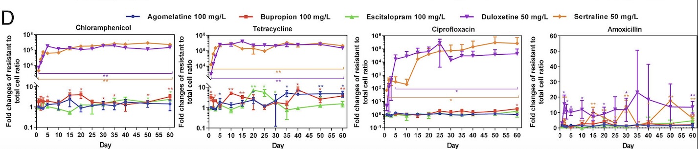
        Collaborating with researchers Zue Wang and Zhigang Yue, Guo’s lab began by choosing five major types of antidepressant medications: sertraline, escitalopram, bupropion, duloxetine, and agomelatine. These medications differ in the ways that they prevent the reuptake of serotonin and norepinephrine in the brain, thereby allowing the researchers to examine the effects of various types of antidepressants that may be prescribed to patients. Then, E. coli bacteria were added to media containing varying concentrations of these five antidepressants. Once these cells were treated with antidepressants, the researchers began to test the cells’ resistance against antibiotics. In order to accurately reflect antibiotic use in the real world, the tested antibiotics covered the six main categories of antibiotic medications available on the market. The antidepressant-treated bacteria were then swabbed onto plates containing one of the tested antibiotics to observe cell growth. Based on the growth present on these plates, the researchers were able to estimate the incidence rate of bacterial resistance of E. coli bacteria treated with different antidepressants.

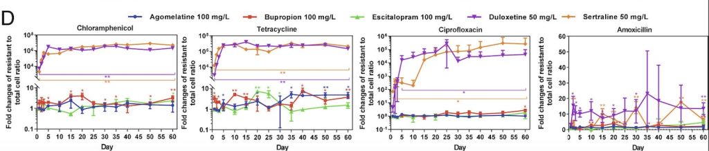
         Through this experiment, the lab observed that E. coli cells grown in sertraline and duloxetine, two antidepressants that inhibit the reuptake of serotonin, exhibited the greatest number of resistant cells across all the tested antibiotics (fig. 1). They also noted that E. coli cells exhibiting resistance to one antibiotic often demonstrate some level of resistance to other antibiotics as well. After detecting a correlation between antibiotic-resistance development and exposure to antidepressants, the lab tested the concentration dependence of this effect. While lowering the concentration of antidepressants seemed to decrease the amount of resistant E. coli cells, resistant cells continued to appear on the plates over time, suggesting that lowering antidepressant dosages only prolongs the process of developing antibiotic resistance.

Figure 1: These graphs showcase the change in the number of antibiotic-resistant E. coli cells after exposure to antidepressants over sixty days. The title of each graph indicates the tested antibiotic while the colored trend lines on the graph represent one of the five, tested antidepressants. On the y-axis of each graph, the fold change measurement is used to describe the change in the number of resistant cells that develop over time. As demonstrated by the purple and yellow trend lines, duloxetine and sertraline are associated with the greatest development of resistant cells to each of the four represented antibiotics. (Adapted from Wang et. al. 2019)

         After analyzing this data, the researchers were confronted with a question: what about anti-depressants led to the development of antibiotic resistance in bacteria? To examine this question, the lab used flow cytometry to examine what was happening within the bacterial cells. This lab technique uses a fluorescent dye that binds to specific intercellular target molecules, thereby allowing these components to be visualized. After applying this dye to resistant cells grown on the antibiotic agar plates, the researchers noticed the presence of specific oxygen compounds known as reactive oxygen species (ROS). Unstable ROS bind to other molecules within a cell, disrupting normal functioning and causing stress. Elevated cellular stress levels have been shown to induce the transcription of specific genes in bacteria that produce proteins to help return the cell to normal functioning (Wang et. al. 2019).

         ROS molecules have been shown to induce the production of efflux pumps in bacteria, leading the lab to investigate if these structures were involved in the antibiotic resistance of E. coli cells. Efflux pumps are structures in the cell membrane of a protein that pump harmful substances out of the cell. The lab mapped the genome to look for activated genes associated with the production of this protein. According to the computer model, more DNA regions in resistance bacteria coding for efflux pumps were active than in the Wild Type. The researchers then concluded that efflux pumps were being produced in response to antidepressant exposure. These additional efflux pumps removed antibiotic molecules in resistant E. coli, thereby allowing them to survive in the presence of lethal drugs.

         The antibiotic resistance uncovered in this study was significant and persistent. Even one day of exposure to antidepressants like sertraline and duloxetine led to the presence of resistant cells. Furthermore, the team demonstrated that these antibiotic-resistant capabilities often do not disappear over time; rather, they are inherited between generations of bacteria, leading to the proliferation of dangerous cells unsusceptible to available treatments. The next logical step towards validating the connection between antidepressants and antibiotic resistance would include studying the gut microbiomes of patients taking anti-depressants to look for antibiotic-resistant bacteria.

         This study reveals a novel issue that must be attended to.  In 2019, 1.27 million deaths worldwide could be directly attributed to antibiotic-resistant microbes, a number expected to grow to 10 million by the year 2050 (O’Neill 2023). These “superbugs” present a dangerously growing reality. If the correlation between antidepressant use and antibiotic resistance is left uninvestigated, superbugs will likely continue to develop even as antibiotic use is regulated and monitored to battle them. Only by taking this connection seriously will researchers be able to fully grapple with and battle the growing antibiotic resistance trends, thereby preventing common infections from becoming death sentences. 

 Sources:

CDC. (2022, July 15). The biggest antibiotic-resistant threats in the U.S. Centers for Disease Control and Prevention. https://www.cdc.gov/drugresistance/biggest-threats.html

Drew, L. (2023). How antidepressants help bacteria resist antibiotics. Nature. https://doi.org/10.1038/d41586-023-00186-y

Jin, M., Lu, J., Chen, Z., Nguyen, S. H., Mao, L., Li, J., Yuan, Z., & Guo, J. (2018). Antidepressant fluoxetine induces multiple antibiotics resistance in Escherichia coli via ROS-mediated mutagenesis. Environment International, 120, 421–430. https://doi.org/10.1016/j.envint.2018.07.046 

Lukić, I., Getselter, D., Ziv, O., Oron, O., Reuveni, E., Koren, O., & Elliott, E. (2019). Antidepressants affect gut microbiota and Ruminococcus flavefaciens is able to abolish their effects on depressive-like behavior. Translational Psychiatry, 9(1), 1–16. https://doi.org/10.1038/s41398-019-0466-x

O’Neill, J. (Ed.). (2016). Tackling Drug-Resistant Infections Globally: Final Report and Recommendations. The Review on Antimicrobial Resistance. https://amr-review.org/sites/default/files/160518_Final%20paper_with%20cover.pdf

Thompson, T. (2022). The staggering death toll of drug-resistant bacteria. Nature. https://doi.org/10.1038/d41586-022-00228-x

Wang, Y., Yu, Z., Ding, P., Lu, J., Mao, L., Ngiam, L., Yuan, Z., Engelstädter, J., Schembri, M. A., & Guo, J. (2023). Antidepressants can induce mutation and enhance persistence toward multiple antibiotics. Proceedings of the National Academy of Sciences, 120(5), e2208344120. https://doi.org/10.1073/pnas.2208344120

Filed Under: Biology, Chemistry and Biochemistry, Science Tagged With: antibiotics, antidepressants, bacteria

Ferroptosis-Related LncRNAs Found in Colon Cancer

April 2, 2023 by Emma Cheung '26

Colon cancer is the malignant growth of tumor cells in the large intestine. It is the third most common cancer and has the fourth highest death rate out of all types of cancer. Current treatments are limited, as they can be painful for the patient by killing healthy cells alongside cancer cells, and there is no guarantee that the treatments will completely eliminate all cancer cells.

Ferroptosis is a type of programmed cell death caused by high intracellular iron levels, which in turn activates cell death pathways. It differs from traditional cell death, apoptosis, since it is triggered by high iron intracellular concentrations. Rather than affecting the cell’s genetic material or plasma membrane, ferroptosis causes cell death through shrinking mitochondria and increasing mitochondrial membrane density. 

Long noncoding RNAs (lncRNAs) are a type of RNA that does not code for protein synthesis. While they don’t code for protein, lncRNAs have other functions, such as controlling gene regulation through unwinding chromatin for transcription and consequent translation and RNA processing. In regards to cancer, lncRNAs have been proven to contribute to proliferation, metastasis, and reproduction of malignant cells, and can therefore be indicators of the disease and its prognosis. Ferroptosis-related lncRNAs (FRLs), which influence the titular cellular process, in particular have been identified as possible indicators of cancer prognosis, yet not much is known.

The purpose of Wu et al (2022)’s study was to determine the molecular functions of FRLs in colon cancer. In this experiment, RNA sequencing data and genes related to ferroptosis were obtained from databases. In addition, human intestinal epithelial cells and various human colon cancer cell lines and colon cancer cell samples taken from patients at the Gastrointestinal Surgery Department of Xiangya 3rd Hospital were tested for cell composition via CIBERSORT, and had their RNA extracted for qRT-PCR and analysis. Malondialdehyde (MDA), Fe2+, reactive oxygen species (ROS), and IC50 levels of various drugs were also tested in these cells, as they all have a role in controlling ferroptosis and the consequent cell death. It was found that 26 different FRLs had some relationship to colon cancer, most of them being risk genes, genes specifically associated with the onset of cancer. Two lncRNAs, AP003555.1 and AC005841.1, had a significant relationship to colon cancer, as seen by the increased MDA, Fe2+, and ROS levels in cells with those two lncRNAs silenced and their knockout inhibiting cell proliferation.


Figure 1: Construction and validation of the ferroptosis-related lncRNA signature model in the training cohort, validation and overall groups. (A–C) The distribution of the risk scores and the distributions of overall survival status and risk score in the training, validation and overall groups. (D–F) The Kaplan–Meier curves for survival status and survival time in the training, validation and overall groups. (G–I) The receiver operating characteristic (ROC) curve shows the potential of the prognostic ferroptosis-related lncRNAs signature in predicting 1-, 2-, and 3-year overall survival (OS) in the training, validation and overall groups. (J–L) AUC of ROC curves comparing the prognostic accuracy of the risk score and other prognostic factors in the training, validation and overall groups.


Sources

Li, Jie, Feng Cao, He-liang Yin, Zi-jian Huang, Zhi-tao Lin, Ning Mao, Bei Sun & Gang Wang (2020), Ferroptosis: past, present and future, Cell Death and Disease, Volume 11, Issue 2, Page 88


Mármol, Inés, Cristina Sánchez-de-Diego, Alberto Pradilla Dieste, Elena Cerrada, and María Jesús Rodriguez Yoldi (2017) Colorectal Carcinoma: A General Overview and Future Perspectives in Colorectal Cancer, International Journal of Molecular Sciences, Volume 18, Issue 1, Pages 197. 


Qian, Yuchen, Lei Shi, and Zhong Luo (2020) Long Non-coding RNAs in Cancer: Implications for Diagnosis, Prognosis, and Therapy, Frontiers in Medicine


Wu, Zhiwei, Zhixing Lu1, Liang Li, Min Ma, Fei Long, Runliu Wu, Lihua Huang, Jing Chou, Kaiyan Yang, Yi Zhang, Xiaorong Li, Gui Hu, Yi Zhang, and Changwei Lin (2022) Identification and Validation of Ferroptosis-Related LncRNA Signatures as a Novel Prognostic Model for Colon Cancer, Sec. Cancer Immunity and Immunotherapy, Volume 12


Yao, Run-Wen, Yang Wang & Ling-Ling Chen (2019) Cellular functions of long noncoding RNAs, Nature Cell Biology, Volume 21, Issue 5, Pages 542-551


Yu, Haitao, Pengyi Guo, Xiaozai Xie, Yi Wang, and Gang Chen (2017) Ferroptosis, a new form of cell death, and its relationships with tumourous diseases, Journal of Cellular and Molecular Medicine, Volume 21, Issue 4, Pages 648–657


Zhang, Kaiming, Liqin Ping, Tian Du, Gehao Liang, Yun Huang, Zhiling Li, Rong Deng, and Jun Tang (2021) A Ferroptosis-Related lncRNAs Signature Predicts Prognosis and Immune Microenvironment for Breast Cancer, Frontiers in Molecular Bioscience

Filed Under: Biology, Chemistry and Biochemistry

Targeting the MYC Proto-Oncogene, BHLH Transcription Factor (MYC) interaction network in B-cell lymphoma via histone deacetylase 6 inhibition

November 11, 2022 by Emma Cheung '26

According to the World Health Organization (WHO), in 2020, cancer was responsible for the deaths of almost ten million people worldwide. Such statistics place cancer as a leading cause of death worldwide, second to heart disease. Cancer is when a series of mutations occurs in a cell, resulting in uncontrollable cellular division that eventually leads to interference in the function of vital organs. One of the more common types of cancer is lymphoma, the malignant growth of tumor cells of the lymphatic system. Current treatments for lymphoma include radiation therapy and chemotherapy, but these treatments can have drawbacks: they can be painful for the patient by killing healthy cells alongside cancer cells, and there is no guarantee that the treatments will completely eliminate all cancer cells. With a treatment that specifically targets the malignant cells, we can better treat lymphoma as well as other types of cancers.

MYC is a gene that when expressed in moderation, is responsible for maintaining cellular functions such as the cell cycle, apoptosis (programmed cell death), and protein production. It does so through “recruiting” enzymes such as histone acetyltransferases p300/CBP or the histone deacetylases (HDACs) to regulate expression of other genes. However, dysregulation of MYC expression can cause these cell functions to lose control as HDACs will have no means of regulation, genes to aid in the increase in cellular processes and pathways that would lead to the cell to become cancerous. MYC has also been found to be overexpressed in other types of cancers, such as uterine leiomyosarcoma.

The purpose of this project was to determine the effect of HDAC6 inhibitor Marbostat-100 (M-100) on oncogenic MYC expression levels in mice with MYC-induced aggressive B-cell lymphoma. In this experiment, mice with B-cell lymphoma as well as human B cell lymphoma cells were treated with various concentrations of M-100. It was found that all experimental concentrations of M-100 caused HDAC inhibition and reduction of MYC expression and protein levels, consequently inducing apoptosis in the murine and human cancer cells and statistically significantly increasing the mice’s survival rates. Therefore, MYC inhibition could be a possible therapeutic treatment for cancers like B-cell lymphoma.

Sources

https://www.nature.com/articles/s41388-022-02450-3

https://www.who.int/news-room/fact-sheets/detail/cancer

https://www.cancer.gov/about-cancer/treatment/types

Filed Under: Biology, Chemistry and Biochemistry

Examining the work of 2022 Nobel Prize in Physiology or Medicine Laureate Svante Pääbo

November 6, 2022 by Luke Taylor '24


Svante Pääbo. Max Planck Institute for Evolutionary Anthropology. Retrieved November 6, 2022. https://www.eva.mpg.de/genetics/staff/paabo/#c28042


            Have you ever wondered how humans lived on Earth before the first major civilizations formed? It turns out that we were not the only hominid species on Earth in those times: other relatives of humans, such as Neanderthals, lived and intermingled with humans. The genetic relationship between humans and these hominids is a subject of great interest to the scientific community, since the traits of our species’s distant relatives can explain genetic phenomena in modern humans. On October 3rd, 2022, the Nobel Prize Committee announced they would be awarding Swedish geneticist Svante Pääbo the prize in Physiology or Medicine “for his discoveries concerning the genomes of extinct hominins and human evolution” (NobelPrize.org, 2022). One of Dr. Pääbo’s most significant achievements leading to his prize was sequencing the Neanderthal genome. A closer look at Dr. Paabo’s work in the field of genetics elucidates how his work led to the foundation of the field of paleogenetics.

            Originally a student of Egyptology, Dr. Pääbo received a Ph.D. in molecular immunology from the University of Uppsala in 1986, but his interest in the former remained: his first Nature publication was about cloning ancient DNA from ancient Egyptian mummies (Gruber Foundation, 2022; Pääbo, 1985). Despite the possibility of degradation or contamination from the mummification process itself and from the millenia that passed since, he found that surface-level tissue samples in a one-year old boy yielded DNA that could be cloned using DNA recombination techniques (Pääbo, 1985). Since publishing this, Dr. Pääbo has made a career of refining techniques that allow the sequencing of genomes from many other types of ancient humans or hominins.
            Dr. Pääbo’s discoveries have advanced the field of paleogenomics, the study of genomes belonging to extinct species. Of primary concern is recovering ancient DNA (aDNA) from specimens in ideal physical conditions, as in dry and high-salinity environments, since in those environments long DNA molecules will not degrade as fast (Lan, 2019). Then, with techniques such as polymerase chain reaction (PCR) and Sanger sequencing, the aDNA molecules can be cloned and amplified to allow scientists to study copies of genes without needing more of the original sample (Lan, 2019). Storing individual genes from these recovered genomes in bacteria allows scientists to form “libraries” of specimen genomes. The contents of these genomic libraries can then be analyzed to fully sequence the genome of the specimen. After sequencing the genome of one species, scientists can then compare the genome to a related species to identify the differences (Genome.gov, 2020).
            In 2010, Dr. Pääbo and colleagues published “A Draft Sequence of the Neandertal Genome,” where they collected and analyzed aDNA from three individual Neanderthal specimens in Europe using the techniques described above (Green, 2010). Using data from this draft genome of the Neanderthal, they identified key genetic differences between modern humans and ancestral species. In particular, there were mutations in certain genes associated with disorders in modern humans, such as in RUNX2. Mutations of the RUNX2 gene can lead to cleidocranial dysplasia, a disorder that causes protruded frontal bones on the cranium and bell-shaped rib cages. These symptoms resemble the known skeletal morphologies of Neanderthals, which gives researchers a clue as to how humans and Neanderthals diverged genetically from each other (Green, 2010).
            The discoveries that Dr. Pääbo made in the field of paleogenomics have brought to light how molecular differences between Neanderthals and humans translate to their defining features as species. With the techniques that he developed, scientists can now examine the genomes of specimens thousands of years old without fear of contamination. Future developments in the field of paleogenomics could be expanding upon the links between Neanderthal DNA in human genomes and risk factors for diseases like COVID-19, which Dr. Pääbo himself has contributed to (Zeberg & Pääbo, 2021). Svante Pääbo’s work will help scientists uncover further links between our distant ancestors and modern humans for decades to come.


Works Cited:

Callaway, E., & Ledford, H. (2022). Geneticist who unmasked lives of ancient humans wins medicine Nobel. Nature, 610(7930), 16–17. https://doi.org/10.1038/d41586-022-03086-9


DNA Sequencing Fact Sheet. (n.d.). Genome.Gov. Retrieved November 6, 2022, from https://www.genome.gov/about-genomics/fact-sheets/DNA-Sequencing-Fact-Sheet


Green, R. E., Krause, J., Briggs, A. W., Maricic, T., Stenzel, U., Kircher, M., Patterson, N., Li, H., Zhai, W., Fritz, M. H.-Y., Hansen, N. F., Durand, E. Y., Malaspinas, A.-S., Jensen, J. D., Marques-Bonet, T., Alkan, C., Prüfer, K., Meyer, M., Burbano, H. A., … Pääbo, S. (2010b). A Draft Sequence of the Neandertal Genome. Science, 328(5979), 710–722. https://doi.org/10.1126/science.1188021


Lan, T., & Lindqvist, C. (2019). Paleogenomics: Genome-Scale Analysis of Ancient DNA and Population and Evolutionary Genomic Inferences. In O. P. Rajora (Ed.), Population Genomics: Concepts, Approaches and Applications (pp. 323–360). Springer International Publishing. https://doi.org/10.1007/13836_2017_7


Pääbo, S. (1985). Molecular cloning of Ancient Egyptian mummy DNA. Nature, 314(6012), Article 6012. https://doi.org/10.1038/314644a0

The Nobel Prize in Physiology or Medicine 2022. (n.d.-a). NobelPrize.Org. Retrieved October 16, 2022, from https://www.nobelprize.org/prizes/medicine/2022/advanced-information/


The Nobel Prize in Physiology or Medicine 2022. (n.d.-b). NobelPrize.Org. Retrieved October 16, 2022, from https://www.nobelprize.org/prizes/medicine/2022/press-release/


The Nobel Prize in Physiology or Medicine 2022. (n.d.-c). NobelPrize.Org. Retrieved October 16, 2022, from https://www.nobelprize.org/prizes/medicine/2022/paabo/facts/

Svante Pääbo | Gruber Foundation. (n.d.). Retrieved October 16, 2022, from https://gruber.yale.edu/genetics/svante-p-bo


Svante Pääbo—Max Planck Institute for Evolutionary Anthropology. (n.d.). Retrieved October 22, 2022, from https://www.eva.mpg.de/genetics/staff/paabo/#c28042


Warren, M. (2018). Mum’s a Neanderthal, Dad’s a Denisovan: First discovery of an ancient-human hybrid. Nature, 560(7719), 417–418. https://doi.org/10.1038/d41586-018-06004-0
Zeberg, H., & Pääbo, S. (2021). A genomic region associated with protection against severe COVID-19 is inherited from Neandertals. Proceedings of the National Academy of Sciences, 118(9), e2026309118. https://doi.org/10.1073/pnas.2026309118

Zeberg, H., & Pääbo, S. (2021). A genomic region associated with protection against severe COVID-19 is inherited from Neandertals. Proceedings of the National Academy of Sciences, 118(9), e2026309118. https://doi.org/10.1073/pnas.2026309118

Filed Under: Biology, Science

From Crystal Balls to Blue Flies: Death Prediction in the Modern Scientific World

November 6, 2022 by Alexa Comess '26

To most people, the phrase “death prediction” conjures distant images of glowing crystal balls, vibrant tarot cards, or the mystical fortune tellers in popular movies like Big and Ghost. Despite the terrifying implications of a finite and predictable death, a societal obsession with it pervades our media, culture, and everyday life. Though death prediction has historically been confined to fiction and spirituality, scientific advances are transforming it into an imminent next step.

Until the early 2010s, death prediction in the scientific sphere was limited to chronological age. The basic understanding that humans are more likely to die as they reach the end of their average life span was, and still is in many ways, the foundation to any scientific attempts to predict mortality (Gaille et al.). However, more recently, researchers have discovered observable markers of “physiological age”, or traits independent of chronological age that indicate when an individual organism is near the end of its life. In a 2012 experiment, scientists Rera et al. found a reliable predictor in the model organism Drosophila melanogaster, otherwise known as the common fruit fly. According to the study, the flies enter an identifiable “pre-death stage” marked by an increase in intestinal permeability, which can accurately predict when they are near the end of their life. Increases in intestinal permeability were tracked by injecting the drosophila flies with a non-digestible blue dye and observing if their intestinal walls allowed the dye to pass through, causing them to externally turn blue (in drosophila with normal levels of intestinal permeability the dye remained confined to the digestive tract). The high intestinal permeability associated with the blue flies and pre-death stage was appropriately dubbed the “Smurf phenotype”.

Drosophila with the Smurf phenotype were observed to have significantly lower remaining life spans than their age-matched non-Smurf counterparts. While the link between intestinal dysfunction and approaching death is still not fully understood, recent data point to changes in immunity-related gene expression and the aging fly’s microbiome as potential causes. These changes can be caused by old age or other afflictions; in the Smurf flies who were significantly below the average lifespan of the species, other morbidities were often observed, such as mitochondrial dysfunction, increased internal bacterial load, and insulin resistance syndrome. Evidently, the flies’ transition to their “pre-death stage”, or the Smurf phenotype, is a more accurate and comprehensive predictor of death than chronological age (Rera et al.). This biological phenomenon was later observed in other animals too, notably zebrafish and nematodes (Gaille et al.). The prevalence of this observable transition in multiple organisms coupled with its accuracy is slowly but surely turning mortality prediction into a reality.

Surprisingly, death prediction in humans is not far off from the developments seen in Drosophila and other model organisms. In a 2019 UCLA clinical trial, scientists tentatively proved that intestinal permeability is linked to approaching mortality in humans. While the trial was small and needs to be replicated, it provided significant evidence that the efficacy of intestinal permeability decline in death prediction has significant potential for human mortality prediction (Angarita et al.).

Outside of intestinal permeability, scientists have discovered alternative ways to predict a pre-death stage in humans. In a 2014 study, Pinto et al. theorized that olfaction could serve as another indicator as it relies heavily on peripheral and central cell regeneration, which tend to degrade near the end of an individual’s life due to old age or other morbidities. In the study, roughly 3,000 adults in the age range of 57-85 were asked to identify five different common odorants via forced choice. After 5 years, scientists collected data on which subjects were still alive, and analyzed the connection between their olfactory capability and mortality within the 5 year span. The findings were startling: the mortality rate was four times higher for adults with complete loss of smell than adults with fully intact senses of smell (Pinto et al.).

  A: Olfactory dysfunction versus 5 year mortality separated by age group

B: Progression of errors in scent identification versus 5 year mortality (Pinto et al.)

While other methods of death prediction such as biomarkers, genetic screenings, and
demographic studies exist, the discovery of pre-death indicators like intestinal permeability and olfactory decline grant us a unique and improved perspective on mortality. As research continues to grow on this subject, we must question the implications these developments have on our society: how will we reckon with the seemingly impossible ability to predict the future? Is it possible to enjoy life with an exact knowledge of its end? How will both health and wealth inequalities affect the commercialization of testing for death prediction? The moral and ethical dilemmas arising from this development are boundless.

Though these questions do not have finite answers, they must remain present in our discussions of death prediction. While scientific innovations like mortality predictors hold great promise in advancing society, they also have the capacity to exacerbate inequity and other social ills. As rapid development continues to occur in the scientific world, we must maintain both an open mind and an understanding of the complex challenges change poses to our world.

Works Cited

Angarita, Stephanie A. K., et al. “Quantitative Measure of Intestinal Permeability Using Blue Food Coloring.” Journal of Surgical Research, vol. 233, Jan. 2019, pp. 20–25. DOI.org (Crossref), https://doi.org/10.1016/j.jss.2018.07.005.

Gaille, Marie, et al. “Ethical and Social Implications of Approaching Death Prediction in Humans – When the Biology of Ageing Meets Existential Issues.” BMC Medical Ethics, vol. 21, no. 1, Dec. 2020, p. 64. DOI.org (Crossref), https://doi.org/10.1186/s12910-020-00502-5.

Pinto, Jayant M., et al. “Olfactory Dysfunction Predicts 5-Year Mortality in Older Adults.” PLoS ONE, edited by Thomas Hummel, vol. 9, no. 10, Oct. 2014, p. e107541. DOI.org (Crossref), https://doi.org/10.1371/journal.pone.0107541.

Rera, Michael, et al. “Intestinal Barrier Dysfunction Links Metabolic and Inflammatory Markers of Aging to Death in Drosophila.” Proceedings of the National Academy of Sciences, vol. 109, no. 52, Dec. 2012, pp. 21528–33. DOI.org (Crossref), https://doi.org/10.1073/pnas.1215849110.

Cover Image Credit: https://www.npr.org/2019/07/26/745361267/hello-brave-new-world

Filed Under: Biology, Science Tagged With: Biology, Death Prediction, Ethics

Immortality: a biological possibility

November 6, 2022 by Anika Sen

Immortality is biologically possible. It has been biologically possible ever since the discovery of the ‘immortal’ jellyfish Turritopsis. dohrnii in the Mediterranean Sea in 1883. This cnidarian is an exception to the normal cycle of life and death; they have an extra stage in their life cycle known as ‘rejuvenation’ where the mature medusa can metamorphose back into its juvenile form – as polyps. This is usually in response to damage or natural deterioration with age. If they are not eaten or killed by predators, they can rejuvenate and live forever. Therefore there could actually be an existing Turritopsis. dohrnii jellyfish that has lived since the time when dinosaurs roamed the Earth, as this species of jellyfish have been floating in the oceans since 66 million years ago. 

Fig 1: The immortal jellyfish: Turritopsis. dohrnii (American Museum of Natural History, 2015)

The diagram on the left of Figure 2 shows what the life cycle of a jellyfish normally is. The fertilized egg first grows into a small larva, known as a planula (Pascual-Torner, 2021). The planula the proceeds to ground itself into a solid surface and form a polyp where it develops a digestive system and reproduces asexually to form a colony (Pascual-Torner, 2021). A section of the polyp within the colony develops a new set of nerves and muscle, which can swim, grow and feed independently; eventually growing into a medusa, a full grown adult jellyfish which can reproduce sexually (Pascual-Torner, 2021). If not by being eaten or injured by a predator, old age is usually what kills these jellyfish (Pascual-Torner, 2021). 

Fig 2: The life cycles of Turritopsis. rubra (‘normal’ lifecycle) and Turritopsis. dohrnii  (Pascual-Torner, 2021)

Aging is generally governed by cellular senescence – formally defined as a state of cell cycle arrest where proliferating cells stop responding towards growth-promoting stimuli (Osterloff). This is usually in response to stresses such as telomere dysfunction and persistent DNA damage (Cell Signaling Technology). The number of senescent cells normally increases with age, negatively impacting other biological processes and impairs any potential for pluripotency – ability of a cell to differentiate into any type of specialized cell – and the possibility of regeneration. But this cnidarian species is able to challenge this trait and reverse their life cycle even after reaching sexual maturity, through a process called ontogeny reversal.

Comparative genomic studies have been conducted in hopes to find the genes involved in ontogeny reversal. In the study conducted by Pascual-Torner et al. (2021), the genes involved in aging and DNA repair were compared between Turritopsis. dohrnii and Turritopsis. rubra, which can’t rejuvenate at mature stages. Some of their findings suggested that Turritopsis. dohrnii may have “more efficient replicative mechanisms and repair systems” (Pascual-Torner et al., 2021). This includes the amplification of the genes POLD1 and POLA2, which encode for the enzyme DNA polymerase. This enzyme is involved in DNA replication, therefore its respective genes being amplified suggest enhanced replication in this cnidarian species. Furthermore, duplications to certain DNA repair genes such as XRCC5, GEN1, RAD51C, and MSH2 suggest more efficient DNA repair mechanisms (Pascual-Torner et al., 2021). A more efficient DNA repair mechanism reduces DNA damage and the triggers for cellular senescence, resulting in the slowing down of aging. In addition, there are also many other genomic differences noted by the study that also contribute to reducing the stressors for senescence that are not mentioned in this article. 

However, the ability Turritopsis. dohrnii has for ontogeny reversal implies that this jellyfish species additionally possesses some sort of cell reprogramming mechanism. To promote dedifferentiation, where cells grow in reverse from a differentiated stage to a less differentiated stage, there should be pathways that target the enzyme PRC2 (polycomb repression complex 2) and pluripotency related genes (Pascual-Torner et al., 2021). PRC2 catalyzes methylation of a specific set of histones to silence specific genes that enhance and maintain pluripotency in embryonic stem cells (Pascual-Torner et al., 2021). According to the same study, the silencing of PCR2 targets and activation of pluripotency targets was observed in Turritopsis. dohrnii (Pascual-Torner et al., 2021). Through these mechanisms, pluripotency is enhanced, leading the jellyfish to be able to form undifferentiated cells and thereby reversing back into its cyst stage, which is similar to its planula stage, as shown in Figure 2. 

It is mind-blowing that a mere floating sea creature is able to reverse its lifecycle and biologically be immortal. There is still a lot unknown about its process to being able to be immortal; scientists have only just started to uncover the basics from their genomic analysis. Maybe as scientists go deeper into uncovering Turritopsis. dohrnii’s strange immortality, we can start to think about whether we can transfer this ability to other creatures, even humans – or if we even want to? Now that immortality is actually a reality, do we want it to be a biological possibility for other creatures, for us? 

References

“Cellular Senescence.” Cell Signaling Technology, https://www.cellsignal.com/science-resources/overview-of-cellular-senescence. 

Matsumoto, Yui, and Maria Pia Miglietta. “Cellular Reprogramming and Immortality: Expression Profiling Reveals Putative Genes Involved in Turritopsis Dohrnii’s Life Cycle Reversal.” Genome Biology and Evolution, edited by Dennis Lavrov, vol. 13, no. 7, July 2021, p. evab136. DOI.org (Crossref), https://doi.org/10.1093/gbe/evab136.

Osterloff, Emily. “Immortal Jellyfish: The Secret to Cheating Death.” Natural History Museum, https://www.nhm.ac.uk/discover/immortal-jellyfish-secret-to-cheating-death.html. 

Pascual-Torner, Maria, et al. “Comparative Genomics of Mortal and Immortal Cnidarians Unveils Novel Keys behind Rejuvenation.” Proceedings of the National Academy of Sciences, vol. 119, no. 36, Sept. 2022, p. e2118763119. DOI.org (Crossref), https://doi.org/10.1073/pnas.2118763119.

“The ‘Immortal’ Jellyfish That Resets When Damaged: AMNH.” American Museum of Natural History, 2015, https://www.amnh.org/explore/news-blogs/on-exhibit-posts/the-immortal-jellyfish.


Filed Under: Biology, Chemistry and Biochemistry, Science

Recently Discovered Fossil Sheds Light on Paired Limb Evolution

November 6, 2022 by Graham Lucas '26

         Evolutionary biologists consistently work with limited information to untangle the complicated web of life’s origin and evolution. However, new evidence constantly emerges and fills gaps in scientific understanding. A well-preserved new species of galeaspid, a clade of extinct jawless fishes, called Tujiaaspis vividus provides novel insight into the evolutionary history of paired appendages. The researchers modeled water flow around the specimen’s paired fin flaps to analyze the function of the non-muscularized fins in improving the mobility of T. vividus (Gai et al., 2022).

The evolution of a jaw is a key development in the history of vertebrates as it marks when paired appendages started to develop and is a shared trait in most living vertebrates. Thus, modern-day jawless fish like hagfish and lampreys are classified into a different clade from all other vertebrates to indicate the divergence. While galeaspids like T. vividus lacked jaws, they were a stepping stone on the evolutionary path to jawed vertebrates and thus can help understand the evolutionary connection between jawless vertebrates and jawed vertebrates (gnathostomes). T. vividus has paired ventrolateral fins that are not muscularized like the fins of a modern bony fish or the appendages of land animals. However, these paired fins flaps are a necessary intermediary in the development of paired fins. Therefore, T. vividus morphology provides insight into how evolution through natural selection can occur gradually (Gai et al., 2022).

The researchers investigated how the development of primitive paired limbs could have improved mobility for galeaspids. This allowed them to hypothesize about the original reasons paired limbs were useful. To do so, they simulated water flow over models of T. vividus with and without paired fins at many angles of attack. Varying the attack angle means that the researchers varied the direction of water flow in their model to gain a more complete understanding of the hydrodynamics of T. vividus. The authors found that ventrolateral fins generated passive lift and improved movability in the water. The surviving ancestors of T. vividus are not sessile, meaning they are not confined to the sea floor like a clam. However, hagfishes and lampreys are mostly benthic, spending their time on the seafloor. Improved lift in galeaspids provides insight into how bony and cartilaginous fishes could move towards being true nekton, which implies total mobility in the water column (Gai et al., 2022).

This analysis is relevant to the understanding of current evolutionary theories. The new-head hypothesis postulates that the mobility of jawed vertebrates enabled by the development of paired appendages allowed them to develop more active feeding practices (Gai et al., 2022). The paired appendages of ancestral gnathostomes that generated lift underwater supports the hypothesis that increased mobility coincided with more active predation practices (Gai et al., 2022). Additionally, the lateral fin fold hypothesis argues that distinct fins emerged from a long fold around the body of early vertebrates (Diogo, 2020). This theory has always been challenging to test, as the remains of many relevant species lack bone structure around the possible fin fold (Gai et al., 2022). The findings of this study suggest a more complex fin evolution where paired fins groups evolved separately, although still in the same chronological order as the fin-fold hypothesis postulates, pectoral fins before pelvic fins (Gai et al., 2022). Overall, this study suggests that paired fins played an important role in creating more mobile life. 

Illustrator: Isabelle Lee ’25 (https://news.wisc.edu/jawless-fish-take-a-bite-out-of-the-blood-brain-barrier/)

Works Cited

Diogo, R. (2020). Cranial or postcranial—Dual origin of the pectoral appendage of vertebrates combining the fin-fold and gill-arch theories? Developmental Dynamics, 249(10), 1182–1200. https://doi.org/10.1002/dvdy.192

Gai, Z., Li, Q., Ferrón, H. G., Keating, J. N., Wang, J., Donoghue, P. C. J., & Zhu, M. (2022). Galeaspid anatomy and the origin of vertebrate paired appendages. Nature, 609(7929), 959–963. https://doi.org/10.1038/s41586-022-04897-6

Filed Under: Biology, Science

Beware the Blob!

November 6, 2022 by Larah Gutierrez-Camano '26

“Beware of the Blob! It creeps, and leaps, and glides and slides across the floor! Indescribable…Indestructible! Nothing Can Stop It! The indestructible creature! Bloated with the blood of its victims!” (The Blob, 1958). Physarum polycephalum, nicknamed the “The Blob” after the 1958 classic from Irvin Yeaworth and Russel Doughten, has taken the scientific community by storm. Its impressive repertoire includes spending a summer in space, modeling the early evolutionary history of eukaryotes, and of course, navigating the reproductive scene with over 720 sexes in one organism. Traveling without legs and healing injuries in just under two minutes, this slime mold “belongs to one of nature’s mysteries” according to Bruno David, director of the Paris Museum of Natural History (The Guardian, 2019). 

Such a creature shrouded in mystery can commonly be located growing within rotting logs searching for food via a long network of thin tendrils. When the organism encounters food it grows over the object, secreting digestive enzymes to “consume” the decaying vegetation or microorganism. Its intricate structure allows for nutrients to be passed around within the network of the organism. 

Toshiyuki Nakagaki,  a mathematical biologist, and colleagues observed Physarum polycephalum’s networking capabilities to predict effective city planning. The laboratory placed the mold into a culture mirroring Tokyo’s infrastructure. Upon placing food in the city’s population centers, the organism’s tendrils uncovered pathways nearly identical to Tokyo’s railway system (Wogan, 2012). The research demonstrated Physarum polycephalum’s incredible ability to solve complex problems, such as uncovering the fastest pathway through a maze, despite having no “brain-like” center (Kramar, 2021).   

The single-celled slime mold’s ability to make intelligent decisions without a central nervous system, “a memory without a brain,” has sparked intrigue into its real-world applications. Within a medical context, the mold’s early growth could be essential to understanding how tumors supply themselves with blood. Slime molds in their early stages of growth begin as a collection of isolated spores that grow in an outwards direction. Next, the spores gather in smaller groupings which release tendrils that connect with other gatherings nearby. This eventually forms a larger single celled organism that can transport nutrients, fluid, etc within itself. This process is called “percolation transition”, when separate networks become interconnected to form a transport system. Tumors subscribe to a similar process.They produce factors that stimulate the creation of blood vessels that supply the components necessary for their growth (NCI, 2018). This process is a highly active subject of research within the field of oncology. “The Blob” may aid in furthering the field’s understanding of tumors. 

Further research on Physarum polycephalum may provide insight into not only understanding but preventing tumor growth. Hans-Gunther Dobereiner, Adrian Fessel, and colleagues from the University of Bremen and Mechanobiology Institute focus their studies on slime mold percolation transition. They observed how the mold’s tendrils grew and joined with one another similar to a subway map system, as seen earlier by Nakagaki. Researchers recorded the connections and discovered that percolation transition always happened when the collection “nodes” of the mold and the tendril lines observed a specific pattern. Regardless of the number of collection “nodes,” there was a constant ratio of tendrils to nodes. Dobereiner hopes that further research into the vascular network formation of the slime mold can lead to techniques of preventing tumor growth using their slime mold-derived mathematical model (Wogan, 2012).

Whether a Mathematician, Puzzle-enthusiast, or Urban planner Physarum polycephalum’s many talents merits many real-world applications. While it may not make a cinema debut quite like its chilling movie-star counterpart, this slime mold is ready for the big screen of the scientific community. What will Physarum polycephalum accomplish next? Certainly something, “Indescribable…indestructible…insatiable” (The Blob, 1958). 

Image Credit: https://www.imdb.com/title/tt0051418/

Works Cited

The Blob. (1958). Movie Quote. Retrieved November 6, 2022, from https://www.moviequotedb.com/movies/blob-the-1958.html.

The Guardian Staff. (2019, October 17). The ‘blob’: Zoo showcases slime mold with 720 sexes that can heal itself in minutes. The Guardian. Retrieved November 6, 2022, from https://www.theguardian.com/world/2019/oct/17/the-blob-zoo-unveils-baffling-new-organism-with-720 sexes#:~:text=The%20slime%20mold%2C%20Physarum%20polycephalum,minutes%20if%20cut%20in%20half.&text=%E2%80%9CThe%20blob%20is%20a%20living,the%20Zoological%20Park%20is%20part.

Mirna Kramar, Karen Alim. Encoding memory in tube diameter hierarchy of living flow network. Proceedings of the National Academy of Sciences, 2021; 118 (10): e2007815118 DOI: 10.1073/pnas.2007815118

National Cancer Institute. (2018). Angiogenesis inhibitors. National Cancer Institute. Retrieved November 6, 2022, from https://www.cancer.gov/about-cancer/treatment/types/immunotherapy/angiogenesis-inhibitors-fact-sheet#:~:text=have%20side%20effects%3F-,What%20is%20angiogenesis%3F,chemical%20signals%20in%20the%20body.

Technical University of Munich (TUM). “A memory without a brain: How a single cell slime mold makes smart decisions without a central nervous system.” ScienceDaily. ScienceDaily, 23 February 2021. <www.sciencedaily.com/releases/2021/02/210223121643.htm>.

Wogan , T. (2012). A slimy insight into treating cancer. Science. Retrieved November 6, 2022, from https://www.science.org/content/article/slimy-insight-treating-cancer.

Filed Under: Biology, Science

Small but Mighty: The Role of Micro-RNAs and Nanotechnology in Revolutionizing Cancer Treatment

November 6, 2022 by Sam Koegler

When you think of cancer, your mind may automatically jump to the terrifying realities of this disease: hair loss, pale skin, constant shivering, and nausea. All of these hallmarks of a cancer patient result from a current popular treatment regimen: chemotherapy. While these medications are effective for many cancer patients, they can be devastatingly hard to endure and are not always an option for every patient. Past cancer history and certain genetic mutations in tumor cells can lead to drug resistance that takes chemotherapy off the table as a treatment option. In an attempt to provide cancer patients with new medication options outside of chemotherapeutics, researchers have turned to an unassuming molecule: micro-RNA. 

Micro-RNAs (miRNAs) are short, non-coding sections of RNA that function in gene regulation cascades. Through binding to a certain region of DNA, these small biomolecules can suppress the translation of that gene into a protein. While this suppression process is a normal function of human gene regulation and protein production, dysregulation of miRNAs can lead to different levels of protein expression throughout a cell, disrupting regular maintenance processes. This dysregulation is often observed in cancer cells, as miRNAs can function as significant influencers of many hallmarks of cancer, such as cell proliferation and immortality (Ferdows, Bijan Emiliano, et al). This ability to influence cancer growth and metastasis makes miRNAs promising targets for treatments that slow the progression of tumors.

Due to the degradation of foreign RNAs in the human body, delivering miRNA treatments to target cells has proven difficult. In order to lessen this challenge, scientists have turned to nanotechnology to increase the efficacy of miRNA-targeted treatments. Nanotechnology encompasses many different organic and synthetic casings that prevent molecules from being degraded by the human body’s natural defense systems. Lipid-based nanoparticles often take the forefront of nanotechnology research. These particles are easily assimilated into the body because of their biological similarity to the lipid-based cell membrane. Cationic lipids are able to bind with the negatively charged phosphate groups in miRNA particles, creating a protective layer around these miRNA particles that can easily bind to target cells (Ferdows, Bijan Emiliano, et al). The biocompatibility found with lipid-based nanoparticles is expanded upon in extracellular vesicles, a type of molecule secreted by cells to facilitate intercellular communication. These lipid pockets are a promising target for miRNA delivery because they share many of the same biological and chemical qualities as their mother cell. Although organic nanoparticles are proving to be effective drug-delivery machines, researchers have also begun to examine the potential of using inorganic compounds to protect miRNAs from degradation. One such compound is gold-iron oxide nanoparticles (GIONS). In addition to the negatively charged GION surface that allows it to bind and transport miRNA, this nanoparticle class can also aid in the diagnosis of tumors. These particles appear on CT and MRI scans, and their appearance can help physicians determine where a tumor is located and how treatment should progress (Ferdows, Bijan Emiliano, et al).

While nanoparticle-based miRNA treatments have yet to hit mainstream cancer treatment plans, current research projects show that these medications have a promising future. In mice with transplanted lung tumors taken from human patients, cationic lipid particles have been used to deliver miRNA particles to study the effect of these treatments on patients with late-diagnosis lung cancer. Researchers used these lipid-based nanoparticles to deliver miRNA-660 to the MIR660, a gene responsible for enabling the activation of the crucial p53 tumor suppressor that results in the killing of cancer cells. In 8 weeks, the mice were shown to have 50% reduced tumor growth compared to controls, a promising result for applying this treatment in human lung cancer patients (Moro, Massimo, et al). 

GION-coated tumor-derived extracellular vesicles (TEVs) have also shown promising results as nanoparticles used in miRNA-based treatments for cancers such as breast cancer. Researchers have bound these particles to a type of naturally occurring miRNAs called anti-miRNA-21. This molecule suppresses oncomiR-21, a type of miRNA associated with assisting cancer development and growth by inhibiting apoptosis, a type of self-programmed cell death that occurs when a cell is functioning abnormally. OncomiR-21 deactivation enables a variety of proteins to regain function, allowing the cellular pathway that signals cell death to resume. When anti-miRNA-21 bound GION-TEVs were administered to breast cancer cells along with low levels of the chemotherapeutic doxorubicin, researchers found that the cells were killed almost three times quicker as compared to cells treated with doxorubicin alone (Bose RJC et al). This experimental result shows significant promise that nanoparticle-based miRNA treatments could be used in combination with chemotherapy in the future to help strengthen tumor cell apoptosis and reduce drug resistance that results from high doses of the same chemotherapeutic (Bose RJC et al). 

While these treatments show extremely significant promise, more research is needed to determine their efficacy in human patients. Specifically, inorganic nanoparticles such as GIONs require additional research to ensure their delivery is minimally toxic to human patients (Ferdows, Bijan Emiliano, et al). Fortunately, the results of current experiments demonstrate that nanoparticle-based miRNA cancer drugs could have a significant role in the future treatment of cancer patients. These treatments are minimally invasive and have the potential to allow physicians and researchers to target miRNAs to patient-specific genetic markers in tumor cells. This individualization could give patients with uniquely mutated tumors a chance for a longer lifespan or remission. In addition, the use of these treatments could reduce reliance on chemotherapy, thereby lessening drug resistance found in cancer patients with tumor recurrences (Ferdows, Bijan Emiliano, et al). Through further funding and research, nanoparticle-based miRNA cancer treatments could become the next big wave of cancer drugs to hit hospitals across the world, giving patients new hope for recovery and life after cancer. 


References

Bose RJC, Uday Kumar S, Zeng Y, Afjei R, Robinson E, Lau K, Bermudez A, Habte F, Pitteri SJ, Sinclair R, Willmann JK, Massoud TF, Gambhir SS, Paulmurugan R. Tumor Cell-Derived Extracellular Vesicle-Coated Nanocarriers: An Efficient Theranostic Platform for the Cancer-Specific Delivery of Anti-miR-21 and Imaging Agents. ACS Nano. 2018 Nov 27;12(11):10817-10832. doi: 10.1021/acsnano.8b02587. Epub 2018 Oct 22. PMID: 30346694; PMCID: PMC6684278.

Ferdows, Bijan Emiliano, et al. “RNA Cancer Nanomedicine: Nanotechnology-Mediated RNA Therapy.” Nanoscale, vol. 14, no. 12, 2022, pp. 4448–55. DOI.org (Crossref), https://doi.org/10.1039/D1NR06991H.

Moro, Massimo, et al. “Coated Cationic Lipid-Nanoparticles Entrapping MiR-660 Inhibit Tumor Growth in Patient-Derived Xenografts Lung Cancer Models.” Journal of Controlled Release, vol. 308, Aug. 2019, pp. 44–56. DOI.org (Crossref), https://doi.org/10.1016/j.jconrel.2019.07.006.

Filed Under: Biology, Chemistry and Biochemistry

Breakthrough in Gene Sequencing and Identification of Leukemia-causing Genes in Iran

November 6, 2022 by Ruby Pollack '25

A research group based in Iran conducted a study to confirm the legitimacy of gene sequencing technology for discovering Chronic Myeloid Leukemia causing genes within three pre-existing cancer patients. Chronic Myeloid Leukemia (CML) is an example of a monoclonal disease, meaning deriving from a single cell in this blood cell (hematopoietic).  CML represents around 15% of Leukemia in adults. Leukemia represents the most common blood cancer for adults older than 55. The blast cycle is the stage in chronic leukemia where tiredness, fever, and enlarged spleen are present. A blast crisis occurs when 20% of all blood or bone marrow has blasts or immature white blood cells. These blasts multiply uncontrollably, causing a blockage and stopping the production of red blood cells and platelets, which are necessary for survival. White cell crowding red blood cells often causes a lower immune system. This article focuses on using integrated genomic sequences to access common gene variants associated with CML, to understand the fundamental mechanisms behind the blast crisis.

Researchers used the Whole Exome Sequences (WES): integrated sequencing, chromosome sequences, and Rna sequences. Using a blood sample from the blast cycle patients, they used WES technology to identify modified, deleted, or incorrectly copied genes. Genes have extraordinary power over the way our body regulates. Sometimes cancer occurs when genes do not work as they are intended to. There are five classes of cancer-causing genes. Mutations in genes change how they activate other cells. A mutation in the process of spreading information through signal pathway components can lead cells to multiply erratically. Another class is transcription factors; proteins bind to a part of DNA to start or stop replication.  If there are modifications within those repressor/activator genes, that could lead to an unregulated abundance of cells—unsuppressed replication results in Tumors due to unregulated cell growth. It is common for some mutations in the replication process, but some proteins repair the genes; sometimes, if a gene gets mis-replicated, it can lead to other classes. 

CML cells are derived from the bone marrow and are progenitor cells, meaning they can turn into white, red blood cells or platelets, depending on the body’s purpose. The researcher’s goal with using WES on these pre-existing blast crisis patients was to discover essential variants and find similarities and differences with their gene makeup.  Researchers then divided their findings into PIF ( potentially significant findings) and PAFs(potentially actionable results). Using WES detected, 16 PIFS affected all five known classes of cancer-causing genes.  

Researchers conducted integrated sequencing on three patients using an in-house filtering algorithm to discover how leukemia develops. Discover that combining Integrated genomic sequencing and Rna sequencing is an accurate way to find and confirm leukemia variants. All patients are based in Iran; patient one was a 66-year-old female whose blast level was 25%. Patient 2 is a 55-year-old female whose blast level is 35%. And the third patient was a 45-year-old male with a 40% blast level. The percentage of blast level indicates how many white blood cells make up entirely of the person’s blood. The higher the blast level, the more immunocompromised you become, as CML’s white blood cells block up the flow of red blood cells stopping your immune system’s response. Using WES and RNA sequencing, researchers discovered multiple patient similarities and differences. Both patients one and two have abnormal karyotypes, or a person’s complete set of chromosomes,  in the form of a Philadelphia Chromosome. A Philadelphia Chromosome is when the 9 and 22 chromosomes break off and pair. A direct consequence is that chromosome 22 will be minimal, resulting in CML. Patients two and three both had multiple chromosome deletions, duplications, and modifications, all with prior research that points to the contribution in Leukemia.


The study affirms the importance of being able to model and analyze CML’s leukemogenesis process in terms of more timely treatment and effective management of blood-borne illness. Using gene-sequencing technology, CML’s transition to the blast phase can be detected more accurately and effectively than in previous studies.  The researchers identified variants in all classes, with an important finding that a  shared deletion was a Transcription Factor 17p, which observed that 45% of blast phase patients have such defect or mutation of a gene, creating a marker of identification and possible treatment. This study points to the importance of identifying and developing more speed-lined processes that make detection more accurate and timely. 


Kazemi-Sefat, Golnaz Ensieh (12/2022). “Integrated genomic sequencing in myeloid blast crisis chronic myeloid leukemia (MBC-CML), identified potentially important findings in the context of leukemogenesis model”. Scientific reports (2045-2322), 12 (1), p. Article.

Filed Under: Biology, Computer Science and Tech, Science

  • « Go to Previous Page
  • Page 1
  • Interim pages omitted …
  • Page 5
  • Page 6
  • Page 7
  • Page 8
  • Go to Next Page »

Primary Sidebar

CATEGORY CLOUD

Biology Chemistry and Biochemistry Computer Science and Tech Environmental Science and EOS Honors Projects Math and Physics Psychology and Neuroscience Science

RECENT POSTS

  • Venom As Medicine: Novel Pathways for Dravet Syndrome Treatment Using Modulatory Peptides from Scorpion Venom January 8, 2026
  • When Distraction Helps: Music as a Tool for Focus in ADHD Cases December 24, 2025
  • Timing Matters: How the menstrual cycle influences ACL injury in Female athletes December 24, 2025

FOLLOW US

  • Facebook
  • Twitter

Footer

TAGS

AI AI ethics Alzheimer's Disease antibiotics artificial intelligence bacteria Bathymetry Biology brain Cancer Biology Cell Biology Chemistry and Biochemistry Chlorofluorocarbons climate change Computer Science and Tech CRISPR Cytoskeleton Depression Dermatology dreams epigenetics Ethics Genes Gut microbiota honors Marine Biology Marine Mammals Marine noise Medicine memory Montreal Protocol Moss neurobiology neuroscience Nutrients Ozone hole Plants Psychology and Neuroscience REM seabirds sleep student Technology therapy Women's health

Copyright © 2026 · students.bowdoin.edu