Global warming is steadily transforming Earth’s oceans. Between 1901 and 2023, sea surface temperatures have increased at an average rate of 0.14℉ per decade (US EPA, 2016). This seemingly small thermal shift is enough to disrupt circulation patterns, alter nutrient availability, and restructure entire marine communities. As oceans absorb over 90% of excess atmospheric heat, they become both a buffer against and a victim of climate change (Climate Change, 2025). Among the many organisms affected by these changes, phytoplankton—the microscopic, photosynthetic organisms that drift near the ocean’s surface—serve as a critical case study. These single-celled producers are responsible for about half of Earth’s oxygen production, and they form the foundation of aquatic food webs, converting sunlight into chemical energy that sustains nearly all marine life (Hook, 2023). Therefore, understanding how phytoplankton respond to warming is essential for predicting the future of marine ecosystems.
Phytoplankton are highly sensitive to temperature fluctuations. Since their metabolic processes, growth rates, and enzymatic activities are temperature-dependent, even minor thermal changes can reshape their abundance and distribution. When waters warm beyond a species’ thermal tolerance, populations may decline or shift toward cooler regions (Barton et al., 2016). At the microscopic level, these shifts can cascade upward through the food web, reducing food availability for zooplankton, fish, and the higher-level predators that feed on them, such as sharks, whales, and seals. However, one key question remains: can phytoplankton adapt to rising temperatures, or will their thermal limits determine the structure of future marine ecosystems?
Huertas et al. (2011) directly addressed this question through controlled laboratory experiments designed to measure the capacity of phytoplankton to evolve under warming. The researchers selected twelve species representing a range of environments—freshwater, coastal, open-ocean, and coral symbiotic systems—to test whether thermal tolerance varied among ecological types. To simulate long-term warming, they employed a “ratchet technique,” in which phytoplankton populations were gradually exposed to higher temperatures. Each population started from a single cloned cell to remove preexisting genetic variation. Then, the cell cultures were repeatedly grown and transferred into warmer conditions, forcing the populations to either adapt to the changes through genetic mutations or face extinction.
The results revealed striking differences among species. Freshwater species, such as Scenedesmus intermedius, exhibited remarkable resilience, adapting to temperatures as high as 40°C. Coastal species like Tetraselmis suecica and Dictyosphaerium chlorelloides tolerated up to 35°C, while open-ocean species such as Emiliania huxleyi and Monochrysis lutheri showed little to no capacity for adaptation. Coral symbionts (Symbiodinium species) demonstrated limited but detectable resistance, reflecting the thermal stress already observed in coral reef environments. Importantly, adaptation was not simply a case of short-term acclimation. The researchers found that resistant populations arose at different times across replicate cultures. This serves as evidence that adaptation stemmed from rare, spontaneous genetic mutations instead of physiological flexibility. Growth rates of adapted populations diverged significantly from their ancestral strains, confirming that true evolutionary change had occurred.
These findings carry major implications for understanding the ecological future of the oceans. If phytoplankton species differ so widely in their ability to adapt, warming will likely reorganize marine communities from the bottom up. Species capable of rapid genetic adaptation may dominate, while others could decline or disappear. This uneven resilience could favor smaller, faster-growing species, altering nutrient cycling and potentially weakening the ocean’s ability to sequester carbon. Because phytoplankton drive roughly half of global primary production, any restructuring of these communities could ripple through food webs, climate regulation, and fisheries.
While Huertas et al. focused on individual species in controlled conditions, Poloczanska et al. (2016) broadens this picture to the scale of global ecosystems. Their review synthesized nearly 2,000 observations of marine organisms responding to climate change, confirming that uneven adaptation is already occurring across taxa and ocean regions. On average, species distributions are shifting towards the north and south poles by about 72 kilometers per decade, and spring life-cycle events such as breeding or migration are advancing by four days per decade. Warm-water species are becoming more abundant, while cold-water species decline. Coral calcification, the process by which corals take in calcium and carbonate ions to build their exoskeletons, is weakening under combined warming and acidification stress. These patterns mirror the interspecific variability observed by Huertas et al.; some organisms adjust successfully to changing conditions, while others falter. Here, the broader conclusion is that climate change does not affect marine life uniformly—it selectively reshapes communities based on biological flexibility, dispersal ability, and evolutionary potential.
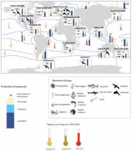
Fig 1. Global distribution of documented marine biological responses to climate change across major ocean regions (Poloczanska et al., 2016). Bars show the proportion of observed responses as consistent (dark blue), equivocal (light blue), or no change (yellow). Numbers indicate total observations per region; symbols identify taxa with ≥10 observations. Background colors represent regional sea-surface warming from 1950–2009 (yellow: low; orange: medium; red: high). Regions are defined by ecological structure and oceanographic features. eveal that climate-driven shifts in abundance, distribution, and phenology vary sharply across ocean basins—mirroring the uneven adaptive capacities described by Huertas et al. (2011).
Together, these studies illustrate both the mechanisms and the consequences of ocean warming. Huertas et al. provides mechanistic insight—showing that adaptation in phytoplankton depends on genetic change, and that some species are inherently more capable than others. Building off of this, Poloczanska et al. reveals how these species-level differences scale up, driving global shifts in abundance, distribution, and ecosystem structure. The two perspectives complement one another; laboratory experiments explain how adaptation might occur, while global syntheses show where and to what extent it already has.
As climate change accelerates, understanding the adaptability of foundational organisms like phytoplankton becomes increasingly urgent. Their evolutionary potential will determine not only the structure of marine ecosystems, but also the ocean’s capacity to regulate the planet’s climate. By linking experimental evidence with global ecological trends, researchers are beginning to map out a future ocean defined by winners and losers—a mosaic of adaptation, migration, and loss. The challenge ahead lies in predicting how these microscopic shifts will ripple through the web of life that depends on them.
References: