• Skip to primary navigation
  • Skip to main content
  • Skip to primary sidebar
  • Skip to footer

Bowdoin Science Journal

  • Home
  • About
    • Our Mission
    • Our Staff
  • Sections
    • Biology
    • Chemistry and Biochemistry
    • Math and Physics
    • Computer Science and Technology
    • Environmental Science and EOS
    • Honors Projects
    • Psychology and Neuroscience
  • Contact Us
  • Fun Links
  • Subscribe

Medicine

POTS vs Atomoxetine: The Unseen Interaction

May 4, 2025 by Martina Tognato Guaqueta

Graph describing the effects of the medication on POTS symptoms

Postural Orthostatic Tachycardia Syndrome (POTS) is a malfunction in the body’s autonomic nervous system. Rather than the blood vessels below their heart compensating by constricting, when a person with POTS goes from a lying to a standing position, a large amount of blood pools in the legs and abdomen. Normally, the blood vessels in the lower extremities constrict to maintain appropriate blood pressure throughout the whole body and help return the blood to the heart and head. The autonomic system (the part of the nervous system that is in charge of the involuntary aspects of the body) responds to low blood pressure by releasing norepinephrine and adrenaline, which cause vasoconstriction and a rise in heart rate. In POTS patients, vessels do not respond to the hormones and remain vasodilated. This combination of high heart rate and insufficient blood flow to the brain causes characteristic dizziness, fainting, and fatigue. POTS can be aggravated by a variety of things, including strenuous exercise, caffeine, hot environments, and certain medications (POTS, n.d.). 

One such class of medications is norepinephrine reuptake inhibitors (NOIs). Used to treat ADHD, major depressive disorder, and narcolepsy, NOIs block the uptake of norepinephrine in the synapses (De Crescenzo et al., 2018). This type of medication allows norepinephrine to stay in the blood longer, elevating mood and energy levels and enhancing focus. A common side effect is an elevated heart rate, which aggravates POTS. 

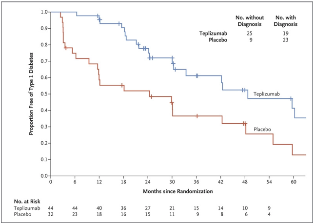
Green et al. conducted the first study examining the acute effects of atomoxetine on POTS patients. The study was composed of 27 patients and a variety of tests. A baseline was created to manage the patients’ diets. This entailed removing methylxanthines from their diet, which includes caffeine among other compounds, and moderating sodium and potassium intake. Additionally, all long-term medications were suspended for at least 5 half-life periods to ensure no hormonal effects would be present. All of these measures were taken to minimize the exacerbation of POTS symptoms (Green et al., 2013). 

All patients received the atomoxetine and the placebo (on different days). During this time, a posture study was done. Measurements of heart rate (HR), systolic blood pressure (SBP), diastolic blood pressure (DBP), mean arterial pressure (MAP), and plasma catecholamines were taken during a lying position and a standing position. This targets the effects of POTS, highlighting the possible impact of the atomoxetine. 

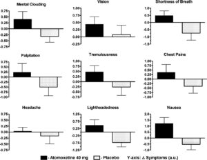
The posture study was paired with the medication study. During the medication study, patients were asked to fill out a symptom feedback form before the experiment, and every hour up to 4 hours after drug administration. This is because peak atomoxetine concentration occurs 1-2 hrs after ingestion. The Vanderbilt Orthostatic Symptom Score (VOSS) was used on the symptom feedback form, where patients are asked to rank the following on a scale from 1-10: mental clouding, brain fog, shortness of breath, palpitations, tremors, headache, tightness in the chest, blurred vision, and nausea. The lowest (1) is no symptom burden, and 10 is the worst. 

Researchers found that when patients took atomoxetine, their symptom burden increased. This presented a statistically significant increase in heart rate and a general upward trend in blood pressure throughout the 4 hours. In the case of the placebo, there was a decrease in symptom burden as the 4-hour period progressed. 

Atomoxetine is a non-stimulant medication used to treat ADHD; unfortunately, the stimulant alternatives are found to have similar effects on POTS patients. Due to a susceptibility to heart rate changes, ADHD medication negatively interacts with the condition and must be administered with exceeding caution. This interaction is important for prescribing professionals to be aware of. As this is a relatively under-researched intersection, consideration of mechanisms and close patient-doctor communication is necessary when considering medication. 

Figure 1: Results of VOSS with and without atomoxetine (Green et al., 2013)

Graph describing the effects of the medication on POTS symptoms

References

De Crescenzo, F., Ziganshina, L. E., Yudina, E. V., Kaplan, Y. C., Ciabattini, M., Wei, Y., & Hoyle, C. H. (2018). Noradrenaline reuptake inhibitors (NRIs) for attention deficit hyperactivity disorder (ADHD) in adults. The Cochrane Database of Systematic Reviews, 2018(6), CD013044. https://doi.org/10.1002/14651858.CD013044

Green, E. A., Raj, V., Shibao, C. A., Biaggioni, I., Black, B. K., Dupont, W. D., Robertson, D., & Raj, S. R. (2013). Effects of norepinephrine reuptake inhibition on postural tachycardia syndrome. Journal of the American Heart Association, 2(5), e000395. https://doi.org/10.1161/JAHA.113.000395

POTS: Causes, Symptoms, Diagnosis & Treatment. (n.d.). Cleveland Clinic. Retrieved April 8, 2025, from https://my.clevelandclinic.org/health/diseases/16560-postural-orthostatic-tachycardia-syndrome-pots


Filed Under: Biology Tagged With: ADHD, Biology, Medicine, POTS

TMJ Arthroscopy: How well does it work in Ehlers Danlos patients?

May 4, 2025 by Martina Tognato Guaqueta

The temporomandibular joint (TMJ) is responsible for the mandible’s chewing, talking, and all movement. As a condylar joint, this joint allows motion in two planes: side to side and up and down. A disc cushions the bone to facilitate this motion and serves as a lubricant. 


Figure 1: TMJ diagram (TMJ Disorders – Symptoms and Causes, n.d.).

 Temporomandibular disorders (TMD) often arise from damage or irritation relating to the disc. To determine the condition of the joint, physicians use the Wilkes scale, which allows for the assessment of internal derangement/damage of the TMJ. TMD is often a symptom of a larger condition, it can arise from connective tissue disorders, injury, teeth grinding, etc.


Figure 2: Wilkes Scale descriptive table (Table 1 . Wilkes Classification of TMJ Internal Derangement, n.d.)


A patient’s placement on this scale indicates the type of treatment they could be a candidate for.  This could range from over-the-counter medications to physical therapy, to Botox injections, and a variety of surgeries. A surgery often seen is a TMJ arthroscopy.  The arthroscopic element refers to the minimally invasive approach assisted by a camera called an arthroscope. Done under general anesthesia, a surgeon will enter the joint space through a small incision. The image of the joint is delivered to a screen through an arthroscope. During the procedure, the surgeon may reposition the disc, flush the joint, and remove scar tissue to alleviate pain (Arthroscopy for Temporomandibular Disorders (TMDs) | Kaiser Permanente, n.d.). This, however, is not the only option, and can only be decided upon looking at the full picture of a patient’s history—for example, a connective tissue disorder. 

Ehlers-Danlos (EDS) is a group of connective tissue disorders that can affect many systems, including the joints and, in turn, the TMJ.  Within the group, each particular variation has a different set of symptoms. For example, vascular EDS causes the blood vessels to rupture, whereas hypermobile EDS (hEDS) causes frequent joint dislocations. Some types of EDS have a clear genetic link, however, the origins of hEDS have yet to be understood. Due to the overly flexible nature of their joints, hEDS patients often encounter issues with their TMJ.  (Ehlers-Danlos Syndrome – Symptoms and Causes, n.d.).

Jerjes et al. conducted a retrospective case study that looked at the outcomes of 18 hEDS patients who underwent arthroscopic surgery to treat their TMDs. All patients were female and between the ages of 23-60 years old. Due to the EDS, TMJ dislocation was a common history within the sample. In turn, the dislocations were linked to the damage and pain, which were visualized during the arthroscopies. This demonstrated a link between the state on the TMJ and hEDS status. Moreover, 12 out of the 18 experienced TMD bilaterally, meaning there was pain on both sides (Jerjes et al., 2010). 

Furthermore, it seemed as though the intervention was most commonly done at/by the Wilkes Stage III (9 patients at Stage III and 5 at Stage II ). As seen in Figure 2, Stage III is characterized by moderate disc deformity and frequent pain/dislocations.  As the Wilkes Scale goes on, the condition becomes more painful and harder to treat. However, although minimally invasive, arthroscopy is considered a more extreme treatment for TMJ. Over the years, the procedure has developed and improved, reaching an 80-90% success rate (Insel et al., 2020). Despite this, the National Institute of Dental and Craniofacial Research brochure still has a negative outlook on the surgical route as a treatment for TMDs (Jerjes et al., 2010). 

Notwithstanding the controversy, Jerjes et al. demonstrate positive post-operative results. The pain subsided quickly (within 1 week post-operative) for 15 out of 18 patients and 5-6 weeks for the remaining 3. Delayed healing occurred in 4 out of 18 patients. Slow healing is a characteristic of hEDS and could be a potential reason in this case for the post-surgical complication. Most importantly, the mouth opening measured 6 months postoperatively increased from an average of approximately 23-28 mm,  with all final postoperative results being positive. 

Overall, the study aims to illustrate the success of TMJ arthroscopy in hEDS patients and suggests it as the first consideration for invasive procedures. Due to hEDS patients often having a difficult time with wound healing, a minimally invasive procedure is favored. Additionally, these patients have previously sought out other forms of treatment before surgery. This study does not suggest that surgery should be an initial consideration, but it should not be forgotten as an option. 


Figure 3: Outside view of an TMJ arthroscopy 

Figure 4: Internal photo is TMJ arthroscopy


The hEDS population is small; therefore, studies that center on them and how treatments interact with their condition are few and far between. Moreover, providers for TMJ-related surgeries are scarce. In turn, this middle ground (as noted by Jerjes et al. ) is a neglected area of research. Further development would focus on EDS patients more and seek to understand the interconnectedness of EDS and joint pain and how it can impact the choice of treatments. 


References

Arthroscopy for Temporomandibular Disorders (TMDs) | Kaiser Permanente. (n.d.). Retrieved April 8, 2025, from https://healthy.kaiserpermanente.org/health-wellness/health-encyclopedia/he.arthroscopy-for-temporomandibular-disorders-tmds.hw209368

Ehlers-Danlos syndrome—Symptoms and causes. (n.d.). Mayo Clinic. Retrieved April 8, 2025, from https://www.mayoclinic.org/diseases-conditions/ehlers-danlos-syndrome/symptoms-causes/syc-20362125

Insel, O., Glickman, A., Reeve, G., Kahan, B., Tran, T., & Israel, H. (2020). New criteria demonstrate successful outcomes following temporomandibular joint (TMJ) arthroscopy. Oral Surgery, Oral Medicine, Oral Pathology and Oral Radiology, 130(1), e20–e21. https://doi.org/10.1016/j.oooo.2019.12.022

Jerjes, W., Upile, T., Shah, P., Abbas, S., Vincent, A., & Hopper, C. (2010). TMJ arthroscopy in patients with Ehlers Danlos syndrome: Case series. Oral Surgery, Oral Medicine, Oral Pathology, Oral Radiology, and Endodontology, 110(2), e12–e20. https://doi.org/10.1016/j.tripleo.2010.03.024

Table 1. Wilkes classification of TMJ internal derangement. (n.d.). ResearchGate. Retrieved April 8, 2025, from https://www.researchgate.net/figure/Wilkes-classification-of-TMJ-internal-derangement_tbl1_7691660

TMJ disorders—Symptoms and causes. (n.d.). Mayo Clinic. Retrieved April 8, 2025, from https://www.mayoclinic.org/diseases-conditions/tmj/symptoms-causes/syc-20350941

Top Five Fun Facts About The TMJ. (2022, July 14). Ladner Village Physiotherapy | Delta BC. https://ladnervillagephysio.com/blog/top-five-fun-facts-about-the-tmj


Filed Under: Biology Tagged With: Biology, Medicine, surgery, TMJ

Asthma and ADHD: How do Pediatricians Approach This Intersection?

December 8, 2024 by Martina Tognato Guaqueta

According to the CDC, 11.4% of children aged 3-17 in the USA are diagnosed with Attention Deficit Hyperactivity Disorder (ADHD) (Data and Statistics on ADHD | Attention-Deficit / Hyperactivity Disorder (ADHD), 2024). ADHD is a developmental disorder characterized by symptoms of hyperactivity, impulsivity, and inattention, as the name suggests. Treating this disorder often requires a variety of approaches including medication, psychotherapy, and workplace or school-based accommodations (Attention-Deficit/Hyperactivity Disorder – National Institute of Mental Health (NIMH), n.d.).  Comorbidities are very common in people with ADHD, this makes it so that it is rarely the only concern during a primary care visit (Silver, 2024). Sleath et al. discuss the communication primary physicians held with families with children that have both ADHD and asthma. There has been found to be a correlation between the severity of ADHD and asthma symptoms (Blackman & Gurka, 2007). In turn, balancing treatment for both primary care visits was a driver for the paper. Asthma is a chronic lung condition that results in the narrowing of the lung pathways. Medication to alleviate symptoms of both ADHD and asthma is often prescribed at primary care visits hence the study of their intersection. 

Figure 1. Happy little girl and pediatrician doing high five after medical checkup. AAP Schedule of Well-Child Care Visits. (2023). Healthy Children.org. https://www.healthychildren.org/English/family-life/health-management/Pages/Well-Child-Care-A-Check-Up-for-Success.aspx

Sleath et al. approach this balance by studying the communication between patients with ADHD and asthma and pediatricians. The study focuses on the communication breakdown when the patient has ADHD during an asthma visit. All of these were pediatric visits. To measure the effectiveness of communication, the American Association of Pediatrics (AAP) guidelines for discussing ADHD were used. The percentage of adherence was measured through the visits using recordings. 

Before data collection eligibility tests were conducted. This made sure that all participants in the study were 8-16 years of age, could speak English, was capable of filling out an assent form, had had at least one prior visit to the clinic, had persistent asthma, and had a guardian present who is over the age of 18 and is competent in English. After the visits concluded, guardians were provided with questionnaires, and children were interviewed. These were used to supplement the recordings. 

The audio taping and coding are the backbone of the data. The audio tapes were transcribed by a coding tool that was flagged for AAP guidelines. To ensure accuracy two research assistants met twice a month to review and refine criteria. The other important aspect of the collection was a thorough socio-demographic data set: gender, age, race, insurance, and tears of asthma. All demographic data but asthma status was also recorded from guardians. 

The results yielded from this were extreme. Throughout the visits 23% of the 296 children had ADHD noted in their medical chart. It was found that boys were more likely to have ADHD diagnoses. It is important to note that it is not because ADHD affects males more, but women are less likely to get diagnosed or are diagnosed later in life due to inattentive presentations (Attoe & Climie, 2023). When understanding the extent of utilization of AAP guidelines, categories were formed; functioning, outcomes, treatment plan, ADHD asthma relationship, chronic and follow-up visits. In all of these categories, the percentage of providers that used AAP guidelines never rose above 40%. In the adherence to medication, only one provider out of the 35 discusses the topic (41 providers participated, but recording forms only 35 were usable). Overall, it was shown that AAP guidelines were more likely to be followed if the visit was unrelated to asthma, highlighting providers’ tendency to neglect proper ADHD management in patients with comorbidities. 

The aim was to highlight the need for better communication practices in the pediatric setting. Particularly in cases where comorbid conditions are present. Future development in this field would be understanding the reason behind the present communication pattern. Approaching the issue from the physician and patient perspective. On the other hand, research on how to remedy the disparity in guideline adherence. 


Article based on ‘Communication about ADHD and its treatment during pediatric asthma visits’

Sleath, B., Sulzer, S. H., Carpenter, D. M., Slota, C., Gillette, C., Sayner, R., Davis, S., & Sandler, A. (2014, Feb). Communication about ADHD and its treatment during pediatric asthma visits. Community Ment Health J ., 50(2), 185-192. 10.1007/s10597-013-9678-3

References

AAP Schedule of Well-Child Care Visits. (2023). Healthy Children.org. https://www.healthychildren.org/English/family-life/health-management/Pages/Well-Child-Care-A-Check-Up-for-Success.aspx

Attention-Deficit/Hyperactivity Disorder – National Institute of Mental Health (NIMH). (n.d.). National Institute of Mental Health. Retrieved November 1, 2024, from https://www.nimh.nih.gov/health/topics/attention-deficit-hyperactivity-disorder-adhd

Attoe, D. E., & Climie, E. A. (2023, March 30). Miss. Diagnosis: A Systematic Review of ADHD in Adult Women. J Atten Disord, 27(7), 645–657. 10.1177/10870547231161533

Blackman, J. A., & Gurka, M. J. (2007). Developmental and Behavioral Comorbidities of Asthma in Children. Journal of Developmental & Behavioral Pediatrics, 28(2), 92-99. 10.1097/01.DBP.0000267557.80834.e

Data and Statistics on ADHD | Attention-Deficit / Hyperactivity Disorder (ADHD). (2024, May 22). CDC. Retrieved November 1, 2024, from https://www.cdc.gov/adhd/data/index.html

Silver, L. (2024, April 3). ADHD Symptoms Or ADHD Comorbidity? Diagnosing Related Conditions. ADDitude. Retrieved November 1, 2024, from https://www.additudemag.com/when-its-not-just-adhd/

Sleath, B., Sulzer, S. H., Carpenter, D. M., Slota, C., Gillette, C., Sayner, R., Davis, S., & Sandler, A. (2014, Feb). Communication about ADHD and its treatment during pediatric asthma visits. Community Ment Health J ., 50(2), 185-192. 10.1007/s10597-013-9678-3

Filed Under: Biology, Psychology and Neuroscience Tagged With: Medicine

Smoke Signals: The Unexpected Long Term Effects of Smoking on the Immune System

April 30, 2024 by Divya Bhargava

Image Source: “Smoking has a Lasting Impact on the Immune System, 2024”

When we get sick, our bodies’ immune systems work to fight off infections by invading pathogens, or organisms like bacteria and viruses that cause disease. However, many factors such as lack of sleep and poor nutrition weaken our immune system, meaning that we are less able to stay healthy. It has been known that smoking is another one of these factors that weaken our immune systems, but a recent study from a group at the Institut Pasteur in France looking at the effects of a variety of factors on the immune system showed that the extent to which smoking plays a role is much higher than many would think. But to understand the results of this study, it is important to first understand the mechanisms the immune system uses to fight infection. 

The immune system has many different moving components, including two distinct branches. The first is the faster, more general innate immune system which has a similar response to all infections. The second is the adaptive immune system which is slower, memory-based, and is involved in pathogen specific response. Although the innate immune system involves general molecules that interact with all cells and the adaptive immune system has specialized molecules that interact with pathogens based on memory of past infection, they share one important class of signaling molecules. These molecules are called cytokines and their role is to coordinate both of these types of immune response. Cytokines are small molecules that are released by immune cells to communicate with other parts of the body and each other. This signaling results in deployment of a response by other immune cells against invading pathogens. However, levels of cytokine production exist in a very fine balance. In order to get the desired immune response, you need the exact right level of cytokines present. If levels are too high or too low, they could cause abnormalities including overactive immune response and inflammation or impaired immune responses. 

To investigate the effects of a variety of different factors on the immune system and cytokine responses of healthy individuals, a project called the Milieu Intérieur put together a cohort of 1000 healthy participants and has been studying variability in the immune system between these individuals (“The Milieu Intérieur Project”). In an investigation of this data, the group from Institut Pasteur, Saint André et al, analyzed 136 variables measured in the Milieu Intérieur Project that could be causing differences in cytokine secretion and immune response (Luo and Stent 2024). These variables included everything from demographics, to diet, to health habits like smoking, to social and environmental characteristics (Saint-André et al. 2024).

After they performed their initial statistical analysis, Saint André et al measured production of 13 disease relevant cytokines as a quantitative measure of immune response in populations with different demographics, health habits, and other characteristics. In the lab, they exposed blood samples from their sample population to 12 different molecules meant to serve as stimulants for the immune system (these molecules included things like viral and bacterial proteins). After this exposure, the authors tested cytokine production in both innate and adaptive immune cells, and once they had that data, they took their results one step further. The group also used epigenetics, or the study of changes in gene expression rather than the DNA code that makes up the genome to investigate possible reasons for variability in immune responses associated with factors tested. Their epigenetic evaluation consisted of analyzing the extent to which one epigenetic process, DNA methylation, occurred at specific regulators of signaling and metabolism (Saint-André et al. 2024) to assess changes associated with smoking. 

As previously stated, one of the authors’ main findings from the initial statistical analysis was that smoking had a large effect on cytokine response. In fact it had the same effect as age, sex, and genetics, three things many would consider much more directly impactful to the immune system than smoking. In their in vitro simulations, they found that smoking had a temporary effect on the ability of the innate immune system to function properly. This result is a relatively intuitive one. If you do something that is considered bad for you, it makes sense that you would get sick more easily. 

However, more surprisingly, they also found that smoking leaves a lasting effect on memory based adaptive immune responses even after cessation of smoking, meaning that even after people quit smoking, their immune systems still are impacted. They found that in samples from individuals who smoked there were higher levels of cytokine expression, especially of an inflammatory cytokine called CXCL5 that is secreted in response to bacterial infection. Secretion of this cytokine is associated with the presence of an inflammatory protein called CEACAM6 in the blood. Consistent upregulation of levels of this protein has been found to have links with multiple cancers such as colon cancer (Wu et al. 2024). In Saint André et al’s epigenetic investigation of this association, they found that DNA methylation, which results in a downregulation of gene expression and in this case an increase in cytokine production, is linked to smoking’s lasting effect on the immune system (Greenberg and Bourc’his 2019). DNA methylation was decreased at many of the sites they tested which are involved in regulation of signaling genes and metabolism. Decreased DNA methylation was likely impacting levels of cytokines in response to detection of pathogens. In these populations, smoking caused lasting changes in gene expression which resulted in long term changes in addition to the expected short term effects on the immune system. 

This study demonstrates that smoking can have lasting negative impacts on your health which are not limited to just lung damage. It is also associated with pro-inflammatory cancer pathways and epigenetic markers that cause increased cytokine production. This overproduction of cytokines can confuse cells and also cause increased inflammation. Over time the extra inflammation can damage tissues and lead to developments of other conditions, like the cancers previously mentioned and complications associated with overproduction of cytokines (“What are Cytokines”). These recent findings emphasize that it is important to consider the possible implications of smoking and all things that we expose ourselves to, and to keep in mind that new data is still being discovered.

Works Cited

The Milieu Intérieur Project Institut Pasteur. Luo,Y. and Stent,S. (2024) Smoking’s lasting effect on the immune system. Nature, 626,  724–725.

Saint-André,V., Charbit,B., Biton,A., Rouilly,V., Possémé,C., Bertrand,A., Rotival,M., Bergstedt,J., Patin,E., Albert,M.L., et al. (2024) Smoking changes adaptive immunity with persistent effects. Nature, 626, 827–835.

Wu,G., Wang,D., Xiong,F., Wang,Q., Liu,W., Chen,J. and Chen,Y. (2024) The emerging roles of CEACAM6 in human cancer (Review). International Journal of Oncology, 64, 1–15.

Greenberg,M.V.C. and Bourc’his,D. (2019) The diverse roles of DNA methylation in mammalian development and disease. Nat Rev Mol Cell Biol, 20, 590–607.

What are Cytokines? Types and Function Cleveland Clinic.

Smoking has a lasting impact on the immune system, a new study finds (2024) Euronews.

Filed Under: Biology Tagged With: Biology, Genes, Medicine

Fine-tuning of Chemotherapeutic Drug Reactions through Ruthenium Organic Complexes

December 4, 2023 by Ahmad Harb '25

The development of cancer treatment reagents aims at optimizing the reactivity of the reagent with the cellular DNA while reducing the reactivity with other bodily sites. This is in order to maximize cytotoxicity to cancer cells while reducing the side effects associated with chemotherapy (Wang, 2005).

Organometallic complexes, organic compounds with one or more metallic central atoms, have been used to control the release of compounds involved in key biological reactions (Renfrew, 2014). In the context of cancer chemotherapy, cisplatin complexes have been successfully developed to react with guanosine 5’ monophosphate, or GMP, as a potential binding site in the cell’s DNA, while avoiding the reactions associated with side effects (Dasari, 2014 & Reedjik, 2003).

The development of chemotherapeutic reagents requires the fine-tuning of the ligand substitution reactions that the organometallic complex can undergo. Ligand substitution reactions are reactions where one or more of the substituents bonded to the metal atom in the organometallic complex are replaced by a compound from the surrounding environment. An example is shown in the figure below.

Fig 1. Example of an organometallic complex undergoing a ligand substitution reaction. When dissolved in pyridine, the chromium complex, [Cr(TPP)(Br)(H2O)], reacts with the solvent, and two of its ligands (Br and OH2) are replaced by pyridine compounds (Py) (Okada, 2012). 

In a 2005 study, a group of chemists tested multiple ligand-substitution reactions of Ruthenium (Ru) complexes to test the possibility of the development of a competitor for cisplatin in chemotherapy (Wang, 2005). The Ruthenium complex studied have “stool”-like structures with an arene upper part and a tetrahedral Ruthenium compound which contains the leaving group. The researchers use X-ray crystallography and other characterization methods to identify the structure of every ruthenium complex involved in the study.

Fig 2. The structure of the Ruthenium organometallic complexes tested in the study. The basic structure is shown on the upper left. The “arene” can be any of the structures shown, while the leaving group, X, can be any of the structures shown as well as a halide or pseudohalide (Wang, 2005). 

Given the high concentration of chloride anions in the bloodstream and the intercellular fluid, the substitution of X by chloride is a main mechanism by which the reagent is lost before it can attack cancer cells (Wang, 2005). Previous research has shown that hydrolysis (substitution of X by a water molecule) is an essential activation step in the reagent’s reaction with GMP (Chen, 2003). The researchers thus investigated how the choice of the arene and the leaving group within the ruthenium complex can affect the reaction rates such that the reagent is cytotoxic but is inactive before reaching its target site. 

The reaction rate with chloride was established by dissolving the complexes in a 104mM NaCl solution, mimicking the high-chloride media within the body, and monitoring the formation of the substitution reaction product using the same characterization methods involved in the identification of the complexes.

The hydrolysis rate was established by allowing the aqueous solutions of the complexes to equilibrate for 24-48 hours at 37°C, mimicking body temperature. The formation of the hydrolysis product was monitored using the same characterization methods. The reaction equilibria were determined using high-performance liquid chromatography. 

The reaction with GMP is believed to be the final step in the reagent’s activityagainst cancer cells. The reaction rate was established by dissolving 0.5mM of each complex in a 0.5mM aqueous GMP solution. The product formation rate was determined using the same methods as the other reactions.

The researchers summarized their key resultsin the table below:

The data show that complex 13, for example, has a faster reaction rate with chloride than GMP, and will, therefore, be lost before attacking cancer cells if it were to be used as a chemotherapyreagent. Data shows that complexes 15 and 17, on the other hand, react faster with water and GMP than chloride, which makes them more suitable for chemotherapy. Complex 1 can undergo hydrolysis and react with GMP at a relatively high rate. A key finding in this research is that complex 21 can bind to GMP without undergoing hydrolysis, skipping a previously thought required first step. 

The overall cytotoxicity of each complex was compared to cisplatin by determining the concentration of the complex which caused at least 50% inhibition in the growth of ovarian cancer cells, IC50 values. As previous research predicted, high hydrolysis rates correlated with high cytotoxicity (Chen, 2003). Cisplatin has IC50 = 0.6µM; chemotherapy reagents are considered to have good cytotoxic activity if they have IC50 < 18µM. Some of the same complexes discussed above, with the exception of complex 17 and 21 had IC50 values competitive with cisplatin (IC50 < 6µM). Furthermore, the studied Ru complexes exhibited cytotoxicity towards cisplatin-resistant ovarian cancer cells (Wang, 2005).

Overall, the results show that the rates of the reactions involved in chemotherapy can be fine tuned by the choice of the ligand within the ruthenium complex. These results can be used in the future development of novel chemotherapy reagents.  

Work Cited:

Chen, H., Parkinson, J. A., Morris, R. E., & Sadler, P. J. (2003). Highly selective binding of organometallic ruthenium ethylenediamine complexes to nucleic acids: novel recognition mechanisms. Journal of the American Chemical Society, 125(1), 173-186.

Dasari, S., & Tchounwou, P. B. (2014). Cisplatin in cancer therapy: molecular mechanisms of action. European journal of pharmacology, 740, 364-378.

Okada, K., Sumida, A., Inagaki, R., & Inamo, M. (2012). Effect of the axial halogen ligand on the substitution reactions of chromium (III) porphyrin complex. Inorganica Chimica Acta, 392, 473-477.

Reedijk, J. (2003). New clues for platinum antitumor chemistry: kinetically controlled metal binding to DNA. Proceedings of the National Academy of Sciences, 100(7), 3611-3616.

Renfrew, A. K. (2014). Transition metal complexes with bioactive ligands: mechanisms for selective ligand release and applications for drug delivery. Metallomics, 6(8), 1324-1335.

Wang, F., Habtemariam, A., van der Geer, E. P., Fernández, R., Melchart, M., Deeth, R. J., … & Sadler, P. J. (2005). Controlling ligand substitution reactions of organometallic complexes: Tuning cancer cell cytotoxicity. Proceedings of the National Academy of Sciences, 102(51), 18269-18274.

Filed Under: Biology, Chemistry and Biochemistry Tagged With: Chemistry and Biochemistry, Medicine

Ending the Biomedical Harvest: Synthetic Alternatives to Horseshoe Crab Blood for Bacterial Endotoxin Detection

December 3, 2023 by Gabe O'Brien

Did you know that horseshoe crabs have incredible immune systems? In fact, horseshoe crabs have the best immune systems out of all living invertebrates. Their secret? Blood. Horseshoe crab blood is very simple in composition, with only a single cell type in general circulation (the granular amebocyte) and only three proteins in the plasma of the blood (hemocyanin, C-reactive proteins, and a2-macroglobulin) (Armstrong et al., 2008). These proteins contribute to the horseshoe crab’s blood clotting system, protecting them from infection. Horseshoe crab blood has been found to be very sensitive to bacterial endotoxins found in illness-causing Gram-negative bacteria (Protecting Health). When horseshoe crab blood cells come into contact with bacterial endotoxin, they clot around it, preventing the bacterium from invading nearby cells (Natural History Museum 2020). 

With the rise of vaccine development, especially in the case of the Covid-19 pandemic, horseshoe crab blood plays an essential role in testing the safety of vaccines due to its endotoxin-detection properties. Additionally, large volumes of horseshoe crab blood can be collected easily, making it a convenient blood source (Armstrong et al., 2008). Despite all the beneficial applications of horseshoe crab blood, horseshoe crab bleeding leaves thousands of horseshoe crabs dead annually, causing their populations to be in decline (Maloney et al., 2018). A 2018 study has promoted a synthetic alternative to horseshoe crab blood, recombinant Factor C (rFC), and proven its efficacy in bacterial endotoxin detection. The use of rFC in vaccine development can eliminate the need for the use of actual horseshoe crab blood, sparing the horseshoe crab and promoting the conservation of this endangered species. 

Typically, horseshoe crab blood is collected by the direct puncture of the heart under sterile conditions that minimize contamination by bacterial endotoxins (Figure 1). A large horseshoe crab can produce between 200 – 400 mL of blood, and the blood clotting system can be studied microscopically. The limitation of contamination by bacterial endotoxins is extremely important in the blood collection process, because cell clotting will compromise the effectiveness of the blood for its intended use of developing vaccines. Only undamaged horseshoe crabs are selected for blood collection, and the animal is bled by the insertion of a needle into the heart through the outer hinge joint of the horseshoe crab (Figure 2). The animal is then squeezed gently so that as much blood as possible can be deposited into the collection tube (Armstrong et al., 2008).

Figure 1: Horseshoe crab bleeding on a larger scale, with precautions taken to ensure sanitary, endotoxin-free conditions for blood collection.

Figure 2: The three major components of the body of a horseshoe crab, including the prosoma (P), the opisthosoma (O), and the telson (T). The hinge (H) is where the prosoma meets the opisthosoma, and that is where the needle is inserted for blood collection. 

Following collection, horseshoe crab blood is ready for use in endotoxin detection (Armstrong et al., 2008). For example, in vaccine development, a Limulus amebocyte lysate (LAL) test detects the levels of clotting in horseshoe crab blood when it comes into contact with different vaccines. Horseshoe crab blood is very precise with detecting even small traces of endotoxin, making it an effective tool to identify small quantities of endotoxin present in potential vaccines (Protecting Health).  

Although horseshoe crab blood is effective in its ability to detect endotoxins, recombinant Factor C (rFC) can do the same job in a way that is more ecologically sustainable. Initially, rFC was discovered by scientists at the National University of Singapore, allowing them to visualize endotoxin detection using animal-free technology. Every year over 500,000 horseshoe crabs are captured and as much as ⅓ of their blood is drained, contributing to high mortality rates. On top of this, around 13% of the horseshoe crabs bled are later sold for bait, resulting in nearly 130,000 horseshoe crab victims to the biomedical industry. A 2018 study confirmed that the biomedical industry could reduce their use of horseshoe crab blood by nearly 90% if they were to employ rFC as a synthetic alternative for endotoxin detection processes. The 2018 study reviews multiple studies that show how rFC is just as effective as actual horseshoe crab blood in endotoxin detection, as rFC has been able to demonstrate the same high rate and sensitivity as horseshoe crab blood in detecting small amounts of endotoxin in a wide range of chemical structures. When endotoxin binds to a synthetic rFC molecule, it causes the rFC to fluoresce directly proportional to the concentration of endotoxin in a substance. rFC has even been able to demonstrate a higher rate of specificity for endotoxin detection (compared to horseshoe crab blood) in some studies (Maloney et al., 2018).

The most important next step of this research is to get synthetic rFC into the hands of the biomedical industry. Exposure to endotoxin can cause serious illness, making endotoxin detection for vaccines an essential part of the vaccine development process.  Even though there is ample evidence that rFC is equivalent to or better than horseshoe crab blood at detecting bacterial endotoxin, there are still limitations to the usage of rFC, as it is difficult for the biomedical industry to adopt new technologies quickly. Endotoxin detection testing is very highly regulated, so many pharmaceutical manufacturers may be hesitant to employ new detection technologies, as they may want to stick to traditional methods instead (Maloney et al., 2018). Despite these limitations, in order to progress towards horseshoe crab conservation, rFC should be produced and employed on a large scale so that the biomedical industry will no longer be solely reliant on the exploitation of horseshoe crabs for bacterial endotoxin detection.

Literature Cited

1. Armstrong, P., Conrad, M. Blood Collection from the American Horseshoe Crab, Limulus Polyphemus. J. Vis. Exp. (20), e958, doi:10.3791/958 (2008). 

2. “Horseshoe Crab Blood: The Miracle Vaccine Ingredient That’s Saved Millions of Lives.” Www.nhm.ac.uk, www.nhm.ac.uk/discover/horseshoe-crab-blood-miracle-vaccine-ingredient.html#:~:text=Horseshoe%20crab%20blood%20is%20bright.  

3. Maloney T, Phelan R, Simmons N. Saving the horseshoe crab: A synthetic alternative to horseshoe crab blood for endotoxin detection. PLoS Biol. 2018 Oct 12;16(10):e2006607. doi: 10.1371/journal.pbio.2006607. PMID: 30312293; PMCID: PMC6200278.  

4. “Protecting Health.” Www.horseshoecrab.org, www.horseshoecrab.org/med/health.html. Accessed 12 Nov. 2023.  


Filed Under: Biology, Chemistry and Biochemistry, Environmental Science and EOS Tagged With: Biology, blood, clinical testing, conservation, horseshoe crabs, Medicine, vaccine development

PedPRM Unveils Promising Treatment for Insomnia in Children with Autism Spectrum Disorder

December 3, 2023 by Fabiola Barocio Prieto

Getting enough sleep is widely considered crucial to our well-being. However, for some individuals with Autism, getting enough quality sleep is not as easy as it sounds. Autism Spectrum Disorder (ASD) is a developmental disorder that widely affects the U.S. population, as 1 in 36 children aged eight has been diagnosed with autism (CDC, 2023). The kind and severity of the symptoms that individuals with ASD may exhibit can vary along the ASD spectrum.  Sleeping, in particular, is a common challenge for those with ASD, as many suffer from problems related to the REM (Rapid Eye Movement) sleep phase, a phase critical for memory consolidation (Devnani & Hedge, 2015). This can quickly become a vicious cycle for some, as lack of sleep can increase the severity of other Autism-related symptoms but also affect both the individual and the quality of life of their family or caretakers. 

To investigate possible treatments, a recent study by Malow et al. focuses on the pharmacological approach of using melatonin to treat sleep disorders. Melatonin is a hormone produced by the body’s pineal gland to regulate circadian rhythm, allowing the body to relax according to the appropriate light-darkness cycles of the day. This study uses a small, long-release tablet (PedPRM) used to mimic endogenous melatonin secretion. It focused mainly on a younger population, from children to adolescents, who met two criteria. The first was that they had confirmed diagnoses of either Autism or Smith-Magenis syndrome and had also experienced sleep abnormalities. Smith-Magenis syndrome is also a developmental disorder that involves symptoms similar to those of ASD, affecting behavior, cognition, and sleep. They also had to have not previously seen improvements when using sleep hygiene treatments to be included in the study, such as establishing a strict bedtime routine and taking steps to provide a calm and comfortable sleep environment (CHOC, 2023).

With the study sample set, the study was conducted over 108 weeks and divided into four phases (Graph I). Throughout the four phases, participants’ caregivers would document sleep quality and total sleep time in a sleep and nap diary to record the efficacy of treatment. The study participants started the first stage with a 2-week period in which they were given placebos. If participants showed improvement while receiving a placebo, they would be removed from the study to reduce the possibility of external factors affecting the results (Scott et al., 2021). After clearing Phase 1, participants entered the second phase, which consisted of a double-blinded 13-week period in which they were randomly placed into either a placebo or treatment group (PedPRM). After this, Phase 3 comprised a longer 91-week open-label period in which both groups were combined. For the final phase of the study, participants were placed again in a 2-week single-blind placebo period to ensure that the drug had been completely removed from the participants with no adverse effects after stopping treatment.

Upon concluding the experimental period, the results to be considered for this study could be divided into three groups: participants’ sleep quality, caregivers’ well-being, and the participants’ growth development. With this data, the researchers found a significant

 decrease in sleep disturbance (Graph II-A) and an increase in caregiver satisfaction (II-B) and quality of life (II-C). These were most pronounced during the first half of the treatment. In the latter half of treatment, sleep disturbance continued to decrease but at a slower pace than the initial treatment phase. Fortunately, there were no reported deaths, and most adverse reactions included daytime fatigue and mood swings. However, the severity and extent of these were not detailed in the results of the study and offer the potential to be analyzed further.  

The study shows compelling initial evidence that PedPRM is an effective treatment for sleep disorders in individuals with ASD. However, as noted by the researchers, it also shows that constant active treatment is required as most sleep quality improvements are removed upon halting treatment. Since medications for children are generally more strictly controlled, PedPRM consistently demonstrates a possibility for effective pediatric treatment, even if for long-term medication. In particular, these findings are essential as it has been found that rapid-release melatonin is not helpful with maintaining sleep a couple of hours after administration, and it had long been considered a challenge to find small, swallowable prolonged-release tablets for children (Fliesler, 2022). As sleep interruption is something that mainly affects those with neurodevelopmental disorders, this is a significant step towards adequate treatment. However, it is essential to note that this pharmacological alternative should only be considered if behavioral interventions and sleep hygiene modifications have been attempted but have been found unsuccessful. 


Sources 

CDC. (2022, December 9). Centers for Disease Control and Prevention. https://www.cdc.gov/ncbddd/autism/facts.html 

CDC Newsroom. (2016, January 1). CDC. https://www.cdc.gov/media/releases/2023/p0323-autism.html 

CHOC – Children’s Hospital of Orange County. (2023, March 2). Autism and Sleep Hygiene – Children’s Hospital of Orange County. Children’s Hospital of Orange County. https://www.choc.org/programs-services/autism-neurodevelopmental/co-occurring-conditions-program/autism-and-sleep/ 

Devnani, P., & Hegde, A. U. (2015). Autism and sleep disorders. Journal of Pediatric Neurosciences, 10(4), 304. https://doi.org/10.4103/1817-1745.174438 

Fliesler, N. (2022, June 13). Melatonin for kids: Is it effective? Is it safe? – Boston Children’s Answers. Boston Children’s Answers. https://answers.childrenshospital.org/melatonin-for-children/#:~:text=There%20is%20some%20evidence%20to,the%20ability%20to%20swallow%20capsules. 

Furfaro, H. (2023, March 10). Sleep problems in autism explained. Spectrum | Autism Research News. https://www.spectrumnews.org/news/sleep-problems-autism-explained/ 

Lemoine, P., Garfinkel, D., Laudon, M., Nir, T., & Zisapel, N. (2011). Prolonged-release melatonin for insomnia &ndash; an open-label long-term study of efficacy, safety, and withdrawal. Therapeutics and Clinical Risk Management, 301. https://doi.org/10.2147/tcrm.s23036 

Malow, B. A., Findling, R. L., Schröder, C., Maras, A., Breddy, J., Nir, T., Zisapel, N., & Gringras, P. (2021b). Sleep, Growth, and puberty after 2 years of Prolonged-Release Melatonin in children with Autism Spectrum Disorder. Journal of the American Academy of Child and Adolescent Psychiatry, 60(2), 252-261.e3. https://doi.org/10.1016/j.jaac.2019.12.007 

Scott, A., Sharpe, L., Quinn, V. F., & Colagiuri, B. (2022). Association of single-blind placebo run-in periods with the placebo response in randomized clinical trials of antidepressants. JAMA Psychiatry, 79(1), 42. https://doi.org/10.1001/jamapsychiatry.2021.3204 

Signs & Symptoms | Autism Spectrum Disorder (ASD) | NCBDDD | CDC. (2023, January 11). Centers for Disease Control and Prevention. https://www.cdc.gov/ncbddd/autism/signs.html

Filed Under: Biology, Chemistry and Biochemistry, Psychology and Neuroscience, Science Tagged With: Autism, Medicine, sleep

The Contraceptive Brain Drain: How Birth Control Alters Women’s Brains

April 2, 2023 by Divya Bhargava

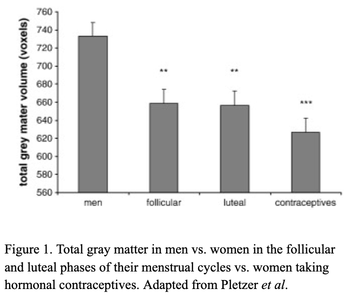
There are millions of women taking steroids every day. But how is this possible? Are they just getting really buff? It feels like we always hear stories about how performance-enhancing drugs, namely steroids, are giving world-class athletes the boost they need to beat out their competition. But women across the globe are taking steroids every day as well, in the form of hormonal birth control. Despite their widespread use, side effects of hormonal contraceptives are largely unstudied, or have been until recently. In the last ten years, several studies have come out about the effect of taking a daily dose of steroids on women’s brains and mental health, which until now has been a severely neglected area where lack of knowledge affects millions of people worldwide. 
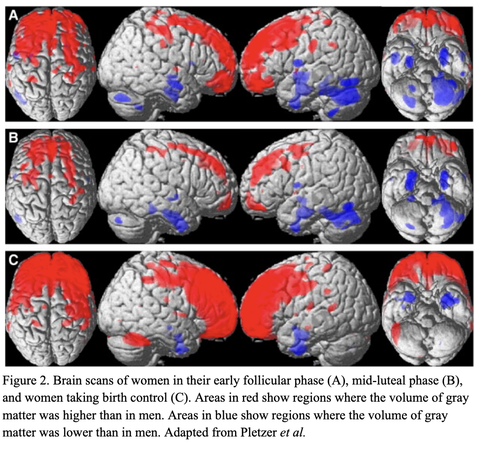
People take hormonal birth control, or hormonal contraceptives, for a myriad of reasons, from the obvious (preventing pregnancy) to the not-so-obvious (lessening iron deficiency) and everything in between. This type of medication simply refers to methods of pregnancy prevention that act on the endocrine system. The endocrine system controls growth, development, metabolism, and reproduction via signaling molecules called hormones. Two hormones in particular, estrogen and progesterone, control the menstrual cycle and are therefore the major components of hormonal birth control. Types of hormonal contraceptives come in many forms including the pill, the patch, the implant, injections, and hormonal intrauterine devices or IUDs, but despite the wide variety in the forms this medication takes, all of them contain one or both of these two hormones. As steroids, both estrogen and progesterone affect other body systems besides the reproductive system.

To study the impacts of taking a daily dose of steroids on other areas of the body, specifically the brain, Dr. Belinda Pletzer and her colleagues conducted a study in 2010. The brain is particularly susceptible to change due to an influx of synthetic hormones because it contains a very high quantity of hormone receptors. The brain needs to act as a “sponge” for these molecules since it plays an important role in creating the appropriate responses in the rest of the body. Pletzer’s study investigated how the sponginess of the brain would affect changes in its structure by comparing images of the brains of adult men, adult women during different stages of their menstrual cycle, and adult women taking hormonal contraceptives. To perform this comparison they used a technique called voxel-based morphology on MRIs of study participants (Pletzer et al., 2010). Voxel-based morphology measures differences in the concentration of tissue and the size and shape of different areas of the brain.

Overall, they found that women taking hormonal birth control had smaller areas of gray matter, or areas of the brain that have a high concentration of the cell bodies of nerve cells, when compared to “naturally cycling women” in both their follicular and luteal menstrual phases (Figure 1). Pletzer’s study also found interesting gendered differences in gray matter volume. While men had greater gray matter overall, the volume of gray matter in the prefrontal cortex, the pre-and postcentral gyri, and the supramarginal gyrus of both naturally cycling women and women taking hormonal contraceptives was higher than the volume of gray matter in these areas in men (Figure 2). These areas are involved in decision-making and problem-solving, controlling motor function, and emotional responses. However, the higher amount of gray matter in women in these areas was overshadowed by the larger volume of gray matter in men in the hippocampus, hypothalamus, parahippocampal and fusiform gyri, putamen, pallidum, amygdala, and temporal regions of the brain during the early follicular phase (A), mid-luteal phase (B), and in women taking hormonal birth control (C) (Figure 2). Many of these areas of reduced gray matter are ones of high importance for neurophysical ability and mental health.

Additionally, a study done by Rush University Medical Center showed an association between higher levels of gray matter and better cognitive function (“Everyday Activities Associated with More Gray Matter in Brains of Older Adults”). These findings suggest that taking birth control, and the associated decrease in gray matter, could be directly causing some of the symptoms women on hormonal contraceptives experience, such as brain fog, mood changes, and even anxiety and depression. For example, a smaller hypothalamus, one of the areas of decreased gray matter, is associated with heightened irritability and depression symptoms (“Study Finds Key Brain Region Smaller in Birth Control Pill Users”). Pletzer’s research and the work of others after her on the impact of birth control on structures of the brain represent important first steps in proving a causative relationship between birth control, symptoms associated with it, and structural changes in the brain.

Although this research has made some crucial preliminary steps into researching how taking a daily dose of steroids affects the brains of women taking hormonal contraceptives, the highly complex nature of the brain and its relationship with the regulation of the rest of the body means that further research is necessary. The sheer number of people that this issue affects means that it is essential to continue researching the impacts of this widely used drug. More importantly, knowing the potentially serious negative side effects enables millions of people to make more informed decisions concerning their health and their bodies.


Works Cited

Rush University Medical Center. (2018, February 14). Everyday activities associated with more gray matter in brains of older adults: Study measured amount of lifestyle physical activity such as house work, dog walking and gardening. ScienceDaily. Retrieved March 11, 2023 from www.sciencedaily.com/releases/2018/02/180214093828.htm.

Lewis, C. A., Kimmig, A. C. S., Zsido, R. G., Jank, A., Derntl, B., & Sacher, J. (2019). Effects of hormonal contraceptives on mood: a focus on emotion recognition and reactivity, reward processing, and stress response. Current psychiatry reports, vol. 21, no.11, 2019, p 115. PubMed Central, https://doi.org/10.1007/s11920-019-1095-z.

Meyer, Craig H., Kinsley, Elizabeth A. “Women’s Brains on Steroids.” Scientific American, https://www.scientificamerican.com/article/womens-brains-on-steroids/. Accessed 7 Mar. 2023.

Nemoto, Kiyotaka. “[Understanding Voxel-Based Morphometry].” Brain and Nerve = Shinkei Kenkyu No Shinpo, vol. 69, no. 5, May 2017, pp. 505–11. PubMed, https://doi.org/10.11477/mf.1416200776.

Pletzer, Belinda, et al. “Menstrual Cycle and Hormonal Contraceptive Use Modulate Human Brain Structure.” Brain Research, vol. 1348, Aug. 2010, pp. 55–62. ScienceDirect, https://doi.org/10.1016/j.brainres.2010.06.019.

Sharma, Rupali, et al. “Use of the Birth Control Pill Affects Stress Reactivity and Brain Structure and Function.” Hormones and Behavior, vol. 124, Aug. 2020, p. 104783. ScienceDirect, https://doi.org/10.1016/j.yhbeh.2020.104783.

“Study Finds Key Brain Region Smaller in Birth Control Pill Users.” ScienceDaily, https://www.sciencedaily.com/releases/2019/12/191204090819.htm. Accessed 7 Mar. 2023.

Filed Under: Biology, Psychology and Neuroscience, Science Tagged With: Biology, Birth control, Medicine, Women's health

Teplizumab: A New Breakthrough in the Treatment of Type 1 Diabetes

April 2, 2023 by Blythe Thompson

     In Ancient Egypt, a diet of whole grains was prescribed to patients with frequent urination and emaciation (History of diabetes, 2020).  This condition, similarly documented by physicians in Ancient Greece, was likely first called “diabetes” by Apollonius of Memphis in the third century BCE. By the fifth century CE, type 1 diabetes had been differentiated from type 2, and in 1776, English physician Matthew Dobson confirmed the presence of excess glucose in the urine of diabetic patients. Canadian physician Frederick Banting and his colleagues successfully used insulin injections to treat a diabetic patient in 1922; this remains the predominant method of treatment today. Another milestone was reached this past November, when the drug Teplizumab (under the name Tzield) gained FDA approval. Teplizumab showed potential in delaying the onset of clinical type 1 diabetes in adults and pediatric patients 8 years and older who have not yet developed the condition (Commissioner of the FDA, 2022). 

     Type 1 diabetes, otherwise known as juvenile diabetes or insulin-dependent diabetes, is a form of diabetes mellitus in which a deficiency of insulin causes hyperglycemia (high blood sugar). It is referred to as “juvenile diabetes” because the symptoms of the condition typically appear in adolescence and is the result of an autoimmune disorder. The onset of type 1 diabetes depends on environmental factors that interact with predisposing genes to induce a long-term autoimmune attack against the pancreatic β cells, the insulin-producing cells of the pancreatic islets of Langerhans (de Beeck and Eizrik, 2018). 

     When left untreated, juvenile diabetes poses serious health risks. The buildup of sorbitol—a sugar alcohol that is manufactured from glucose—within the eye can result in cataracts and blindness (CDC, 2022).  Excess sorbitol is also associated with blood vessel lesions and gangrene. Other significant health risks for individuals with juvenile diabetes include ketoacidosis, the buildup of acidic ketones that can cause diabetic comas and diminished brain function, and hypoglycemia, otherwise known as “insulin shock,” resulting from an overdose of insulin or the failure to eat and potentially causing nerve damage and death (CDC, 2022). Approximately 1.6 million Americans live with type 1 diabetes, including 200,000 youth. Furthermore, despite significant developments in the treatment of type 1 diabetes, desired glycemic targets are rarely achieved in patients, who continue to face a higher risk of complications and death because of their condition (Herold et. al., 2019). 

     In those who are genetically susceptible to type 1 diabetes, there are two asymptomatic stages prior to the development of overt hyperglycemia, the clinical disease which requires insulin treatment. Stage 1 is characterized by the appearance of autoantibodies targeting pancreatic cells and stage 2 involves dysglycemia, an abnormality in blood sugar stability (Herold et. al., 2019). In this case, metabolic responses to high levels of glucose could be impaired, but other metabolic indexes, such as the level of glycosylated hemoglobin, are normal. During these stages, insulin treatment is not required. The goal of Teplizumab is to delay the development of clinical (stage 3) diabetes in those currently in stages 1 and 2 (Herold et. al., 2019). 

     Teplizumab is an Fc receptor–nonbinding anti-CD3 monoclonal antibody that modifies CD8+ T lymphocytes, which are part of the body’s adaptive immune response and thought to be important effector cells that kill β cells in the pancreas (Herold et. al., 2019). Evidence indicates that type 1 diabetes is initiated by both CD4+ and CD8+ T cells (Li and Qin, 2014). Autoreactive T cells differentiate into effector (CD4+) cells by engaging β-cell antigens on local antigen-presenting cells; these effector CD4+ T cells stimulate other immune cells to target β cells, whereas the cytotoxic CD8+ T cells can directly kill β cells via cell-to-cell contact (Gearty et. al., 2022). The CD3 protein complex is involved in activating both the cytotoxic and helper T-cells (Yang et. al., 2005); thus, the anti-CD3 properties of Teplizumab can inhibit the T cell–mediated damage to β-cells. 

     In the Teplizumab trial, which was conducted over a 7-year period and led by Dr. Kevan Herold of Yale University, patients were randomly assigned to a single 14-day course of Teplizumab or placebo. Seventy-two percent of the participants were under the age of 18, and the majority had siblings with clinical type 1 diabetes, meaning they were at a high risk of developing the condition themselves (Herold et. al., 2019). Furthermore, of the 55 patients who were under 18, 47 had a confirmed dysglycemic oral glucose-tolerance test, one of the hallmarks of stage 2 type 1 diabetes, before undergoing randomization in the trial. Follow-up for progression to clinical type 1 diabetes was performed via oral glucose-tolerance tests every 6 months. The study indicated that a 2-week course of treatment with Teplizumab delayed the diagnosis of clinical type 1 diabetes in high-risk participants: following the completion of the trial, 57% of individuals in the Teplizumab trial group were diabetes-free compared to 28% in the placebo group, with a median delay in the diagnosis of clinical diabetes of 2 years (Figure 1). These results also reinforced prior findings that type 1 diabetes is a T-cell mediated condition and showed that immunomodulation before the onset of clinical type 1 diabetes is a promising development in the treatment of this disease (Herold et. al., 2019).


Figure 1. From the time of randomization until the clinical diagnosis of type 1 diabetes, the recipients of the Teplizumab infusion experienced a longer median time until diagnosis than those in the placebo group (adapted from K. C. Herold, et. al.). 

     Given the prevalence of patients with clinical type 1 diabetes, these results of this study are highly promising and a significant step forward in the mitigation of the harmful side effects of this condition. However, there are several areas which, in an effort to make the benefits of this trial widely applicable, require further research. Since the participants in the study were all relatives of patients with type 1 diabetes, it is currently unknown whether the findings can be applied to those who seem to be at risk for the type 1 diabetes and lack first-degree relatives with the condition (Evans-Molina and Oram, 2023). Furthermore, while patients can be carefully screened for the immunological or metabolic markers of preclinical type 1 diabetes in the research setting, a lack of infrastructure prevents a larger-scale screening of the general public and high-risk populations are typically the only groups surveyed for these symptoms (Evans-Molina and Oram, 2023). Finally, despite the promising results of the newly-approved drug, the current cost of treatment presents a significant barrier regarding access to care: one vial of Tzield costs $13,850, amounting to $193,000 over the 14-day infusion (Rodriguez, n.d.).  The exorbitant sum is not surprising, given the high cost of insulin medications and their widely-reported underuse (Herkert et. al., 2019). As a result, further efforts must be taken to ensure that those who are eligible for this new and potentially life-saving medication are able to access it. 


Sources: 

CDC. (2022, November 3). Prevent diabetes complications. Centers for Disease Control and Prevention. https://www.cdc.gov/diabetes/managing/problems.html 

Commissioner, O. of the. (2022, November 18). FDA approves first drug that can delay onset of type 1 diabetes. FDA. https://www.fda.gov/news-events/press-announcements/fda-approves-first-drug-can-delay-onset-type-1-diabetes 

de Beeck, A. O., & Eizirik, D. L. (2016). Viral infections in type 1 diabetes mellitus—Why the β cells? Nature Reviews. Endocrinology, 12(5), 263–273. https://doi.org/10.1038/nrendo.2016.30 

Dolgin, E. (2023). How a pioneering diabetes drug offers hope for preventing autoimmune disorders. Nature, 614(7948), 404–406. https://doi.org/10.1038/d41586-023-00400-x 

Evans-Molina, C., & Oram, R. A. (2023). Teplizumab approval for type 1 diabetes in the USA. The Lancet Diabetes & Endocrinology, 11(2), 76–77. https://doi.org/10.1016/S2213-8587(22)00390-4 

Gearty, S. V., Dündar, F., Zumbo, P., Espinosa-Carrasco, G., Shakiba, M., Sanchez-Rivera, F. J., Socci, N. D., Trivedi, P., Lowe, S. W., Lauer, P., Mohibullah, N., Viale, A., DiLorenzo, T. P., Betel, D., & Schietinger, A. (2022). An autoimmune stem-like CD8 T cell population drives type 1 diabetes. Nature, 602(7895), 156–161.  https://doi.org/10.1038/s41586-021-04248-x 

Herkert, D., Vijayakumar, P., Luo, J., Schwartz, J. I., Rabin, T. L., DeFilippo, E., & Lipska, K. J. (2019). Cost-related insulin underuse among patients with diabetes. JAMA Internal Medicine, 179(1), 112–114. https://doi.org/10.1001/jamainternmed.2018.5008 

Herold, K. C., Bundy, B. N., Long, S. A., Bluestone, J. A., DiMeglio, L. A., Dufort, M. J., Gitelman, S. E., Gottlieb, P. A., Krischer, J. P., Linsley, P. S., Marks, J. B., Moore, W., Moran, A., Rodriguez, H., Russell, W. E., Schatz, D., Skyler, J. S., Tsalikian, E., Wherrett, D. K., … Greenbaum, C. J. (2019). An anti-cd3 antibody, teplizumab, in relatives at risk for type 1 diabetes. New England Journal of Medicine, 381(7), 603–613. https://doi.org/10.1056/NEJMoa1902226 

History of diabetes: Early science, early treatment, insulin. (2020, June 17). https://www.medicalnewstoday.com/articles/317484 

Li, M., Song, L.-J., & Qin, X.-Y. (2014). Advances in the cellular immunological pathogenesis of type 1 diabetes. Journal of Cellular and Molecular Medicine, 18(5), 749–758. https://doi.org/10.1111/jcmm.12270 

Masharani, U. B., & Becker, J. (2010). Teplizumab therapy for type 1 diabetes. Expert Opinion on Biological Therapy, 10(3), 459–465. https://doi.org/10.1517/14712591003598843 

Rodriguez, A. (n.d.). FDA approves first treatment that delays Type 1 diabetes. Why it could be “game changing.” USA TODAY. Retrieved April 2, 2023, from https://www.usatoday.com/story/news/health/2022/11/18/fda-approves-teplizumab-delays-onset-diabetes/10721707002/ 

Yang, H., Parkhouse, R. M. E., & Wileman, T. (2005). Monoclonal antibodies that identify the CD3 molecules expressed specifically at the surface of porcine γδ-T cells. Immunology, 115(2), 189–196. https://doi.org/10.1111/j.1365-2567.2005.02137.x 


Filed Under: Biology Tagged With: Diabetes, Immunology, Immunotherapy, Medicine

Primary Sidebar

CATEGORY CLOUD

Biology Chemistry and Biochemistry Computer Science and Tech Environmental Science and EOS Honors Projects Math and Physics Psychology and Neuroscience Science

RECENT POSTS

  • Floating Systems: Jellyfish and Evolving Nervous Systems May 22, 2025
  • Biological ChatGPT: Rewriting Life With Evo 2 May 4, 2025
  • Unsupervised Thematic Clustering for Genre Classification in Literary Texts May 4, 2025

FOLLOW US

  • Facebook
  • Twitter

Footer

TAGS

AI AI ethics Alzheimer's Disease antibiotics artificial intelligence bacteria Bathymetry Beavers Biology brain Cancer Biology Cell Biology Chemistry and Biochemistry Chlorofluorocarbons climate change Computer Science and Tech CRISPR Cytoskeleton Depression dreams epigenetics Ethics Genes honors Luis Vidali Marine Biology Marine Mammals Marine noise Medicine memory Montreal Protocol Moss neurobiology neuroscience Nutrients Ozone hole Plants Psychology and Neuroscience REM seabirds sleep student superintelligence Technology therapy

Copyright © 2025 · students.bowdoin.edu