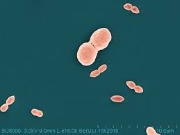
• Skip to primary navigation
  • Skip to main content
  • Skip to primary sidebar
  • Skip to footer

Bowdoin Science Journal

  • Home
  • About
    • Our Mission
    • Our Staff
  • Sections
    • Biology
    • Chemistry and Biochemistry
    • Math and Physics
    • Computer Science and Technology
    • Environmental Science and EOS
    • Honors Projects
    • Psychology and Neuroscience
  • Contact Us
  • Fun Links
  • Subscribe

Biology

Treating allergic asthma with bacteria

April 21, 2024 by Mauricio Cuba Almeida

The prevalence of allergic diseases increased globally following the 1960s. Between 1982 and 1997, the prevalence of asthma and hay fever in Australian schoolchildren rose from 12.9 to 38.6% and 22.5 to 44.0%, respectively (Downs et al., 2001). Similar trends are observed globally (Thomsen, 2015; Turke, 2017). Allergic asthma occurs in about 12 million individuals in the U.S. and its prevalence continues to rise (Gutowska-Ślesik et al., 2023; GenenTech, n.d.).  The immune system is the body’s form of defense against pathogens like viruses and bacteria, and is also where allergies begin. When the immune system regularly overreacts to a harmless substance, one is said to have an allergic disease (Allergies and the Immune System, 2021). Allergies are the subject of many immunological studies due to their health effects. Asthma, for example, is characterized by minor or life-threatening inflammation in the airways.

Theories have surfaced in order to explain the dramatic increase in allergic diseases. One leading theory is the Hygiene Hypothesis. The hypothesis claims that lack of exposure to certain microbial species, like bacteria, is important for the proper development of our immune system (Bloomfield et al., 2006). Therefore, researchers have investigated the mechanisms by which allergic disease, particularly asthma, is deterred by these species. A 2023 study by Yao and colleagues focuses on PepN—a bacterial protein that has shown promise in previous studies—to uncover how the immune system changes when exposed to allergy-reducing disease.

Alveolar macrophages (AMs) are a kind of macrophage found in the lungs that substantially influence the development of asthma (macrophages are a type of immune cell). AMs produce signalers that either encourage or inhibit inflammation in the lungs, which makes them targets for asthma treatments. In fact, AMs, when activated, undergo reprogramming that transforms them into a pro-infammatory or anti-infammatory macrophage (referred to as a CD11chigh macrophage). Yao and colleagues believe this process to be the theoretical foundation of the Hygiene Hypothesis, suggesting that asthma can be treated or prevented by deliberately transforming macrophages to be anti-inflammatory.

Yao and colleagues induced allergic asthma in mice through the use of intranasally injected allergens. To serve as a baseline, the control group was given no further treatment; in the experimental group, mice were exposed to bacterial protein PepN multiple times before and after being inflicted with asthma. Yao and colleagues then dissected the mice, investigating the CD11chigh macrophages and other forces at play.

After comparing the control group of mice to the experimental group, PepN was found to recruit macrophages from the bone marrow into the respiratory tract and transform them to be anti-inflammatory through changes in the macrophages’ metabolism. PepN also encouraged the proliferation of already existing CD11chigh macrophages that reside in the lungs. These forces culminated in a protective effect against allergic asthma (see fig. 1). 

Figure 1. The proposed mechanism by which PepN reduces inflammation in allergic asthma. PepN encourages the proliferation of CD11chigh macrophages in the lungs and recruits additional macrophages which also differentiate into CD11chigh macrophages. Monocytes and CD11int macrophages are earlier forms of the CD11chigh macrophage. Adapted from Yao et al. (2023).

For the future, Yao and colleagues believe more research is necessary to determine other mechanisms of the Hygiene Hypothesis. Though there are limitations to their current study, Yao and colleagues provide a new idea for the prevention and treatment of allergic asthma: targeting CD11chigh macrophages to combat asthmatic inflammation.


References

Allergies and the immune system. (2021, August 8). Johns Hopkins Medicine. https://www.hopkinsmedicine.org/health/conditions-and-diseases/allergies-and-the-immune-system

Bloomfield, S. F., Stanwell‐Smith, R., Crevel, R., & Pickup, J. C. (2006). Too clean, or not too clean: the Hygiene Hypothesis and home hygiene. Clinical & Experimental Allergy/Clinical and Experimental Allergy, 36(4), 402–425. https://doi.org/10.1111/j.1365-2222.2006.02463.x

Downs, S. H., Marks, G. B., Sporik, R., Belosouva, E. G., Car, N., & Peat, J. K. (2001). Continued increase in the prevalence of asthma and atopy. Archives of Disease in Childhood, 84(1), 20–23. https://doi.org/10.1136/adc.84.1.20

GenenTech: Asthma Statistics. (n.d.). https://www.gene.com/patients/disease-education/asthma-statistics#:~:text=Prevalence,asthma%20sufferers%20in%20the%20U.S

Gutowska-Ślesik, J., Samoliński, B., & Krzych‐Fałta, E. (2023). The increase in allergic conditions based on a review of literature. Postępy Dermatologii I Alergologii, 40(1), 1–7. https://doi.org/10.5114/ada.2022.119009

Thomsen, S. F. (2015). Epidemiology and natural history of atopic diseases. European Clinical Respiratory Journal, 2(1), 24642. https://doi.org/10.3402/ecrj.v2.24642

Turke, P. W. (2017). Childhood food allergies. Evolution, Medicine and Public Health, 2017(1), 154–160. https://doi.org/10.1093/emph/eox014

Yao, S., Weng, D., Wang, Y., Zhang, Y., Huang, Q., Wu, K., Li, H., Zhang, X., Yin, Y., & Xu, W. (2023). The preprogrammed anti-inflammatory phenotypes of CD11chigh macrophages by Streptococcus pneumoniae aminopeptidase N safeguard from allergic asthma. Journal of Translational Medicine, 21(1). https://doi.org/10.1186/s12967-023-04768-2

Filed Under: Biology

Your DNA Remembers: Correlating Epigenetics and Early Childhood Trauma through DNA Methylation

April 21, 2024 by Lia Scharnau

What’s your earliest memory? Can you remember everything that happened between the ages of three to five? No? Me neither. Despite the gaps in your memory, your body—specifically your DNA—knows what happened to you. It shouldn’t be surprising that DNA has such a good memory. After all, it stores genetic information tracing back thousands of years. This of course prompts several questions, namely the following; how does DNA store my memories and how does that affect me?

Let’s start from the beginning. Epigenetics encompasses all heritable changes in gene activity that do not stem from a change in DNA sequences (Moore et al, 2013). DNA methylation introduces a particularly salient new epigenetic mechanism for gene regulation and cell differentiation. The key to DNA methylation lies within the special enzymes that modify the cytosine DNA base by adding a methyl group (Moore et al, 2013; Suelves, 2016). Interestingly, cell differentiation is partly driven by differing levels of DNA methylation. An overall increase in DNA methylation occurs during the differentiation process, while a decrease in it at cell-specific loci helps define cellular identity (Suelves et al, 2016). Additionally, the progressive decrease in overall DNA methylation can contribute to physiological aging and the development of cancer (Suelves et al, 2016). Overall, these altered cytosines play key roles in human development and health issues (Moore et al, 2013).  

This brings us to the second question: how does my DNA’s photographic memory affect me? Well, it turns out that choices such as diet, drinking, exercise, illness, and environmental conditions can all have an impact on genomic stability and gene-specific DNA methylation (Lim et al, 2012). So while you might not remember what happened as a kid, your DNA may already be internalizing those experiences. The Avon Longitudinal Study of Parents and Children patiently waited for years so they could specifically study how childhood experiences affect epigenetic markers in adolescence. A cohort of 13,988 children with due dates between April 1991 and 1992 were monitored for exposure to childhood adversity from birth to the age of eleven. Using changes in DNA methylation at the age of fifteen, researchers investigated whether the timing of adversity has epigenetic consequences across childhood and adolescence (Lussier et al, 2023). 

Each mother reported whether their child faced any of the following seven types of childhood adversity; caregiver physical or emotional abuse, sexual or physical abuse, maternal psychopathology, one-adult households, family instability, and neighborhood disadvantage as well as the timing that the adversity was present. Out of the 13,988 children, 609-665 showed signs of both adversity and a decrease in DNA methylation at 15 years old (Lussier et al, 2023). 

Within this sample from the original cohort, further DNA analysis identified twenty-two loci that showed significant associations between exposure to adversity and altered DNA methylation at the age of 15 (Lussier et al, 2023). Of the loci identified to be associated with decreased DNA methylation, the highest percent of loci were correlated with growing up in one-adult households (Lussier et al, 2023). None of the identified loci indicated that adversity may alter DNA methylation at birth or the age of seven, but instead only emerged in adolescence (Lussier et al, 2023).  

Researchers concluded that the ages between three and five years old are when children are vulnerable to adversity and the consequences of this adversity may biologically embed itself and later manifest itself in adolescence (Lussier et al, 2023). Additionally, the adversity-associated decrease in DNA methylation is correlated to have effects on the central nervous system (Lussier et al, 2023). Of course, it’s great timing that the altered DNA methylation becomes an issue during puberty. Especially since it’s associated with poor self-esteem and increased depressive symptoms. 

What’s the upside? There has to be a silver lining or else this article is just a doomsday proclamation. The ability to finally trace health complications back to epigenetic mechanisms due to latent childhood trauma provides an important piece of the puzzle of understanding complex diseases. However, the puzzle is far from solved. The cohort in this study was predominantly of European descent and further research into how the complexities of race factor into DNA methylation and childhood adversity is the next step in this journey.

Literature Cited:

Lim , U., & Song, M. (2012). Dietary and Lifestyle Factors of DNA Methylation. In Cancer Epigenetics (Vol. 863, pp. 359–376). Humana Press.

 Lussier, A. A., Zhu, Y., Smith B. J., Cerutti J., Fisher J., Melton P. E., Wood N. M., Cohen-Woods S., Huang R., Mitchell C., et al. (2023). Association between the timing of childhood adversity and epigenetic patterns across childhood and adolescence: findings from the Avon Longitudinal Study of Parents and Children (ALSPAC) prospective cohort. Lancet Child Adolesc Health. 7(8), 532-43. https://doi.org/10.1016/S2352-4642(23)00127-X

Moore, L. D., Le, T., & Fan, G. (2013). DNA methylation and its basic function. Neuropsychopharmacology, 38(1), 23–38. https://doi.org/10.1038/npp.2012.112

Suelves, M., Carrió, E., Núñez-Álvarez, Y., & Peinado, M. A. (2016). DNA methylation dynamics in cellular commitment and differentiation. Briefings in Functional Genomics, elw017. https://doi.org/10.1093/bfgp/elw017

Filed Under: Biology Tagged With: epigenetics, Genes, Trauma

Unveiling the True Potential of Telomeres in Cellular Health

April 21, 2024 by Jared Lynch

Telomeres have long been known as repetitive DNA elements protecting the ends of chromosomes, but recently, reports of their transcription and translation have revealed an emerging pathological role upon abnormal expression.

When studying the nuclear genome of eukaryotes, one of the most basic characteristics is the organization of DNA into chromosomes that condense and duplicate during mitosis and meiosis. Within each chromosome, a critical protective feature is a string of repeated sequences at each end known as telomeres. These repeat sequences exist as (TTAGGG)n in mammals, which serve to prevent chromosome fusing and shortening. In fact, the shortening of telomeres throughout one’s lifetime is linked closely to aging, reflecting one of the many important health implications of telomeres [1].

More recently, these implications have grown with the discovery that telomeres are both transcribed and translated into repeat sequences (Figure 1, top) [2]. Transcription creates what is known as telomeric repeat-containing RNA (TERRA), and while most of these molecules never leave the nucleus, some escape to the cytoplasm [3]. Initially, TERRA was believed to have no substantial effect on cell functioning due to the absence of AUG start codons. However, its unique 3D structure is able to bypass this roadblock and initiate AUG-independent translation [4]. The result is one of two repeated protein sequences, depending on which reading frame translation begins at: valine-arginine (VR) and glycine-leucine (GL) [4]. In appreciable quantities, these are expected to impact cell functioning, but in what way is largely unknown, particularly in humans.

Al-Turki and Griffith sought to fill this knowledge gap by assessing the properties of VR repeats and GL repeats in vitro and in human cells [5]. To start, the researchers hypothesized that VR would bind nucleic acids, which are negative charged, due to its positively-charged arginines. By contrast, GL was predicted to form large aggregates known as amyloids due to its hydrophobic properties. In both cases, these molecules could severely impact cell functioning, so determining the exact behavior of VR and GL was an essential start.

For their first experiment, Al-Turki and Griffith synthesized VR and GL, then added them to a salt buffer to mimic how they would self-interact in the cytoplasm. Visualization revealed that VR was unable to aggregate due to the repulsion of repeated arginines. However, GL demonstrated mild aggregation with the potential to form amyloids and induce inflammatory responses in extreme cases (Figure 1). 

Next, the binding of VR to nucleic acids was tested by adding the peptide to a solution of RNA or DNA. In support of their hypothesis, the authors found that VR bound to both with a high affinity. They then took this a step further and added DNA plasmids containing a replication fork (i.e., the structure that forms during DNA replication) to test the interaction with VR. Intriguingly, VR bound with high preference to the replication fork itself which altered its geometry, possessing the ability to disrupt the replication process (Figure 1). 

Finally, an antibody for VR was developed which allowed the authors to visualize its presence in human cells. VR levels were found to positively correlate with the abundance of TERRA, and in osteosarcoma (“U2OS”) cells specifically, these were both at unusually high levels. Moreover, VR was often found in the nucleus as discrete clumps or “foci,” contrary to the in vitro experiment, due to stabilization by the negatively-charged DNA. The authors then attempted to alter VR levels by either knocking down TERRA or promoting its transcription. Intriguingly, VR aggregates increased in size for both cases, which may result in a cytotoxic effect that has yet to be explored.

Telomeres were initially viewed as protective elements with no function beyond the DNA level. Yet, the discovery of TERRA and its translated peptide sequences have completely flipped this narrative. Al-Turki and Griffith expand on this by showing that VR and GL peptides generated by translation could have significant physiological effects on cells including altered DNA replication, inflammation, and cytotoxicity. In particular, VR can not only bind DNA but also aggregate in the nucleus. These aggregates increased in size when altering TERRA levels, indicating that dysfunctional telomeres capable of promoting TERRA could have negative consequences for cell functioning.

In spite of these discoveries, future work remains in order to understand the full picture of VR and GL. For one, GL was unable to be assessed in live cells as the authors were unable to create an antibody for it. Additionally, these peptides are likely to have other functions beyond aggregating and killing cells. Understanding these abilities is crucial for not only characterizing its pathological potential but also for developing treatments down the line. Nevertheless, the present study creates a strong foundation for exploring this underappreciated chromosomal feature.

References

1. Ren F et al. (2009). Estimation of human age according to telomere shortening in peripheral blood leukocytes of Tibetan. Am J Forensic Med Pathol, 30(3):252-5. https://doi.org/10.1097/PAF.0b013e318187df8e.

2. Azzalin CM et al. (2007). Telomeric repeat containing RNA and RNA surveillance factors at mammalian chromosome ends. Science, 318(5851):798-801. https://doi.org/10.1126/science.1147182. 

3. Schoeftner S, Blasco MA (2008). Developmentally regulated transcription of mammalian telomeres by DNA-dependent RNA polymerase II. Nat Cell Biol, 10(2):228-36. https://doi.org/10.1038/ncb1685.

4. Zu T et al. (2010). Non-ATG-initiated translation directed by microsatellite expansions. Proc Natl Acad Sci U S A, 108(1):260-5. https://doi.org/10.1073/pnas.1013343108. 

5. Al-Turki TM, Griffith JD (2023). Mammalian telomeric RNA (TERRA) can be translated to produce valine-arginine and glycine-leucine dipeptide repeat proteins. Proc Natl Acad SciU  S A, 120(9): e2221529120. https://doi.org/10.1073/pnas.2221529120.

Filed Under: Biology, Science

Pollinator-mediated viral infections in stoned fruits; are the bees to blame?

December 10, 2023 by Henry Anderson

PNRV Effect on Plants

The neverending fight against fruit viruses has gotten even muddier. A recent study based out of Clemson University and the greater southern South Carolina area has made significant discoveries toward a better understanding of the role that pollinators play in pollen-borne plant viral transmissions. Prunus necrotic ringspot virus (PNRSV) and Prune dwarf virus (PDV) are pollen-borne viral infections that attack stone fruit crops worldwide, often greatly impacting the actual fruit yield.  In the southeastern United States, the prevalence of PNRSV and PDV in peach crops and their mechanisms of virus spread are relatively unknown and under-studied. The researchers, Mandeep Tayal, Christopher Wilson, and Elizabeth Cieniewicz, studied the number of bees and thrips, small pollen-carrying insects, containing virus-positive pollen in peach orchards and the relationship to virus-positive pollen-carrying bees/thrips and their genus type.

The researchers selected two orchards (see Figure 1), one with a higher suspected incidence of both PDV and PNRSV. Trees in each orchard were divided into blocks and then tested for PNRSV and PDV. Bees and thrips, two major pollinators of peach trees, are primarily active during peach bloom (from early March to early April). During the two years studied, bees and thrips were trapped using blue vane traps and sticky cards, respectively. Reverse Transcriptase-Polymerase Chain Reaction (RT-PCR) of RNA extracted from bee pollen and thrips was then used to detect either PNRSV or PDV.

Figure 1. Map of orchard and peach block location, with PNRSV-positive trees labeled.

The researchers found different blocks of peaches had contrasting relative prevalences of the two viruses.  In areas with largely PDV-infected trees, the majority of bees carried PDV-positive pollen, a phenomenon consistent throughout the different blocks. The virus and scope to which a block of trees was infected were directly proportionate to the number of bees carrying each virus-positive pollen. The researchers concluded that pollinators play an important role in spreading these viruses. From an ecological standpoint, they extrapolated that larger bees, such as Bumble and Blueberry Bees, could carry greater amounts of pollen and thus may have a larger impact on pollen-mediated viral infections (see Figure 2).

Figure 2. Relationship between bee genus and virus carried during peach bloom

This is an important development for farmers and researchers alike. Both PNRSV and PDV have been previously known to be vertically integrated in stoned fruit generations; meaning viruses are passed on from parent to offspring. Given this, the primary strategy for combatting these viruses has been to cut down any infected trees. In theory, by sourcing uninfected juvenile saplings and cutting down the diseased trees, the spread and effect should be contained. Unfortunately, this tactic deteriorates with tree to tree transmission. The prevalence of horizontal integration adds a whole new dimension to the issue. As the virus is too common to be eradicated, a treatment that accurately targets and kills these viruses will likely be the only effective long term solution. 

Pollinator-mediated viral infections, especially involving the physical mechanisms and driving ecological factors, are a highly under-researched field of biology. The aforementioned research simply scrapes the surface of all the possible driving forces and nuances surrounding this minimally understood process. Moreover, this information is concerning to both the peach industry and beyond. PDV and PNRSV not only diminish the product yield of stoned fruits, but they are also incredibly illusive and hard to treat. If infected pollen can not only be stored and transmitted by trees but also by pollinators, it makes containing the spread of such viruses remarkably challenging. Given the over half-billion industry that is just peaches alone, pollen-borne viruses represent a pressing matter. With farmers and biologists feeling the pressure to find a solution, it seems they will need to turn to new, innovative treatments. 


References: 

Amari K, Burgos L, Pallás V, Sánchez-Pina MA. Vertical transmission of Prunus necrotic ringspot virus: hitch-hiking from gametes to seedling. J Gen Virol. 2009 Jul;90(Pt 7):1767-1774. doi: 10.1099/vir.0.009647-0. Epub 2009 Mar 12. PMID: 19282434.

(Title Image) – Garvey, K. K. (2011, March 14). Peachy Keen – Bug Squad – ANR Blogs. UC ANR. Retrieved December 10, 2023, from https://ucanr.edu/blogs/blogcore/postdetail.cfm?postnum=4390

Hampson, CR, et al. “Prunus necrotic ringspot virus (almond bud failure) | CABI Compendium.” CABI Digital Library, 18 December 2021, https://www.cabidigitallibrary.org/doi/10.1079/cabicompendium.42426. Accessed 10 December 2023.

Mandeep Tayal, Christopher Wilson, and Elizabeth Cieniewicz “Bees and thrips carry virus-positive pollen in peach orchards in South Carolina, United States,” Journal of Economic Entomology 116(4), 1091-1101, (4 July 2023).

Peaches. (2023, February). Agricultural Marketing Resource Center. Retrieved November 12, 2023, from https://www.agmrc.org/commodities-products/fruits/peaches


Filed Under: Biology

ProCAR-T Cells: An Innovative Treatment for Solid Cancers

December 9, 2023 by Alice Fang '27

There is an ongoing search for better and more effective treatments for cancer. The reason cancer is difficult to treat is because many cancer cells can evade the immune system. Chimeric antigen receptor (CAR) T cell therapy uses the patient’s own immune cells to treat their cancer. During CAR-T therapy, T cells, which are immune system cells that help kill off pathogens, are extracted from a patient’s blood. These T cells are then mixed with a virus that allows the T cells to develop a chimeric antigen receptor (CAR), an artificial receptor that will navigate toward the location of the cancer. Then, the CAR-T cells are released back into the patient’s bloodstream intravenously. The CAR-T cells track and home in on a tumor-associated antigen (TAA) that is specific to the patient’s cancer. The CAR-T cells then release molecules that trigger cell death pathways within the tumor cells. This process is illustrated in Figure 1. 

T-cell Transfer Therapy - Immunotherapy - NCI

Figure 1 CAR-T cell therapy process from the National Cancer Institute. 

 CAR-T cells are shown to be effective at treating blood cancers such as leukemia and lymphoma. Despite the success CAR-T cells have with these cancer types, this therapy is much less effective for treating solid tumors. CAR-T cells can only induce lysis once they bind to the cell surface. However, solid tumors have a small surface area to volume ratio, making it difficult for CAR-T cells to infiltrate and reach all the tumor cells. Even if it does, the rate at which the tumor cells are killed by this process is much slower than the rate at which the tumor cells replicate, rendering the system inefficient. Another challenge lies in the identification of markers that are “specifically and uniformly expressed on heterogeneous solid tumors” (Vincent et al., 2023). Heterogeneous solid tumors are ones that have a sub-population of cells with different features to that of the tumor cells. TAAs identified are rarely tumor-specific, meaning most antigens are present in healthy cells too. Thus, the use of these TAAs carries a high risk of fatal on-target, off-tumor toxicity that kills the vital, healthy cells of the patient.   

To better treat solid tumors, Vincent et al. from Columbia University took a slightly different approach whereby they created probiotic-guided CAR-T cells (ProCARs) that responded to synthetic CAR targets released by tumor-colonizing colonizing probiotic bacteria. The researchers engineered a probiotic strain, Escherichia coli Nissle 1917 (EcN), which is used as a delivery vesicle for synthetic CAR targets called Tags. EcN is able to colonize tumor cells and grow in tumor microenvironments (TMEs) and contains quorum-regulated circuits, meaning bacteria will die after reaching a certain population density threshold and release Tags within the tumor. When Tags are released, they broadly bind to surface and matrix proteins of the tumor cell. CAR-T cells are engineered with GFP28z receptors, which will bind to Tags and activate the CAR-T cell. Thus, activation of CAR-T cells allows for Tag-mediated in situ lysis of the tumor cells.  

Overall, this system overcomes the two previous challenges faced when treating solid tumors. By using Tag, this system allows for antigen-agnostic immunotherapy, meaning eliciting an immune response against tumor cells without the identification of TAAs. Furthermore, since the probiotic bacteria migrates to the tumor location, this treatment mechanism minimizes off-tumor toxicity. Last but not least, the advantage of using bacteria in this system lies in its ability to colonize tumor cells and grow within TMEs, enabling treatment options for difficult-to-infiltrate tumors.  

This system has shown significant lysis across seven human cancer cell lines. Empirical data reflect no significant weight loss shown in mice, no bacterial growth outside of tumor cells or in any healthy organs, and Tag plasmids were well maintained in vivo. While this treatment has been shown to be effective, the potential toxicity from repeated dosing of bacterial therapy in humans is still an important concern for clinical translation, since humans are more sensitive to toxins inside the bacterial cell than mice. Overall, the researchers conclude that probiotic-guided CAR-T cells may provide a novel and safe treatment method for solid tumors.  


Works Cited: 

T-cell Transfer Therapy—Immunotherapy—NCI (nciglobal,ncienterprise). (2022, April 1). [cgvArticle]. https://www.cancer.gov/about-cancer/treatment/types/immunotherapy/t-cell-transfer-therapy 

Vincent, R. L., Gurbatri, C. R., Li, F., Vardoshvili, A., Coker, C., Im, J., Ballister, E. R., Rouanne, M., Savage, T., de los Santos-Alexis, K., Redenti, A., Brockmann, L., Komaranchath, M., Arpaia, N., & Danino, T. (2023). Probiotic-guided CAR-T cells for solid tumor targeting. Science, 382(6667), 211–218. https://doi.org/10.1126/science.add7034 

Filed Under: Biology

Fine-tuning of Chemotherapeutic Drug Reactions through Ruthenium Organic Complexes

December 4, 2023 by Ahmad Harb '25

The development of cancer treatment reagents aims at optimizing the reactivity of the reagent with the cellular DNA while reducing the reactivity with other bodily sites. This is in order to maximize cytotoxicity to cancer cells while reducing the side effects associated with chemotherapy (Wang, 2005).

Organometallic complexes, organic compounds with one or more metallic central atoms, have been used to control the release of compounds involved in key biological reactions (Renfrew, 2014). In the context of cancer chemotherapy, cisplatin complexes have been successfully developed to react with guanosine 5’ monophosphate, or GMP, as a potential binding site in the cell’s DNA, while avoiding the reactions associated with side effects (Dasari, 2014 & Reedjik, 2003).

The development of chemotherapeutic reagents requires the fine-tuning of the ligand substitution reactions that the organometallic complex can undergo. Ligand substitution reactions are reactions where one or more of the substituents bonded to the metal atom in the organometallic complex are replaced by a compound from the surrounding environment. An example is shown in the figure below.

Fig 1. Example of an organometallic complex undergoing a ligand substitution reaction. When dissolved in pyridine, the chromium complex, [Cr(TPP)(Br)(H2O)], reacts with the solvent, and two of its ligands (Br and OH2) are replaced by pyridine compounds (Py) (Okada, 2012). 

In a 2005 study, a group of chemists tested multiple ligand-substitution reactions of Ruthenium (Ru) complexes to test the possibility of the development of a competitor for cisplatin in chemotherapy (Wang, 2005). The Ruthenium complex studied have “stool”-like structures with an arene upper part and a tetrahedral Ruthenium compound which contains the leaving group. The researchers use X-ray crystallography and other characterization methods to identify the structure of every ruthenium complex involved in the study.

Fig 2. The structure of the Ruthenium organometallic complexes tested in the study. The basic structure is shown on the upper left. The “arene” can be any of the structures shown, while the leaving group, X, can be any of the structures shown as well as a halide or pseudohalide (Wang, 2005). 

Given the high concentration of chloride anions in the bloodstream and the intercellular fluid, the substitution of X by chloride is a main mechanism by which the reagent is lost before it can attack cancer cells (Wang, 2005). Previous research has shown that hydrolysis (substitution of X by a water molecule) is an essential activation step in the reagent’s reaction with GMP (Chen, 2003). The researchers thus investigated how the choice of the arene and the leaving group within the ruthenium complex can affect the reaction rates such that the reagent is cytotoxic but is inactive before reaching its target site. 

The reaction rate with chloride was established by dissolving the complexes in a 104mM NaCl solution, mimicking the high-chloride media within the body, and monitoring the formation of the substitution reaction product using the same characterization methods involved in the identification of the complexes.

The hydrolysis rate was established by allowing the aqueous solutions of the complexes to equilibrate for 24-48 hours at 37°C, mimicking body temperature. The formation of the hydrolysis product was monitored using the same characterization methods. The reaction equilibria were determined using high-performance liquid chromatography. 

The reaction with GMP is believed to be the final step in the reagent’s activityagainst cancer cells. The reaction rate was established by dissolving 0.5mM of each complex in a 0.5mM aqueous GMP solution. The product formation rate was determined using the same methods as the other reactions.

The researchers summarized their key resultsin the table below:

The data show that complex 13, for example, has a faster reaction rate with chloride than GMP, and will, therefore, be lost before attacking cancer cells if it were to be used as a chemotherapyreagent. Data shows that complexes 15 and 17, on the other hand, react faster with water and GMP than chloride, which makes them more suitable for chemotherapy. Complex 1 can undergo hydrolysis and react with GMP at a relatively high rate. A key finding in this research is that complex 21 can bind to GMP without undergoing hydrolysis, skipping a previously thought required first step. 

The overall cytotoxicity of each complex was compared to cisplatin by determining the concentration of the complex which caused at least 50% inhibition in the growth of ovarian cancer cells, IC50 values. As previous research predicted, high hydrolysis rates correlated with high cytotoxicity (Chen, 2003). Cisplatin has IC50 = 0.6µM; chemotherapy reagents are considered to have good cytotoxic activity if they have IC50 < 18µM. Some of the same complexes discussed above, with the exception of complex 17 and 21 had IC50 values competitive with cisplatin (IC50 < 6µM). Furthermore, the studied Ru complexes exhibited cytotoxicity towards cisplatin-resistant ovarian cancer cells (Wang, 2005).

Overall, the results show that the rates of the reactions involved in chemotherapy can be fine tuned by the choice of the ligand within the ruthenium complex. These results can be used in the future development of novel chemotherapy reagents.  

Work Cited:

Chen, H., Parkinson, J. A., Morris, R. E., & Sadler, P. J. (2003). Highly selective binding of organometallic ruthenium ethylenediamine complexes to nucleic acids: novel recognition mechanisms. Journal of the American Chemical Society, 125(1), 173-186.

Dasari, S., & Tchounwou, P. B. (2014). Cisplatin in cancer therapy: molecular mechanisms of action. European journal of pharmacology, 740, 364-378.

Okada, K., Sumida, A., Inagaki, R., & Inamo, M. (2012). Effect of the axial halogen ligand on the substitution reactions of chromium (III) porphyrin complex. Inorganica Chimica Acta, 392, 473-477.

Reedijk, J. (2003). New clues for platinum antitumor chemistry: kinetically controlled metal binding to DNA. Proceedings of the National Academy of Sciences, 100(7), 3611-3616.

Renfrew, A. K. (2014). Transition metal complexes with bioactive ligands: mechanisms for selective ligand release and applications for drug delivery. Metallomics, 6(8), 1324-1335.

Wang, F., Habtemariam, A., van der Geer, E. P., Fernández, R., Melchart, M., Deeth, R. J., … & Sadler, P. J. (2005). Controlling ligand substitution reactions of organometallic complexes: Tuning cancer cell cytotoxicity. Proceedings of the National Academy of Sciences, 102(51), 18269-18274.

Filed Under: Biology, Chemistry and Biochemistry Tagged With: Chemistry and Biochemistry, Medicine

Unpacking the Ethical Implications of Human Germline Editing

December 3, 2023 by Kennedy Kirkland

On December 30th, 2019, biophysicist Dr. He Jiankui was sentenced to three years in prison for forging ethical review documents and misleading doctors into unknowingly implanting gene-edited embryos into two women in China (Normile 2019). One of these women had a set of twins and the other had a single child. These children are now the first genetically modified humans in history to be resistant to HIV. They can pass this modification to the next generation, and their whereabouts are still unknown (Greely 2019). Jiankui used CRISPR/Cas9, a gene editing tool, to modify their germlines and edit the CCR5 gene, a contributor to broad immune responses (his focus was its importance to HIV viruses).

Based on the National Natural Science Foundation of China, there has been a total of 3.7 billion yuan (roughly $576 million) government spending on embryonic stem cell research from 1997 to 2019 (Lou 2021). Germline gene editing research is allowed, but establishing a pregnancy with genetically modified embryos has been outlawed by multiple regulations (Xinqing 2014). Similarly to China, the National Institutes of Health within the US estimates that human embryonic stem cell research has received $1.48 billion in government funding since 2009. There are no current laws or regulations that ban germline gene editing conducted through private funding within the US, but it would have to be approved by the FDA for marketing and clinical studies. No proposals have been submitted (​​Genetic Literacy Project 2019). Dr. Jiankui has since been disgraced by the scientific community around the world for his actions. Some scientists believe that germline gene editing is not only an unethical practice but a potentially dangerous one that could lead to a new era of eugenics with irreversible harm (Genetic Literacy Project 2019). Little is known about the intricate details of Dr. Jiankui’s experiment, but what has been revealed poses several hard-hitting ethical questions. Should we manipulate the next generation of humans? What are the ethical dilemmas in the advancement of this technology? To create an informed opinion on the matter, it is helpful to first understand the intricacies of Dr. Jiankui’s experiment, and how exactly he made the first genetically modified children.

Dr. Jiankui conducted human germline genome editing, which refers to the technique of modifying not only the genetic information of a subject but also what can be passed down to the next generation (Normile 2019). Germ cells can create a new generation (ex. sperm and eggs). So germ cells, and cells that produce germ cells, are known as the germline. These are different from somatic cells, which are body cells that continuously divide throughout a person’s lifetime and play a minimal role in gene inheritance (Greely 2019). Certain alleles or alterations to CCR5 within the human genome can provide resistance to HIV. Thus, Jiankui used CRISPR to alter CCR5 in HIV-susceptible patients to make them resistant to the disease.

CRISPR, which stands for clustered regularly interspaced short palindromic repeats, is a technique originally found in bacteria as a defense system to render inserted viral DNA ineffective. It also allows for the modification of DNA (Rapini 2023). The enzyme Cas-9 can cut pieces of DNA and is guided to its location based on an attached strand of RNA (called the guide RNA strand). By cutting a targeted sequence of DNA, it can render protein-coding genes inactive or disfunctional. After Cas-9 has cut the target DNA, researchers can choose to place a modified DNA sequence in the vacant space (Fig 1).

Figure 1. The editing technique of CRISPR involves the guide RNA strand, guide RNA sequence, Cas-9 enzyme, and target DNA (Roach 2015).

While little is known about Dr. Jiankui’s specific technique in using the CRISPR/Cas-9 system, we know that he modified the gene CCR5 (Greely 2019). CCR5 is a gene on chromosome 3 that encodes for a protein called C-C chemokine receptor type 5 (also shortened to CCR5) (Normile 2019). CCR5, along with another receptor called CD4, is utilized by the HIV virus to bind to bacteria-detecting/destruction cells called macrophages (white blood cells) and infect them. The HIV virus protein envelope binds to the primary receptor CD4 on the macrophage with the gp120 protein, which are exterior “protective” protein around the HIV cell. If the CCR5 coreceptor is present as well, it can successfully enter the macrophage, release viral RNA and enzymes, and infect the macrophage. This infection alters the function of the macrophage, causing it to assemble and release viruses. Eventually, the gene that codes for the gp120 receptor is altered by a mutation and can now bind to a different co-receptor called CXCR4 which is found on the CD4 plus T-cells. (Prakash 2019). The same process of virus construction occurs within T-cells, but as the viruses leave the T-cell it ruptures the plasma membrane. This kills the T-cells, causing a weakened immune response and eventually resulting in the onset of AIDS.

Figure 2. The mechanism by which the HIV viral envelope infects the macrophage creates mutated viral cells with different gp120 receptors and destroys T-cells. (Prakash 2019) 

It is believed that Dr. Jiankui used the CRISPR/Cas-9 system to delete 32 base pairs of the CCR5 gene, therefore making it produce non-functional copies of the CCR5 protein. This was done in the hope that HIV would be unable to infect the white blood cells of the babies born from the embryos (Normile 2019). If Jiankui only modified somatic cells using this technique, the children may or may not have gained protection against HIV and could not pass it down to the next generation.

In 2015 (before He’s experiment was released) Jennifer Doudna, one of two researchers who published their findings on CRISPR, convened with the U.C Berkeley’s Institute for Genome Innovation, which was composed of Nobel laureates and esteemed professors of bioethics. They concluded that conducting germline editing would be irresponsible until matters such as balancing the potential risks and benefits have been concluded and that there is a broad societal consensus about the appropriate use of proposed applications of this technology (Cohan, 2018). Jennifer Doudna and Emmanuelle Charpentier created this technology to find ways to edit out faulty genes and cure diseases at the source—within our genetic code (Doudna 2019). However, after the release of He’s experiment, Doudna publicly stated her evolving concerns relating to this application of her discovery. During her national press tour in 2019, she shared a frightening dream she had in which a colleague of her’s asked about CRISPR but later turned out to be Hitler (Than 2019). This likely stems from the horrifying history of Nazis and the eugenics movement, which stemmed from the idea of improving the genetic quality of humans and sterilizing and or eliminating those with unfavorable traits. She also voiced that her opinion on germline genome editing has been evolving from an outright ban to warranting it in certain circumstances. However, her underlying opinion remains that it would have to be under transparent and safe circumstances and concern a medical need that was unmet by any technology. In addition to Doudna, countless other scientists have come out in disagreement with He Jianku’s work including researchers from Stanford University, Harvard Medical School, and the NIH (Cohen 2018). Specifically, a journalist from Science magazine states that representatives from eight countries who attended the International Summit on Human Genome Editing in Hong Kong came to a consensus that his actions were irresponsible, violated international norms, lacked transparency, and did not have sufficient medical justification (Cohen 2018).

Jennifer Doudna stated that her goal was to edit out faulty genes, but that definition leaves a lot to interpretation. How can we as a society come together to define the parameters of a faulty gene? From treating HIV to Down Syndrome, these conditions hold more than just health implications, but social ones as well. If the scientific community deems it accessible to prevent Down syndrome using human germline genome editing, are they stating that humans that currently possess this condition are “wrong” and have “faulty genes” in need of correction? Whoever holds the power to manipulate this technology essentially has the power to decide what the next generation of humans will be capable of and what genes are deemed “faulty” within our society. Jianku’s reckless use of technology was majorly deemed wrong for its lack of transparency, safety, and following set regulations upheld by currency bioethical standards. However, in situations like parents wanting their children to be healthy, it is completely understandable that human germline editing sounds like an intriguing way to protect their children. For these reasons, it is imperative to familiarize ourselves with the development of this technology, and its continued push for regulations across the world to maintain ethical standards. Further, it is also important to ask ourselves hard-hitting questions such as: should one person, scientist, or government have the power to determine what the next generation will possess, look like, or become? We as members of society must continue to stay informed in a world with the capacity to genetically modify human beings.

References:

  1. Bhanu Prakash. “How the HIV Infection Cycle Works.” Proceum Pvt. Ltd. January 2019. https://www.youtube.com/watch?v=GyofqO1TRjU
  2. Brianna Rapini, Sarina Peterson, “Genetic Engineering.” September 2023. https://www.youtube.com/watch?v=CDw4WPng2iE
  3. Dennis Normile. “Chinese scientist who produced genetically altered babies sentenced to 3 years in jail.” Science. December 2019.https://www.science.org/content/article/chinese- scientist-who-produced-genetically-altered-babies-sentenced-3-years-jail
  4. Deng Luo, Zihui Xu, Zhongjing Wang, Wenzhuo Ran. “China’s Stem Cell Research and Knowledge Levels of Medical Practitioners and Students.” Stem Cells International. 2021. https://doi.org/10.1155/2021/6667743.
  5. Doudna J. 2019. Faculty Research Page. Department of Molecular & Cell Biology. http://mcb.berkeley.edu/faculty/BMB/doudnaj.html.
  6. Dylan Roach, Tanya Lewis. “CRISPR, the gene-editing tech that’s making headlines, explained in one graphic.” Insider. December 2015. https://www.businessinsider.com /crispr-gene-editing-explained-2015-12
  7. Genetic Literacy Project. 2019 July 23. United States: Germline / Embryonic. Global Gene Editing Regulation Tracker. https://crispr-gene-editing-regs-tracker.geneticliteracy project.org/united-states-embryonic-germline-gene-editing/.
  8. Henry T Greely. “CRISPR’d babies: human germline genome editing in the ‘He Jiankui affair.’” Journal of Law and the Biosciences. Volume 6, Issue 1. October 2019, Pages 111–183, https://doi.org/10.1093/jlb/lsz010
  9. Indra Mani. “CRISPR-Cas9 for treating hereditary diseases.” Progress in molecular biology and translational science. Volume 181. February 2017. https://pubmed.ncbi.nlm.nih.gov/34127193/
  10. John Cohen. “After last week’s shock, scientists scramble to prevent more gene-edited babies.” Science. December 2018.https://www.science.org/content/article/after-last- weeks-shock-scientists-scramble-prevet-more-gene-edited-babies
    Join David Ignatius. “Walter Isaacson & Jennifer Doudna join Washington Post Live to discuss CRISPR.” Washington Post. March 2021. https://www.youtube.com/ watch?v=cKHuuALENZk
  11. Than K. 2019 Nov 12. AI and gene-editing pioneers to discuss ethics. Stanford News. [accessed 2023 Dec 3]. https://news.stanford.edu/2019/11/12/ai-gene-editing-pioneers- discuss-ethics/#:~:text=Doudna%20decried%20the%20act%20but.
  12. Yi Zheng et al. “Structure of CC Chemokine Receptor 5 with a Potent Chemokine Antagonist Reveals Mechanisms of Chemokine Recognition and Molecular Mimicry by HIV.” Immunity. Volume 46, Issue 6. June 2017. https://www.cell.com/immunity /pdf/S1074-7613(17)30218-2.pdf
  13. ‌Zhang Xinqing, Zhang Wenxia, Zhao Yandong. “The Chinese Ethical Review System and its Compliance Mechanisms.” TRUST. September 2014. https://trust-project.eu/ wp-content/uploads/2016/03/Chinese-Ethics-Review-System.pdf

Filed Under: Biology, Chemistry and Biochemistry, Science Tagged With: Bioethics, Genes, HIV

Inverse Vaccines: A New Way to Treat Autoimmune Disorders

December 3, 2023 by Divya Bhargava

Williams, 2023

Of the 8.1 billion people in the world, 1 in 10 have an autoimmune disorder. There are 8.1 billion people in the world, and 810 million of these people, or 1 in 10, have an autoimmune disorder (“1 in 10 people”, 2023). Autoimmune disorders are a category of conditions in which the body attacks itself. Although management systems for most of these types of conditions have been developed, autoimmune diseases still cannot be cured (“Autoimmune disorders”). In fact, even with management of their symptoms, up to 50% of patients with autoimmune disorders still experience impairment in their health-related quality of life (Pryce and Fontana, 2017). The development of so-called “inverse vaccines” may provide the much needed mechanism to help find a cure for this class of conditions by teaching the body not to attack itself. 

A traditional vaccine works because it helps the body learn to recognize parts of foreign pathogens and builds up the body’s immune response against these pathogens so that the response is stronger and happens more quickly after recognition. In people with autoimmune diseases, the body also forms an immune response against self molecules because it mistakenly identifies them as foreign antigens, or pathogenic molecules (usually proteins or sugars) that induce an immune response. The idea behind “inverse vaccines” is that instead of building up the immune system’s response to foreign antigens, they could suppress the response to self-antigens by helping to teach the body to recognize these misidentified molecules as self. 

A study led by Andrew Tremain at the University of Chicago’s Pritzker School of Medicine is one of the groups who are involved in this novel inverse vaccine research. The inverse vaccines they developed contain modified copies of the self-antigens that are targeted by the immune system which are attached to long chains of sugars called polysaccharides (Tremain et al., 2023). These polysaccharide chains guide the self-antigens to the liver, which plays an important role in the establishment of tolerance to these molecules. Once these modified self-antigens arrive at the liver, specialized immune cells pick them up and then inhibit the action of T cells against them through T cell uptake (Leslie, 2023). T cells are a type of immune cell that carry out part of the typical response against molecules identified as foreign or invaders through either cytotoxic or signaling based immune responses. The inhibition of the T cell response against misidentified self-antigens reduces or prevents the autoimmune response that causes the body to attack itself, thereby acting as a kind of “inverse vaccine”. 

Once Tremain et al. developed their inverse vaccine, they conducted testing to determine its efficacy and viability as a method for increasing tolerance to self-antigens in those with autoimmune disorders. First, they wanted to ascertain whether inverse vaccines could truly provide inhibition of an immune response. To do this, they injected an egg white protein into mice as an experimental foreign antigen to trigger a strong immune response. Then an inverse vaccine against the egg white protein was injected to suppress the response to the original dose of the protein. In their analysis, they found that the T cells that would’ve responded to the egg white protein were not present. These results suggested that the inverse vaccine blocked the typical immune response, demonstrating viability as a treatment method against stimulated immune responses (Tremain et al. 2023).

However, Tremain and his colleagues still had to demonstrate the efficacy of inverse vaccines in inhibiting an autoimmune response rather than one caused by a foreign antigen. To do this, they induced an autoimmune disease called experimental autoimmune encephalomyelitis (EAE) in mice. In EAE, the immune system attacks myelin, the substance responsible for forming insulation around nerve cell axons. EAE is a particularly informative experimental model in mice because it mimics multiple sclerosis (MS), a human autoimmune disorder. Once they induced EAE, Tremain et al. injected an inverse vaccine made up of a polysaccharide carrying part of a myelin protein. Physiological analysis after injection suggested that this treatment had stopped mice from developing EAE. Furthermore, injection of a different inverse vaccine targeting an alternate form of EAE showed prevention of symptom relapse. This means that Tremain et al. were able to demonstrate inhibition of autoimmune responses against two types of EAE in mice, representing two different types of MS, providing a heartening outlook on this research (Tremain et al., 2023). 

In summary, inverse vaccines had the ability to turn off immune responses to particular antigens in mice. These results are a promising sign for the ability of inverse vaccines to combat autoimmune diseases. Additionally, initial clinical trials testing the safety and efficacy of inverse vaccine strategy to increase tolerance to self-antigens in humans have had positive results so far for immune disorders such as multiple sclerosis and celiac disease where the misidentified self-antigens are known (Leslie, 2023). However research into tolerance-increasing strategies tends to stall both because we do not know which self-antigens are attacked in several autoimmune disorders, and because the mechanisms that produce tolerance after antigens are brought to the liver are not well understood (“Immune Tolerance”). This means that even if autoimmune vaccines prove to be a viable form of treatment for autoimmune disorders, they will only be able to treat diseases with known self-antigens until further research into the antigen and tolerance mechanisms is conducted. Nevertheless, inverse vaccine research is incredibly promising and has the potential to help hundreds of millions of people worldwide.

Works Cited

Pryce, CR., and Fontana, A. (2017). Depression in autoimmune diseases. Current Topics in Behavioral Neurosciences, 31. https://doi.org/10.1007/7854_2016_7 

1 in 10 people suffer from autoimmune diseases. (2023). Neuroscience News.  https://neurosciencenews.com/population-autoimmune-disease-23198/ 

Autoimmune disorders. (n.d.). Retrieved November 5, 2023, from  http://www.betterhealth.vic.gov.au/health/conditionsandtreatments/autoimmune-disorders

Tremain, A.C., Wallace, R.P., Lorentz, K.M. et al. Synthetically glycosylated antigens for the antigen-specific suppression of established immune responses. Nat. Biomed. Eng 7, 1142–1155 (2023). https://doi.org/10.1038/s41551-023-01086-2 

Leslie, M. (2023). ‘Inverse vaccine’ could help tame autoimmune diseases, Science. https://www.science.org/content/article/inverse-vaccine-could-help-tame-autoimmune-diseases 

Immune Tolerance in Autoimmune Disease, Immune Tolerance Network. (n.d). Retrieved November 5, 2023, from https://www.immunetolerance.org/researchers/clinical-trials/autoimmune-disease 

Understanding Multiple Sclerosis, Oregon Health and Science University. Retrieved November 5, 2023, from  https://www.ohsu.edu/brain-institute/understanding-multiple-sclerosis 

Williams, S. (2023). “Inverse vaccine” shows potential to treat multiple sclerosis and other autoimmune diseases. Pritzker School of Molecular Engineering,  The University of Chicago. https://pme.uchicago.edu/news/inverse-vaccine-shows-potential-treat-multiple-sclerosis-and-other-autoimmune-diseases

Filed Under: Biology, Chemistry and Biochemistry, Science Tagged With: autoimmune disease, autoimmune disorder, inverse vaccine, multiple sclerosis, Vaccine

Plant Talk: Eavesdropping on Underground Plant Communication

December 3, 2023 by Riley Simon '26

Have you ever looked at a tiny sapling, a winding vine, or a massive oak tree and felt like they have some sort of personality? With the rustle of some leaves or the snap of a twig it might seem like these plants are talking to each other. As it turns out, these fantasies aren’t too far from the truth. Vascular plants (which consist of most plants other than mosses and algae) can actually communicate. These plants can exchange messages through their root systems with the help of mycorrhizal fungi. These fungi exist in a mutualistic relationship with plants and, along with acting as a living walkie talkie, they provide many survival benefits to the plants they live with. 

To be clear, vascular plants aren’t chatting in some sort of plant language in the same way that we talk to each other. Instead, they communicate through the transfer of infochemicals. “Infochemical” is an umbrella term for substances released by one plant and detected by another (Chen 2018). Infochemicals can take the form of plant hormones or nutrients and are passed between plants through the soil. The problem with this system is that transport through the soil is incredibly inefficient. When infochemicals move from plant to plant, they can quickly be absorbed by organic material or degrade in the soil such that they do not reach the intended “listener” plant. This is where mycorrhizal fungi come into play.

Mycorrhizal fungi (MF) are distinguished from other fungi by the symbiotic relationship that they have with plant roots. MF attach to plant roots and perform beneficial services for the plant in return for the carbon necessary for MF’s survival. MF networks add large amounts of surface area to plant root systems, allowing for the more efficient uptake of nutrients to the plants such as nitrogen, phosphorus, and carbon. The MF relationship increases efficiency of water collection, enhances photosynthesis, and improves resistance to pathogens. (Barto 2012). 

When it comes to plant communication, MF act as “superhighways” for the infochemicals to travel from plant to plant. Instead of having to travel through the soil, infochemicals can be safely transported between plants through common mycorrhizal networks (CMNs). CMNs are made up of interconnected networks of fungal branches, which span the distance between plant roots (Chen 2018). These networks are not exclusive to one species of plant because MF are not host specific and therefore can associate with multiple species at the same time. This allows for messages, in the form of infochemicals, to be passed efficiently between plants of varying species. This method is exponentially more efficient than infochemical transport through the soil, allowing plants to communicate much easier.

You might be wondering, what do these plants have to talk about? It turns out, they have a whole lot to discuss. The world can be a dangerous place and plants use these CMN superhighways as an emergency warning system to let neighboring plants know about potential threats. A plant that experiences a disturbance, such as infection by a pathogen or herbivore attack, can send signals to surrounding plants to let them know of the potential danger. The plants receiving the message can then increase their defense to better prepare for the threat. This exact phenomenon has been observed in neighboring plants where one plant is infected with a pathogen, and then surrounding uninfected plants respond to infochemical signals by activating defense proteins (Chen 2018).

Beyond plant defense, there is still a lot to learn about how plants are communicating and what kinds of things they are “talking” about. There are still questions to be answered such as how plant relatedness impacts infochemical transfer or how far these networks can span underground. If we continue to eavesdrop on this “plant talk” then we can start to understand the interconnected nature of plant communities even better.

Works Cited

Barto, E. K., Weidenhamer, J. D., Cipollini, D., & Rillig, M. C. (2012). Fungal superhighways: do common mycorrhizal networks enhance below ground communication?. Trends in plant science, 17(11), 633–637. https://doi.org/10.1016/j.tplants.2012.06.007

Chen, M., Arato, M., Borghi, L., Nouri, E., & Reinhardt, D. (2018). Beneficial Services of Arbuscular Mycorrhizal Fungi – From Ecology to Application. Frontiers in plant science, 9, 1270. https://doi.org/10.3389/fpls.2018.01270

Bonazzi, D. (2021). The secret underground life of trees. Weizmann Compass. Retrieved December 3, 2023, from https://www.weizmann.ac.il/WeizmannCompass/sections/features/the-secret-underground-life-of-trees.


Filed Under: Biology, Chemistry and Biochemistry Tagged With: Biology, Communication, Fungi, Nutrients, Trees

Ending the Biomedical Harvest: Synthetic Alternatives to Horseshoe Crab Blood for Bacterial Endotoxin Detection

December 3, 2023 by Gabe O'Brien

Did you know that horseshoe crabs have incredible immune systems? In fact, horseshoe crabs have the best immune systems out of all living invertebrates. Their secret? Blood. Horseshoe crab blood is very simple in composition, with only a single cell type in general circulation (the granular amebocyte) and only three proteins in the plasma of the blood (hemocyanin, C-reactive proteins, and a2-macroglobulin) (Armstrong et al., 2008). These proteins contribute to the horseshoe crab’s blood clotting system, protecting them from infection. Horseshoe crab blood has been found to be very sensitive to bacterial endotoxins found in illness-causing Gram-negative bacteria (Protecting Health). When horseshoe crab blood cells come into contact with bacterial endotoxin, they clot around it, preventing the bacterium from invading nearby cells (Natural History Museum 2020). 

With the rise of vaccine development, especially in the case of the Covid-19 pandemic, horseshoe crab blood plays an essential role in testing the safety of vaccines due to its endotoxin-detection properties. Additionally, large volumes of horseshoe crab blood can be collected easily, making it a convenient blood source (Armstrong et al., 2008). Despite all the beneficial applications of horseshoe crab blood, horseshoe crab bleeding leaves thousands of horseshoe crabs dead annually, causing their populations to be in decline (Maloney et al., 2018). A 2018 study has promoted a synthetic alternative to horseshoe crab blood, recombinant Factor C (rFC), and proven its efficacy in bacterial endotoxin detection. The use of rFC in vaccine development can eliminate the need for the use of actual horseshoe crab blood, sparing the horseshoe crab and promoting the conservation of this endangered species. 

Typically, horseshoe crab blood is collected by the direct puncture of the heart under sterile conditions that minimize contamination by bacterial endotoxins (Figure 1). A large horseshoe crab can produce between 200 – 400 mL of blood, and the blood clotting system can be studied microscopically. The limitation of contamination by bacterial endotoxins is extremely important in the blood collection process, because cell clotting will compromise the effectiveness of the blood for its intended use of developing vaccines. Only undamaged horseshoe crabs are selected for blood collection, and the animal is bled by the insertion of a needle into the heart through the outer hinge joint of the horseshoe crab (Figure 2). The animal is then squeezed gently so that as much blood as possible can be deposited into the collection tube (Armstrong et al., 2008).

Figure 1: Horseshoe crab bleeding on a larger scale, with precautions taken to ensure sanitary, endotoxin-free conditions for blood collection.

Figure 2: The three major components of the body of a horseshoe crab, including the prosoma (P), the opisthosoma (O), and the telson (T). The hinge (H) is where the prosoma meets the opisthosoma, and that is where the needle is inserted for blood collection. 

Following collection, horseshoe crab blood is ready for use in endotoxin detection (Armstrong et al., 2008). For example, in vaccine development, a Limulus amebocyte lysate (LAL) test detects the levels of clotting in horseshoe crab blood when it comes into contact with different vaccines. Horseshoe crab blood is very precise with detecting even small traces of endotoxin, making it an effective tool to identify small quantities of endotoxin present in potential vaccines (Protecting Health).  

Although horseshoe crab blood is effective in its ability to detect endotoxins, recombinant Factor C (rFC) can do the same job in a way that is more ecologically sustainable. Initially, rFC was discovered by scientists at the National University of Singapore, allowing them to visualize endotoxin detection using animal-free technology. Every year over 500,000 horseshoe crabs are captured and as much as ⅓ of their blood is drained, contributing to high mortality rates. On top of this, around 13% of the horseshoe crabs bled are later sold for bait, resulting in nearly 130,000 horseshoe crab victims to the biomedical industry. A 2018 study confirmed that the biomedical industry could reduce their use of horseshoe crab blood by nearly 90% if they were to employ rFC as a synthetic alternative for endotoxin detection processes. The 2018 study reviews multiple studies that show how rFC is just as effective as actual horseshoe crab blood in endotoxin detection, as rFC has been able to demonstrate the same high rate and sensitivity as horseshoe crab blood in detecting small amounts of endotoxin in a wide range of chemical structures. When endotoxin binds to a synthetic rFC molecule, it causes the rFC to fluoresce directly proportional to the concentration of endotoxin in a substance. rFC has even been able to demonstrate a higher rate of specificity for endotoxin detection (compared to horseshoe crab blood) in some studies (Maloney et al., 2018).

The most important next step of this research is to get synthetic rFC into the hands of the biomedical industry. Exposure to endotoxin can cause serious illness, making endotoxin detection for vaccines an essential part of the vaccine development process.  Even though there is ample evidence that rFC is equivalent to or better than horseshoe crab blood at detecting bacterial endotoxin, there are still limitations to the usage of rFC, as it is difficult for the biomedical industry to adopt new technologies quickly. Endotoxin detection testing is very highly regulated, so many pharmaceutical manufacturers may be hesitant to employ new detection technologies, as they may want to stick to traditional methods instead (Maloney et al., 2018). Despite these limitations, in order to progress towards horseshoe crab conservation, rFC should be produced and employed on a large scale so that the biomedical industry will no longer be solely reliant on the exploitation of horseshoe crabs for bacterial endotoxin detection.

Literature Cited

1. Armstrong, P., Conrad, M. Blood Collection from the American Horseshoe Crab, Limulus Polyphemus. J. Vis. Exp. (20), e958, doi:10.3791/958 (2008). 

2. “Horseshoe Crab Blood: The Miracle Vaccine Ingredient That’s Saved Millions of Lives.” Www.nhm.ac.uk, www.nhm.ac.uk/discover/horseshoe-crab-blood-miracle-vaccine-ingredient.html#:~:text=Horseshoe%20crab%20blood%20is%20bright.  

3. Maloney T, Phelan R, Simmons N. Saving the horseshoe crab: A synthetic alternative to horseshoe crab blood for endotoxin detection. PLoS Biol. 2018 Oct 12;16(10):e2006607. doi: 10.1371/journal.pbio.2006607. PMID: 30312293; PMCID: PMC6200278.  

4. “Protecting Health.” Www.horseshoecrab.org, www.horseshoecrab.org/med/health.html. Accessed 12 Nov. 2023.  


Filed Under: Biology, Chemistry and Biochemistry, Environmental Science and EOS Tagged With: Biology, blood, clinical testing, conservation, horseshoe crabs, Medicine, vaccine development

  • « Go to Previous Page
  • Page 1
  • Page 2
  • Page 3
  • Page 4
  • Page 5
  • Page 6
  • Page 7
  • Go to Next Page »

Primary Sidebar

CATEGORY CLOUD

Biology Chemistry and Biochemistry Computer Science and Tech Environmental Science and EOS Honors Projects Math and Physics Psychology and Neuroscience Science

RECENT POSTS

  • Floating Systems: Jellyfish and Evolving Nervous Systems May 22, 2025
  • Biological ChatGPT: Rewriting Life With Evo 2 May 4, 2025
  • Unsupervised Thematic Clustering for Genre Classification in Literary Texts May 4, 2025

FOLLOW US

  • Facebook
  • Twitter

Footer

TAGS

AI AI ethics Alzheimer's Disease antibiotics artificial intelligence bacteria Bathymetry Beavers Biology brain Cancer Biology Cell Biology Chemistry and Biochemistry Chlorofluorocarbons climate change Computer Science and Tech CRISPR Cytoskeleton Depression dreams epigenetics Ethics Genes honors Luis Vidali Marine Biology Marine Mammals Marine noise Medicine memory Montreal Protocol Moss neurobiology neuroscience Nutrients Ozone hole Plants Psychology and Neuroscience REM seabirds sleep student superintelligence Technology therapy

Copyright © 2025 · students.bowdoin.edu