• Skip to primary navigation
  • Skip to main content
  • Skip to primary sidebar
  • Skip to footer

Bowdoin Science Journal

  • Home
  • About
    • Our Mission
    • Our Staff
  • Sections
    • Biology
    • Chemistry and Biochemistry
    • Math and Physics
    • Computer Science and Technology
    • Environmental Science and EOS
    • Honors Projects
    • Psychology and Neuroscience
  • Contact Us
  • Fun Links
  • Subscribe

Biology

Pupil Mimicry Strengthens Infant-Parent Bonding

December 3, 2023 by Mercy Kim '27

Sometimes, it is a wonder how something so small can connect human beings on a deeper level, but that is what pupil mimicry does. Pupil mimicry describes the changes in pupil size that occur in both participants during eye contact, which can help with social bonding. It also reflects the different cognitive and emotional processes that occur during eye contact and socialization, such as showing social interest (Aktar et al., 2020). When pupil size synchronously dilates, meaning the pupil expands when eye contact is made, there is a promotion of trust and bonding between the two responders. The opposite is true for synchronous pupil constriction, which diminishes a positive social bond between the two responders.  

Pupil mimicry is an old, robust phenomenon (Prochazkova et al., 2018) that is modulated by oxytocin. This evolutionarily conserved neuropeptide acts as a hormone and neurotransmitter and facilitates social bonding (Aktar et al., 2020). Pupil mimicry has been observed in monkeys and chimpanzees, where it also increases trust and social familiarity (Kret et al., 2014). This effect occurs in infants as well, which suggests that pupil mimicry may help facilitate bonding between infants and their parents. But how can scientists measure that? 

It has been shown that young infants can differentiate between their own-race faces and other-race faces. Therefore, scientists hypothesized that infants would have quicker pupillary responses to pupils belonging to the same race as their parents when compared to other races. Researchers Aktar, Raijmakers, and Kret conducted a study with three aims to test this hypothesis: 

  1.  Do infants’ pupils react to dynamic videos of eyes with pupil sizes that change realistically? 
  2. Do parents and infants have the same speed in matching pupil size? 
  3. Do both the parents’ and infants’ pupils have differing rates of pupil mimicry between own-race faces and other-race faces

For the first aim, infants and parents watched black-and-white dynamic videos of same-race models (Dutch male and female) that had constricting, static, or dilating pupils while their pupillary reactions were tracked (Figure 1; Aktar et al., 2020). For the second and third aims, infants and parents watched black-and-white dynamic videos with two races: Dutch for the same-race category and Japanese for the other-race category (Aktar et al., 2020). The researchers compared the parents’ pupil mimicry speed to the infants’. 

Figure 1: Experimental set-up of infants and parents as they observe the stimuli (Aktar et al., 2020).

The researchers confirmed that both infants and parents were able to perform pupil mimicry. They also found that parents had quicker pupil response to dilated or constricted pupils than infants, possibly due to adults being more cognitively advanced than infants. Finally, they concluded that there was no significant difference in pupil mimicry response between own race and other races, but there were slight pupil mimicry delays. The researchers have several explanations for the slight delays. For instance, the infants’ pupils tend to stay dilated when they see a dilated pupil, regardless of race, since infants are still developing their pupil mimicry control. For adults, pupil mimicry tends to take about 2.5 milliseconds longer when given other-race stimuli. This may be from greater cognitive effort used to process other-race faces than own-race faces (Aktar et al., 2020). 

The key finding of the research is race does not affect the participants’ rate of pupil mimicry during emotionally neutral interactions (Aktar et al., 2020). So, though pupil mimicry helps strengthen parent-infant relationships, infants also have the skills to establish trust and awareness with strangers regardless of race. However, when infants are not in a neutral setting, meaning an environment where they feel unsafe and discontent, they are more likely to seek out their parents and less likely to make eye contact (Aktar et al., 2020). That is why, if the infants felt fussy or frightened, the researchers sat the parents right next to them to provide a feeling of safety (Figure 1). Eye contact conveys a great deal of information. Maybe the next time you make eye contact with someone, stare at them to see how their pupil responds to you!

References

Aktar, E., Raijmakers, M. E. J., & Kret, M. E. (2020). Pupil mimicry in infants and parents. Cognition and Emotion, 34(6), 1160–1170. https://doi.org/10.1080/02699931.2020.1732875

Kret, M. E., Tomonaga, M., & Matsuzawa, T. (2014). Chimpanzees and humans mimic pupil-size of conspecifics. PloS one, 9(8), e104886. https://doi.org/10.1371/journal.pone.0104886

Prochazkova, E., Prochazkova, L., Giffin, M. R., Scholte, H. S., De Dreu, C. K. W., & Kret, M. E. (2018, July 16). Pupil mimicry promotes trust through the theory-of-mind network – PNAS. Proceedings of the National Academy of Sciences. https://www.pnas.org/doi/10.1073/pnas.1803916115

Filed Under: Biology, Psychology and Neuroscience, Science Tagged With: bonding, infant-parent relationship, infants, neurobiology, parents, Psychology and Neuroscience, pupil mimicry

CRISPR-izing the Mitochondrial Genome Is Here

December 3, 2023 by Jared Lynch

CRISPR-Cas9 has become a standard method for editing the nuclear genome, but researchers have long debated whether the mitochondrial genome could be edited too. At last, we have an answer: yes.

When you hear the word “genome,” what comes to mind? The collection of DNA present in the nucleus? While technically correct, our old friend, the “powerhouse of the cell,” would like to object: it too has a genome! In humans, the mitochondrial genome is a circularized strand of DNA that is around 16,500 base pairs long, and within a single cell, it can have hundreds to thousands of copies [1]. At first glance, mitochondrial DNA (mtDNA) doesn’t seem that impressive, especially compared to the 3.4 billion base pairs of the nuclear genome. However, it has significant relevance to human health: mtDNA mutations are associated with numerous diseases from the cardiomyopathy of Barth syndrome to the neurodegeneration of Leigh syndrome [2]. Thus, efforts have been made to “fix” these defects, particularly through CRISPR-Cas9 gene editing.

The CRISPR-Cas9 system consists of an enzyme (Cas9) and a guide RNA (sgRNA) that can locate a specific region of the genome and induce a double-stranded break in the DNA. Using DNA repair mechanisms, this can be exploited to delete or insert a new sequence. For instance, homology-directed repair (HDR) uses a DNA template, either natural or supplied by researchers, to rebuild the broken strand with that template’s sequence. This has been especially powerful for editing nuclear genomes, but it has been debated whether this method could be used for mtDNA as well [3].

This is where Bi et al. enter the scene with their recent study [4]. Their goal was simple: to demonstrate that the mitochondrial genome can, in fact, be “CRISPR-ized.” The first step was to design a CRISPR-Cas9 system capable of entering the mitochondrion. To accomplish this, they took an existing system for nuclear editing and inserted “localization signals” that tell the cell where the protein needs to go. One of these was the mitochondrial targeting signal (MTS) derived from a nuclear-encoded gene, COX8A, that allows the protein to travel to the mitochondrion. Next, an sgRNA was designed that targets the ND4 gene of the mitochondrial genome. Combining these, Bi et al. created a system that could be tested in human cells. Indeed, with the use of fluorescent markers and microscopy, they demonstrated that both the Cas9 protein and sgRNA could enter the mitochondrion (Figure 1).

Next, the researchers needed to determine whether this system could edit mtDNA once inside the organelle. In addressing this, they added the CRISPR-Cas9 system alongside a template strand (called an ssODN) that contained an inserted sequence, GAATTC, that they could screen for (Figure 1). Once the cells were treated, they confirmed the insertion through two methods: PCR and direct sequencing. The PCR method used a primer that could only bind to the inserted sequence, so if the mtDNA was amplified, that indicated a successful “knockin.” The sequencing method, “PCR-free 3rd generation sequencing,” provided additional confirmation without the risk of false-positives from PCR artifacts (Figure 1).

Finally, as a proof of concept, Bi et al. altered the sgRNA and template strand to insert a pathogenic mtDNA mutation (changing ACCTTGC to GCAAGGT on the ND1 gene). This is associated with mitochondrial myopathy (a disease that causes muscular problems), and if successfully inserted, it could provide researchers with a new cell that models this mutant. Indeed, PCR and sequencing confirmed its knockin, demonstrating the power of editing mtDNA.

The authors of this paper directly confirmed that mtDNA could be edited via CRISPR-Cas9 for the first time. This is in contrast to previous studies which have shown mitochondrial localization of the Cas9-sgRNA complex, but with no clear indication of knockin because of PCR-based artifacts or indirect screening [5]. However, the present study is not without its own limitations, particularly in the method’s feasibility at a clinical level. First and foremost, an individual cell has many more copies of mtDNA than nuclear DNA, so editing every strand is more difficult. Indeed, the “knockin efficiency,” or proportion of DNA with the GAATTC insertion, was only 0.03-0.05% (compared to the 20-30% seen in some nuclear genome editing studies) [6]. Furthermore, Bi et al. reported an increase of reactive oxygen species (ROS) formation upon the addition of CRISPR-Cas9, a class of reactive molecules that can degrade DNA and induce cell death. 

Work remains to be done, whether it is through improving the efficacy of the CRISPR-CAS9 system or confirming the method’s safety amidst ROS production. Nonetheless, Bi et al. give us a powerful starting point for treating mitochondrial dysfunction in ways never previously thought possible. In the coming years, CRISPR-izing mtDNA may even become a standard technique, just as we saw with nuclear DNA in the last decade.

References:

  1. Falkenberg M, Larsson NG, Gustafsson CM. (2007). DNA replication and transcription in mammalian mitochondria. Annu Rev Biochem, 76: 679-99. https://doi.org/10.1146/annurev.biochem.76.060305.152028. 
  2. Stenton SL, Prokisch H. (2020). Genetics of mitochondrial diseases: Identifying mutations to help diagnosis. EBioMedicine, 56: 102784. https://doi.org/10.1016/j.ebiom.2020.102784.
  3. Gammage PA, Moraes CT, Minczuk M. (2018). Mitochondrial Genome Engineering: The Revolution May Not Be CRISPR-Ized. Trends Genet, 34(2): 101-110. https://doi.org/10.1016/j.tig.2017.11.001. 
  4. Bi R et al. (2022). Direct evidence of CRISPR-Cas9-mediated mitochondrial genome editing. Innovation (Camb), 3(6): 100329. https://doi.org/10.1016/j.xinn.2022.100329. 
  5. Antón Z et al. (2020). Mitochondrial import, health and mtDNA copy number variability seen when using type II and type V CRISPR effectors. J Cell Sci, 133(18): jcs248468. https://doi.org/10.1242/jcs.248468. 
  6. Zhang JP et al. (2017). Efficient precise knockin with a double cut HDR donor after CRISPR/Cas9-mediated double-stranded DNA cleavage. Genome Biol, 18(1): 35. https://doi.org/10.1186/s13059-017-1164-8. 

Filed Under: Biology Tagged With: CRISPR, Gene editing, Mitochondrial DNA, Powerhouse of the cell

Retrieving Forgotten Memories With Optogenetics

December 3, 2023 by Hailey Ryan '26

Alzheimer’s disease is a neurodegenerative disorder in which patients experience a progressive decline in memory (Roy et. al, 2016). The early stages involve struggles to remember mostly episodic memories, which are memories of personal experiences that are linked to activity in the hippocampus. To discover potential treatments, scientists have tried to better understand the biological mechanisms behind the disease. Optogenetic activation of engram cells and calculation of dendritic spine density are two methods to research the disease. Dendritic spines are extensions from dendrites, which are the structures that receive signals from other neurons and pass them to the cell body. Memory is associated with changes in dendritic spines or the formation of new ones, which makes scientists believe that they serve as sites for memory formation and storage (Roy et. al, 2016). Engram cells are biological traces of established memories that store the information and are reactivated when memories are retrieved (Ortega-de San Luis et al., 2022). Optogenetics is a technique in which genes for light sensitive proteins are injected into specific neurons in the brain so that they can be activated by laser lights that open ion channels (Figure 1). 

Figure 1. The process of optogenetics (Adapted from Buchen, 2010). 

A 2016 study affiliated with the Massachusetts Institute of Technology used optogenetics to activate engram cells in the hippocampus of mouse models of early Alzheimer’s disease (AD) (Roy et al., 2016). With direct activation of hippocampal cells, the mice were able to retrieve the memories they had forgotten, indicating that the issue is retrieving memories; the memories are still available in the brain, but there is a problem in recalling them. Amnesia in early AD mice is also correlated with a reduction of spine density of engram cells in the hippocampus. The study showed that restoring dendritic spine density with optogenetic activation allows for the retrieval of long-term memory. Spine density was restored as the optogenetic activation allowed sodium ions to flow into the neurons and consequently depolarized them (meaning the cell is less negatively charged). This depolarization triggers long term potentiation, which is the process of strengthening connections between neurons that underlies memory and learning. Thus, restoration of spine density could lead to an effective strategy for treating memory loss in patients with early Alzheimer’s disease. 

In the experiment, the scientists labeled engram cells in the mouse models of Early Alzheimer’s disease and tested memory recall with contextual fear conditioning and long-term memory testing. Contextual fear conditioning is when mice are trained to associate a particular context (a cage) with a shock, creating a memory of the association in the hippocampus. Long-term memory was tested by placing them back into the cage and measuring their freezing behavior, a commonly used measure of fear. They also calculated dendritic spine density. 

The researchers found that optogenetic activation of memory engrams restores fear memory in early AD mice and that reversal of engram-specific spine deficits rescues memory in early AD mice. The diagram below shows the process of fear conditioning (panel t): the mice were trained to associate a shock with a certain context, and then their long term memory was tested by placing them back into the cage and observing their “freeze response,” which demonstrates that they feel fear in the cage because of the memory of the shock in the cage. The graphs show that when the engrams were activated optogenetically, the mice froze more than they did when the engrams were not activated, indicating that the optogenetic activation of the engram cells restored the mice’s memory of the fearful context. 

Figure 2. Optogenetic activation of memory engrams restores fear memory in early AD mice (Adapted from Roy et al., 2016, Fig. 1). 

The results of this study provide directions and hope for future treatments for Alzheimer’s Disease. If the amnesia is due to retrieval impairments, memory could be restored by technologies involving brain stimulation, like the optogenetic activation did in the mouse models. However, optogenetics is currently a technique only possible in animals since it is invasive, so further research will have to be done to discover a technique employing the principle in a more plausible way for humans, perhaps by finding a way to restore spine density in engram cells. There are other limitations when it comes to future treatments for Alzheimer’s because of the fact that they studied only early AD mice and episodic memory. Even though they showed that amnesia in early AD mice impairs memory retrieval, long-term memory storage in advanced stages of AD may also be impaired and eventually lost as neuronal degeneration progresses. Also, humans with early AD often exhibit non-episodic memory deficits as well, which involve brain structures outside of the medial temporal lobe that the current study did not investigate. However, overall, the findings contribute to a better understanding of memory retrieval deficits in early cases of AD, which may also apply to other neurological diseases in which patients have difficulty with memory retrieval, such as Huntington’s Disease. 


Literature Cited

Buchen, L. (2010). Neuroscience: Illuminating the brain. Nature, 465(7294), Article 7294. https://doi.org/10.1038/465026a

Ortega-de San Luis, C., & Ryan, T. J. (2022b). Understanding the physical basis of memory: Molecular mechanisms of the engram. The Journal of Biological Chemistry, 298(5), 101866. https://doi.org/10.1016/j.jbc.2022.101866

Roy, D. S., Arons, A., Mitchell, T. I., Pignatelli, M., Ryan, T. J., & Tonegawa, S. (2016d). Memory retrieval by activating engram cells in mouse models of early Alzheimer’s disease. Nature, 531(7595), 508–512. https://doi.org/10.1038/nature17172

Yuhas, D. (n.d.). Forgotten Memories May Remain Intact in the Brain. Scientific American. Retrieved November 4, 2023, from https://www.scientificamerican.com/article/forgotten-memories-may-remain-intact-in-the-brain/

Filed Under: Biology, Psychology and Neuroscience Tagged With: memory, optogenetics

PedPRM Unveils Promising Treatment for Insomnia in Children with Autism Spectrum Disorder

December 3, 2023 by Fabiola Barocio Prieto

Getting enough sleep is widely considered crucial to our well-being. However, for some individuals with Autism, getting enough quality sleep is not as easy as it sounds. Autism Spectrum Disorder (ASD) is a developmental disorder that widely affects the U.S. population, as 1 in 36 children aged eight has been diagnosed with autism (CDC, 2023). The kind and severity of the symptoms that individuals with ASD may exhibit can vary along the ASD spectrum.  Sleeping, in particular, is a common challenge for those with ASD, as many suffer from problems related to the REM (Rapid Eye Movement) sleep phase, a phase critical for memory consolidation (Devnani & Hedge, 2015). This can quickly become a vicious cycle for some, as lack of sleep can increase the severity of other Autism-related symptoms but also affect both the individual and the quality of life of their family or caretakers. 

To investigate possible treatments, a recent study by Malow et al. focuses on the pharmacological approach of using melatonin to treat sleep disorders. Melatonin is a hormone produced by the body’s pineal gland to regulate circadian rhythm, allowing the body to relax according to the appropriate light-darkness cycles of the day. This study uses a small, long-release tablet (PedPRM) used to mimic endogenous melatonin secretion. It focused mainly on a younger population, from children to adolescents, who met two criteria. The first was that they had confirmed diagnoses of either Autism or Smith-Magenis syndrome and had also experienced sleep abnormalities. Smith-Magenis syndrome is also a developmental disorder that involves symptoms similar to those of ASD, affecting behavior, cognition, and sleep. They also had to have not previously seen improvements when using sleep hygiene treatments to be included in the study, such as establishing a strict bedtime routine and taking steps to provide a calm and comfortable sleep environment (CHOC, 2023).

With the study sample set, the study was conducted over 108 weeks and divided into four phases (Graph I). Throughout the four phases, participants’ caregivers would document sleep quality and total sleep time in a sleep and nap diary to record the efficacy of treatment. The study participants started the first stage with a 2-week period in which they were given placebos. If participants showed improvement while receiving a placebo, they would be removed from the study to reduce the possibility of external factors affecting the results (Scott et al., 2021). After clearing Phase 1, participants entered the second phase, which consisted of a double-blinded 13-week period in which they were randomly placed into either a placebo or treatment group (PedPRM). After this, Phase 3 comprised a longer 91-week open-label period in which both groups were combined. For the final phase of the study, participants were placed again in a 2-week single-blind placebo period to ensure that the drug had been completely removed from the participants with no adverse effects after stopping treatment.

Upon concluding the experimental period, the results to be considered for this study could be divided into three groups: participants’ sleep quality, caregivers’ well-being, and the participants’ growth development. With this data, the researchers found a significant

 decrease in sleep disturbance (Graph II-A) and an increase in caregiver satisfaction (II-B) and quality of life (II-C). These were most pronounced during the first half of the treatment. In the latter half of treatment, sleep disturbance continued to decrease but at a slower pace than the initial treatment phase. Fortunately, there were no reported deaths, and most adverse reactions included daytime fatigue and mood swings. However, the severity and extent of these were not detailed in the results of the study and offer the potential to be analyzed further.  

The study shows compelling initial evidence that PedPRM is an effective treatment for sleep disorders in individuals with ASD. However, as noted by the researchers, it also shows that constant active treatment is required as most sleep quality improvements are removed upon halting treatment. Since medications for children are generally more strictly controlled, PedPRM consistently demonstrates a possibility for effective pediatric treatment, even if for long-term medication. In particular, these findings are essential as it has been found that rapid-release melatonin is not helpful with maintaining sleep a couple of hours after administration, and it had long been considered a challenge to find small, swallowable prolonged-release tablets for children (Fliesler, 2022). As sleep interruption is something that mainly affects those with neurodevelopmental disorders, this is a significant step towards adequate treatment. However, it is essential to note that this pharmacological alternative should only be considered if behavioral interventions and sleep hygiene modifications have been attempted but have been found unsuccessful. 


Sources 

CDC. (2022, December 9). Centers for Disease Control and Prevention. https://www.cdc.gov/ncbddd/autism/facts.html 

CDC Newsroom. (2016, January 1). CDC. https://www.cdc.gov/media/releases/2023/p0323-autism.html 

CHOC – Children’s Hospital of Orange County. (2023, March 2). Autism and Sleep Hygiene – Children’s Hospital of Orange County. Children’s Hospital of Orange County. https://www.choc.org/programs-services/autism-neurodevelopmental/co-occurring-conditions-program/autism-and-sleep/ 

Devnani, P., & Hegde, A. U. (2015). Autism and sleep disorders. Journal of Pediatric Neurosciences, 10(4), 304. https://doi.org/10.4103/1817-1745.174438 

Fliesler, N. (2022, June 13). Melatonin for kids: Is it effective? Is it safe? – Boston Children’s Answers. Boston Children’s Answers. https://answers.childrenshospital.org/melatonin-for-children/#:~:text=There%20is%20some%20evidence%20to,the%20ability%20to%20swallow%20capsules. 

Furfaro, H. (2023, March 10). Sleep problems in autism explained. Spectrum | Autism Research News. https://www.spectrumnews.org/news/sleep-problems-autism-explained/ 

Lemoine, P., Garfinkel, D., Laudon, M., Nir, T., & Zisapel, N. (2011). Prolonged-release melatonin for insomnia – an open-label long-term study of efficacy, safety, and withdrawal. Therapeutics and Clinical Risk Management, 301. https://doi.org/10.2147/tcrm.s23036 

Malow, B. A., Findling, R. L., Schröder, C., Maras, A., Breddy, J., Nir, T., Zisapel, N., & Gringras, P. (2021b). Sleep, Growth, and puberty after 2 years of Prolonged-Release Melatonin in children with Autism Spectrum Disorder. Journal of the American Academy of Child and Adolescent Psychiatry, 60(2), 252-261.e3. https://doi.org/10.1016/j.jaac.2019.12.007 

Scott, A., Sharpe, L., Quinn, V. F., & Colagiuri, B. (2022). Association of single-blind placebo run-in periods with the placebo response in randomized clinical trials of antidepressants. JAMA Psychiatry, 79(1), 42. https://doi.org/10.1001/jamapsychiatry.2021.3204 

Signs & Symptoms | Autism Spectrum Disorder (ASD) | NCBDDD | CDC. (2023, January 11). Centers for Disease Control and Prevention. https://www.cdc.gov/ncbddd/autism/signs.html

Filed Under: Biology, Chemistry and Biochemistry, Psychology and Neuroscience, Science Tagged With: Autism, Medicine, sleep

mRNA: Unpacking the Research that led to the COVID-19 Vaccines and the 2023 Nobel Prize in Medicine

November 19, 2023 by Maren Cooper '27

        By 2023, 81.4% of the United States population have received at least one dose of the COVID-19 mRNA vaccine, and just about everyone has heard of these lifesaving shots (CDC, 2023). But how many of us actually know how they work? In general, vaccinations expose the body to a weakened strain of a virus that initiates an immune response so that the virus can be recognized, and quickly neutralized, in the future (Jain, 2021). This works because of the structure of our immune system, which is split up into innate immunity and adaptive immunity. Innate immunity is the immediate response that our bodies put up when exposed to a pathogen; it sends out macrophages to gobble up infected cells, creates an inflammatory response to kill off the infection, and uses a host of molecules and pathways to destroy the virus. Adaptive immunity involves the presence of B and T cells, which arise in a viral infection and create immunological memory through antibodies and memory cells so that a future infection of the same virus is immediately recognized and shut down. Vaccines work to stimulate this adaptive immunity, while minimizing innate immunity.

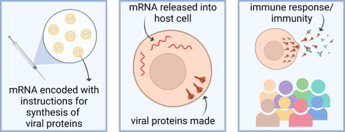
        mRNA vaccines use this same basic principle, but instead of injecting a weakened strain of the actual virus into someone, they instead inject a piece of mRNA, or messenger RNA, which codes for a viral protein that stimulates this immune response (Jain, 2021).

Figure 1: The methodology of mRNA vaccines. Adapted from Jain, et. al., 2021

mRNA vaccines are much easier to make than traditional vaccines because they don’t require growing and inactivating a virus, and they don’t carry the risk that live vaccines have of infecting the subject, but up until recently, they weren’t even remotely possible because in vitro mRNA would create a huge inflammatory response when entering the body (Karikó, 2005). However, Hungarian biochemist Katalin Karikó and her colleague Drew Weissman were undeterred, and their research to create mRNA without an inflammatory response paved the way for mRNA vaccines and won them the Nobel Prize in Medicine in 2023 (Karikó, 2005).

        Karikó and Weissman’s research focused on in vitro mRNA’s stimulation of Toll-like receptors (TLRs), which recognize pathogens and activate signaling pathways leading to an inflammatory response (Karikó, 2005). They discovered that mammalian mRNA, which doesn’t stimulate an immune response, contains many base pair modifications, whereas bacterial RNA, which is known to stimulate a response, has no modifications (Karikó, 2005). In vitro mRNA, grown in a lab rather than from mammalian cell cultures, also has no, or very few, modifications. This led the researchers to question whether base pair modifications were how cells distinguished between foreign and non-foreign RNA, and thus which pieces of RNA stimulate an immune response (Karikó, 2005). They discovered that in vitro RNA activates three main TLRs known for activating an inflammatory response, but that nucleoside modifications limit that activation. Most importantly, they discovered that the suppressed immune response is proportional to the number of nucleotide modifications, and that these modifications also increase the stability of in vitro mRNA in the body, another original roadblock in the usage of mRNA for vaccines. (Karikó, 2005).

Figure 2: The use of base modifications to suppress inflammatory response of RNA. Adapted from Nobelprize.org

        Combined with later research also by Karikó and Weissman discovering that modified base-pairs also create increased protein production and other research designing more stable lipid carriers to deposit the mRNA into human cells, mRNA became a promising new vaccine technology, eventually allowing the COVID-19 vaccines to be produced in record times, shortening the pandemic and granting all of us our lives back. But the road to Karikó’s research being recognized, and especially the road to the Nobel Prize, was not predetermined. Karikó’s research was long overlooked, in part because of her identity as a Hungarian immigrant. She was “demoted four times” at UPenn, being told that her research didn’t measure up to their standards, but she continued to persevere, making her both a scientific success story, and a personal one (Shrikant, 2023). The COVID-19 vaccines created by Pfizer and Moderna were the first mRNA vaccines to be released, but future research is focusing on creating a universal flu vaccine and possibly even an HIV vaccine, all using mRNA technologies, which could drastically improve the landscape of viral infections, both in the US and across the globe.  


Literature Cited

Karikó, K., Buckstein, M., Ni, H., & Weissman, D. (2005). Suppression of RNA Recognition by Toll-Like Receptors: The Impact of Nucleoside Modification and the Evolutionary Origin of RNA. Immunity, 23(2), 165–175. https://doi.org/10.1016/j.immuni.2005.06.008

 Karikó, K., Muramatsu, H., Welsh, F. A., Ludwig, J., Kato, H., Akira, S., & Weissman, D. (2008). Incorporation of Pseudouridine into mRNA yields Superior Nonimmunogenic Vector with Increased Translational Capacity and Biological Stability. Molecular Therapy, 16(11), 1833–1840. https://doi.org/10.1038/mt.2008.200

 Jain, S., Venkataraman, A., Wechsler, M. E., & Peppas, N. A. (2021). Messenger RNA-Based Vaccines: Past, Present, and Future Directions in the Context of the COVID-19 Pandemic. Advanced Drug Delivery Reviews, 179, 114000. https://doi.org/10.1016/j.addr.2021.114000

 The Nobel Prize in Physiology or Medicine 2023. (n.d.). NobelPrize.Org. Retrieved October 22, 2023, from https://www.nobelprize.org/prizes/medicine/2023/press-release/

 CDC. (2020, March 28). Covid Data Tracker. Centers for Disease Control and Prevention. https://covid.cdc.gov/covid-data-tracker

Shrikant, A. (2023, October 6). Nobel Prize Winner Katalin Karikó was “demoted 4 times” at her old job. How she persisted: “You have to focus on what’s next.” CNBC. https://www.cnbc.com/2023/10/06/nobel-prize-winner-katalin-karik-on-being-demoted-perseverance-.html 


Filed Under: Biology, Chemistry and Biochemistry, Science

The Possibilities of the CRISPR-Cas9 System

April 2, 2023 by Stephanie Christianson '26

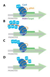
          As humans continue to further explore the fields of science, they deepen their understanding of convoluted subjects through the use of advanced technologies. One of the notable technologies today is the CRISPR-Cas9 system, a highly precise DNA editing tool that allows for genome manipulation in humans, animals, and plants. CRISPR (Clustered Regularly Interspaced Short Palindromic Repeats) works with a CRISPR-associated (Cas) nuclease, which was originally derived from a genome editing system used as immune defense by bacteria. To put it simply, if infected by a virus, the bacteria captures a small piece of the invading viruses’ DNA and inserts their own DNA to create a CRISPR segment (Tavakoli et al., 2021). With this segment, the bacteria can produce RNA segments that will attach to parts of the viruses’ DNA if it attacks again. The bacteria use the Cas9 enzyme to cut the viruses’ DNA apart and incapacitate it. 

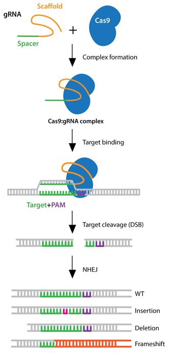
          Scientists adapted the system so it could be inputted into the human genome and edit DNA. Researchers created small segments of RNA that have a “guide” sequence which binds to a target sequence in the DNA and attaches to the Cas9 enzyme (Tavakoli et al., 2021). The guide RNA (gRNA) identifies the intended DNA sequence and the Cas9 enzyme cuts the specific location, similar to how the process occurs in bacteria. The cell’s own DNA machinery is then used to add or delete pieces of the genetic material, known as gene knock-in and gene knockout, respectively. However, it can also alter the DNA by replacing segments with more customized sequences. CRISPR activation (CRISPRa) is used to up-regulate the expression of a gene while CRISPR interference (CRISPRi) down-regulates the expression of a cell (CRISPR Guide).

Figure 1. Gene Knockout
Figure 2. Activation of Target  Gene with CRISPRa

                                       Note. Adapted from CRISPR Guide, Addgene, 2022.

          There are many potential applications in cancer immunotherapy, treatment of genetic diseases, improving plant genetic variation, and more. For example, there have been advances towards mitigating the effects of blood disorders such as sickle cell disease (SCD). In those with SCD, the misshapen red blood cells can block blood vessels, thus slowing or stopping blood flow. Effects of this fatal disease are anemia, chronic pain, strokes, and organ damage. In this instance, CRISPR technology is not used to directly rectify the disease-inducing gene variant. Instead of restoring adult hemoglobin, it works to increase the levels of fetal hemoglobin, an oxygen carrier protein that only fetuses make in the womb (Henderson, 2022). Since it’s not affected by the sickle cell mutation, it can be a substitute for the defective adult hemoglobin in red blood cells (Henderson, 2022).

Figure 3. Hemoglobin Switching in Infants

Note. Adapted from CRISPR Clinical Trials: A 2022 Update, Henderson, 2022, Copyright 2022 by Innovative Genomics Institute.

         As seen in the diagram above, SCD symptoms begin during infancy when levels of fetal hemoglobin decrease. The blood stem cells are harvested from the blood and then the genomes are edited to activate the fetal hemoglobin gene. Next, chemotherapy eliminates the disease-inducing blood stem cells from the person’s body, and the genome-edited stem cells are put back into the bloodstream through an IV, creating a new blood stem cell population that produces fetal hemoglobin (Henderson, 2022). This technique is ex vivo genome editing, where the editing occurs outside the body to prevent the risk of lingering and unwanted CRISPR components in the body. Those treated for SCD in CRISPR clinical trials show normal to near-normal hemoglobin levels with at least 30% as fetal hemoglobin (Henderson, 2022).

          It raises the following questions: is it possible to select certain features in humans that are seen as favorable or desirable? Is it possible to have almost complete manipulation of physical features or to enhance the body’s biological processes to unlock superhuman abilities only seen in movies? These human experimentations would be best measured if implemented during the early stages of life so as to let the body integrate the changes and develop properly. Perhaps we could alter ourselves to increase muscle mass, heighten athletic ability, or augment intelligence. In regard to altering the human genome, there are certain limitations as to what can and can’t be done without violating certain ethical boundaries. 

          This can be seen in the case of He Jianku, a Chinese scientist who used CRISPR to edit two embryos, both of which are now living babies. Though there was little information that only came from He himself and the Chinese government, it has been said that sometime in late 2017, He injected the two embryos with a CRISPR construct that would delete a 32-base-pair in the CCR5 gene on chromosome 3, leading to a non-functional CCR5 protein (Greely, 2019). He sought to achieve humans who could not contract AIDS because they don’t have this functioning protein (Greely, 2019). Though this theory was later proved wrong and the CRISPR construct did not fulfill his intentions, he managed to induce never before seen changes that led to the production of non-functional proteins. His actions prompted the Chinese court to convict him on the basis of deliberately violating medical regulations (Greely, 2019). Could it be argued that pushing the boundaries are necessary for scientific growth and greater human evolution? How far is too far? As CRISPR and other gene editing tools continue to develop, we can keep imagining the unnerving yet exciting possibilities that are awaiting.


Literature Cited

CRISPR Guide. (n.d.). Addgene. 

Greely, H. T. (2019). CRISPR’d babies: human germline genome editing in the ‘He Jiankui affair’. Journal of Law and Biosciences, 6(1), 111-183.

Henderson, H. (2022). CRISPR Clinical Trials: A 2022 Update. Innovative Genomics Institute.

Tavakoli, K., Pour-Aboughadareh, A., Kianersi, F., Poczai, P., Etminan, A., & Shooshtari, L. (2021). Applications of CRISPR-Cas9 as an Advanced Genome Editing System in Life Sciences. BioTech, 10(3), 14. 

What are genome editing and CRISPR-Cas9? (2022). MedlinePlus.

Filed Under: Biology, Science

The Chronic Lyme Debate

April 2, 2023 by Sophie Nigrovic '24

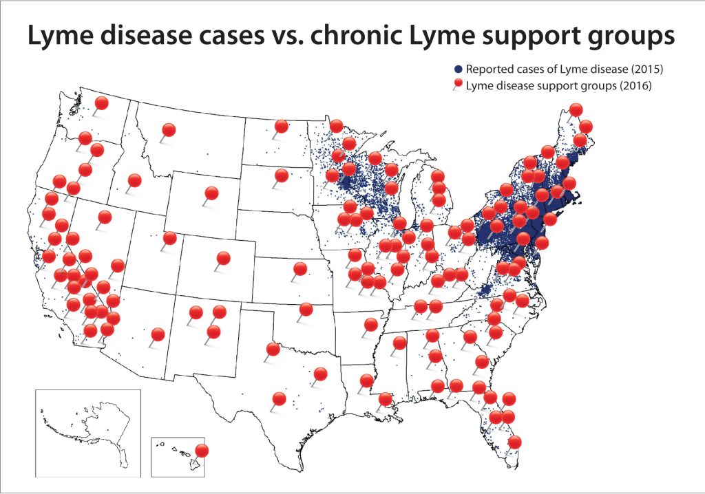
Second possibly only to mosquitos, ticks are the most reviled insect found in New England nature. Like mosquitos, which are notorious vehicles for viruses such as Zika and West Nile, blacklegged ticks (deer ticks) spread Borrelia burgdorferi infection, resulting in Lyme disease. Affecting over 30,000 people a year in the United States, mostly in the northeastern states, Lyme disease is a bacterial infection which causes both local and global symptoms. Early symptoms include fever, muscle fatigue, swollen lymph nodes, and, mostly notably, an erythema migrans rash. Left untreated, patients may develop facial palsy (partial facial paralysis), arthritis, central nervous system inflammation, and heart palpitations. Diagnosis of Lyme disease is based primarily on symptoms and the possibility of exposure to Lyme-carrying ticks, although laboratory tests of patient serum for anti-B. Burgdorferi antibodies may also be considered. Treatments usually consist of a several week course of antibiotics. 

Although the majority of Lyme disease patients recover after initial treatment, 5-20% of patients continue feeling symptoms such as fatigue, muscle pain, and difficulty concentrating. This disorder, called post-treatment Lyme disease syndrome, falls under a broader category of disorders referred to as “chronic Lyme disease.” Also included in this category are patients without a history of Lyme disease but with Lyme-like symptoms; patients with other recognizable disorders seeking an alternative diagnosis; and patients with positive serological results for anti-B. Burgdorferi antibodies but no past exposure to ticks or other routes of infection. 

The majority of chronic Lyme patients fall into the middle two categories: those with symptoms but no history and the misdiagnosed. Moreover, in clinical trials, traditional treatment routes for Lyme disease such as antibiotics have not been effective in alleviating symptoms. Because many of those diagnosed with chronic Lyme do not match the diagnostic criteria for traditional Lyme disease, most scientists and physicians reject the diagnosis. However, a small but very vocal group of patients and physicians fervently believe in the existence of chronic Lyme disease. They are represented by powerful advocacy groups such as the International Lyme and Associated Diseases Society (ILADS) and the Lyme Disease Association (LDA). These groups have been effective at lobbying politicians and regulators to get protections for chronic Lyme sufferers, even as the physician community become increasingly convinced of its inaccuracy.

Although Lyme disease may seem a relatively simple disease with a clear cause (tick-borne bacterial infection) and treatment plan (antibiotics), the controversy over chronic Lyme disease reveals complexities. And this particular controversy doesn’t seem to be abating. It seems that the concept of chronic Lyme disease, like the disorder it purports to describe, is here to stay.


Works Cited:

CDC. (2022, January 19). Lyme disease home | CDC. Centers for Disease Control and Prevention. https://www.cdc.gov/lyme/index.html 

Chronic symptoms. (2018, April 11). Lyme Disease. https://www.columbia-lyme.org/chronic-symptoms 

Feder, H. M., Johnson, B. J. B., O’Connell, S., Shapiro, E. D., Steere, A. C., & Wormser, G. P. (2007). A critical appraisal of “chronic lyme disease.” New England Journal of Medicine, 357(14), 1422–1430. https://doi.org/10.1056/NEJMra072023 

Lantos, P. M. (2011). Chronic Lyme disease: The controversies and the science. Expert Review of Anti-Infective Therapy, 9(7), 787–797. https://doi.org/10.1586/eri.11.63 

Mosquito-borne diseases | niosh | cdc. (2020, February 21). https://www.cdc.gov/niosh/topics/outdoor/mosquito-borne/default.html 

Whelan, D. (n.d.). Lyme inc. Forbes. Retrieved April 2, 2023, from https://www.forbes.com/forbes/2007/0312/096.html 

Filed Under: Biology, Science Tagged With: chronic lyme, lyme disease, ticks

Mercury, Contaminating Our Oceans and Your Food

April 2, 2023 by Riley Simon '26

Mercury, you may know it as the solar system’s smallest planet or as the “red stuff” in old thermometers. You may have even heard of its toxic effects on people if exposure occurs. However, you may not know that mercury is significant outside of the realm of toxic thermometers and astronomy. The chemical element mercury is a contaminant that is being pumped into the atmosphere at an alarming rate and is poisoning aquatic environments. Mercury is becoming an increasingly common pollutant in our oceans and lakes and its toxic effects are causing harm to marine life and creating an imbalance in our marine ecosystems.

Before mercury can enter aquatic environments, it is released in large quantities by anthropogenic sources. Mercury can enter the atmosphere through natural sources such as volcanoes or forest fires, but it is primarily released through the burning of fossil fuels and small-scale gold mining (Montes 287). The release of mercury is problematic because it is very easily transported through the atmosphere. Mercury is a volatile element, which means that it evaporates at low temperatures (mercury can even evaporate at room temperature) and easily enters its gaseous state to be carried long distances through the air (Pollet 860).

After mercury is transported through the atmosphere and enters aquatic environments, it is transformed into its more toxic state, methylmercury (CH3Hg or MeHg). Mercury is transformed into methylmercury through the process of mercury methylation when Hg incorporates CH3, making it into CH3Hg (or MeHg). In the ocean, methylation of mercury is carried out by bacteria. Essentially, bacteria that are present in aquatic environments absorb mercury and perform the methylation reaction before releasing methylmercury back into the ecosystem (Poulain 1280-1281).

Once organisms ingest methylmercury, they experience detrimental effects on their function and, ultimately, their survival. For example, seabirds with more than 0.2 μg (micrograms) of mercury in their blood per gram of wet weight have observed negative effects on their bodies’ systems and their function. An approximately equivalent concentration can be represented by one person out of the entire state of Alabama, which has a population of 5.04 million. At this level or greater, birds experience detrimental effects on their nervous and reproductive systems as well as changes to their hormonal makeup and trouble with motor and behavioral skills (Pollet 860). 

Another interesting observation of mercury contamination is its differing distribution of concentrations among populations. The methylmercury concentration in seabirds was explored between 2013 and 2019 when egg and blood samples were taken of Leach’s storm petrels along with the GPS tracking of foraging petrels. By comparing measured mercury concentration in blood and eggs to ocean depth of foraging locations, a correlation was found. The study concluded that the water depth had a significant effect on the methylmercury levels measured in Leach’s storm petrels. Storm petrels who foraged in deep waters had higher methylmercury concentrations in their blood than storm petrels who forage in shallow or coastal waters. The positive correlation between ocean depth and mercury concentration is likely due to differences in diet based on foraging location. This could also be related to the fact that mercury methylation is most efficient in deep water (Pollet 860).

These negative effects on seabirds also have broad effects on the entire ecosystem that they are a part of. This is because methylmercury biomagnifies up the food web. As the methylmercury moves up trophic levels in a food chain, its concentration in a given organism increases. This increase in concentration is dramatic. In fact, measured mercury concentrations in predator species can be millions of times greater than the concentration observed in surface waters. The problem of biomagnification is even more dramatic in Maine because of its latitude. While the phenomenon is not entirely understood, ecosystems located at higher latitudes have been observed to be more susceptible to biomagnification than tropical regions (Lavoie 13385-13394). 

Other than affecting our Maine wildlife, mercury contamination could have a negative impact on human health. Mercury is not only a problem for seabirds, but fish are also just as susceptible to contamination. This can become a major problem for commercial fisheries because the seafood they produce for our consumption have the potential for mercury contamination. In fact, there was an incident in Japan in 1956 when people who consumed contaminated seafood became severely ill or died. Over the course of 36 years after the incident, 2252 people were infected 1034 people were killed in relation to the initial methylmercury contamination (Harada 1-24). While this level of contamination is an anomaly, a 2018 study projected that approximately 38% of countries experience some level of human exposure to mercury due to contaminated seafood consumption. There are steps that can be taken to prevent this. For example, setting stricter regulations on mercury content limits, applying proper culinary treatments, or updating fishing practices could all diminish the probability of mercury exposure to humans (Jinadasa 112710). 

That being said, addressing the root of the problem is the only way to most effectively diminish mercury exposure for marine organisms and people. Mercury contamination is a problem that has a dramatic domino effect beginning in our atmosphere and ending in human consumption. It is an issue that is often forgotten in discussions of the environmental impact of burning fossil fuels. However, considering its impact on marine life and eventually human life, it is a byproduct that cannot be overlooked.


Works Cited

da Silva Montes, C., Ferreira, M. A. P., Giarrizzo, T., Amado, L. L., & Rocha, R. M. (2022). The
legacy of artisanal gold mining and its impact on fish health from Tapajós Amazonian
region: A multi-biomarker approach. Chemosphere, 287, 132263.

Harada, M. (1995). Minamata disease: methylmercury poisoning in Japan caused by
environmental pollution. Critical reviews in toxicology, 25(1), 1-24.

Jinadasa, B. K. K. K., Jayasinghe, G. D. T. M., Pohl, P., & Fowler, S. W. (2021). Mitigating the
impact of mercury contaminants in fish and other seafood—A review. Marine Pollution
Bulletin, 171, 112710.

Lavoie, R. A., Jardine, T. D., Chumchal, M. M., Kidd, K. A., & Campbell, L. M. (2013).
Biomagnification of mercury in aquatic food webs: a worldwide meta-analysis.
Environmental science & technology, 47(23), 13385-13394.

Pollet, I. L., McFarlane-Tranquilla, L., Burgess, N. M., Diamond, A. W., Gjerdrum, C., Hedd, A.,
… & Mallory, M. L. (2023). Factors influencing mercury levels in Leach’s storm-petrels at
northwest Atlantic colonies. Science of The Total Environment, 860, 160464.

Poulain, A. J., & Barkay, T. (2013). Cracking the mercury methylation code. Science, 339(6125),
1280-1281.

Filed Under: Biology, Environmental Science and EOS Tagged With: contaminants, fossil fuels, Marine Biology, mercury, seabirds

The Contraceptive Brain Drain: How Birth Control Alters Women’s Brains

April 2, 2023 by Divya Bhargava

There are millions of women taking steroids every day. But how is this possible? Are they just getting really buff? It feels like we always hear stories about how performance-enhancing drugs, namely steroids, are giving world-class athletes the boost they need to beat out their competition. But women across the globe are taking steroids every day as well, in the form of hormonal birth control. Despite their widespread use, side effects of hormonal contraceptives are largely unstudied, or have been until recently. In the last ten years, several studies have come out about the effect of taking a daily dose of steroids on women’s brains and mental health, which until now has been a severely neglected area where lack of knowledge affects millions of people worldwide. 

People take hormonal birth control, or hormonal contraceptives, for a myriad of reasons, from the obvious (preventing pregnancy) to the not-so-obvious (lessening iron deficiency) and everything in between. This type of medication simply refers to methods of pregnancy prevention that act on the endocrine system. The endocrine system controls growth, development, metabolism, and reproduction via signaling molecules called hormones. Two hormones in particular, estrogen and progesterone, control the menstrual cycle and are therefore the major components of hormonal birth control. Types of hormonal contraceptives come in many forms including the pill, the patch, the implant, injections, and hormonal intrauterine devices or IUDs, but despite the wide variety in the forms this medication takes, all of them contain one or both of these two hormones. As steroids, both estrogen and progesterone affect other body systems besides the reproductive system.

To study the impacts of taking a daily dose of steroids on other areas of the body, specifically the brain, Dr. Belinda Pletzer and her colleagues conducted a study in 2010. The brain is particularly susceptible to change due to an influx of synthetic hormones because it contains a very high quantity of hormone receptors. The brain needs to act as a “sponge” for these molecules since it plays an important role in creating the appropriate responses in the rest of the body. Pletzer’s study investigated how the sponginess of the brain would affect changes in its structure by comparing images of the brains of adult men, adult women during different stages of their menstrual cycle, and adult women taking hormonal contraceptives. To perform this comparison they used a technique called voxel-based morphology on MRIs of study participants (Pletzer et al., 2010). Voxel-based morphology measures differences in the concentration of tissue and the size and shape of different areas of the brain.

Overall, they found that women taking hormonal birth control had smaller areas of gray matter, or areas of the brain that have a high concentration of the cell bodies of nerve cells, when compared to “naturally cycling women” in both their follicular and luteal menstrual phases (Figure 1). Pletzer’s study also found interesting gendered differences in gray matter volume. While men had greater gray matter overall, the volume of gray matter in the prefrontal cortex, the pre-and postcentral gyri, and the supramarginal gyrus of both naturally cycling women and women taking hormonal contraceptives was higher than the volume of gray matter in these areas in men (Figure 2). These areas are involved in decision-making and problem-solving, controlling motor function, and emotional responses. However, the higher amount of gray matter in women in these areas was overshadowed by the larger volume of gray matter in men in the hippocampus, hypothalamus, parahippocampal and fusiform gyri, putamen, pallidum, amygdala, and temporal regions of the brain during the early follicular phase (A), mid-luteal phase (B), and in women taking hormonal birth control (C) (Figure 2). Many of these areas of reduced gray matter are ones of high importance for neurophysical ability and mental health.

Additionally, a study done by Rush University Medical Center showed an association between higher levels of gray matter and better cognitive function (“Everyday Activities Associated with More Gray Matter in Brains of Older Adults”). These findings suggest that taking birth control, and the associated decrease in gray matter, could be directly causing some of the symptoms women on hormonal contraceptives experience, such as brain fog, mood changes, and even anxiety and depression. For example, a smaller hypothalamus, one of the areas of decreased gray matter, is associated with heightened irritability and depression symptoms (“Study Finds Key Brain Region Smaller in Birth Control Pill Users”). Pletzer’s research and the work of others after her on the impact of birth control on structures of the brain represent important first steps in proving a causative relationship between birth control, symptoms associated with it, and structural changes in the brain.

Although this research has made some crucial preliminary steps into researching how taking a daily dose of steroids affects the brains of women taking hormonal contraceptives, the highly complex nature of the brain and its relationship with the regulation of the rest of the body means that further research is necessary. The sheer number of people that this issue affects means that it is essential to continue researching the impacts of this widely used drug. More importantly, knowing the potentially serious negative side effects enables millions of people to make more informed decisions concerning their health and their bodies.


Works Cited

Rush University Medical Center. (2018, February 14). Everyday activities associated with more gray matter in brains of older adults: Study measured amount of lifestyle physical activity such as house work, dog walking and gardening. ScienceDaily. Retrieved March 11, 2023 from www.sciencedaily.com/releases/2018/02/180214093828.htm.

Lewis, C. A., Kimmig, A. C. S., Zsido, R. G., Jank, A., Derntl, B., & Sacher, J. (2019). Effects of hormonal contraceptives on mood: a focus on emotion recognition and reactivity, reward processing, and stress response. Current psychiatry reports, vol. 21, no.11, 2019, p 115. PubMed Central, https://doi.org/10.1007/s11920-019-1095-z.

Meyer, Craig H., Kinsley, Elizabeth A. “Women’s Brains on Steroids.” Scientific American, https://www.scientificamerican.com/article/womens-brains-on-steroids/. Accessed 7 Mar. 2023.

Nemoto, Kiyotaka. “[Understanding Voxel-Based Morphometry].” Brain and Nerve = Shinkei Kenkyu No Shinpo, vol. 69, no. 5, May 2017, pp. 505–11. PubMed, https://doi.org/10.11477/mf.1416200776.

Pletzer, Belinda, et al. “Menstrual Cycle and Hormonal Contraceptive Use Modulate Human Brain Structure.” Brain Research, vol. 1348, Aug. 2010, pp. 55–62. ScienceDirect, https://doi.org/10.1016/j.brainres.2010.06.019.

Sharma, Rupali, et al. “Use of the Birth Control Pill Affects Stress Reactivity and Brain Structure and Function.” Hormones and Behavior, vol. 124, Aug. 2020, p. 104783. ScienceDirect, https://doi.org/10.1016/j.yhbeh.2020.104783.

“Study Finds Key Brain Region Smaller in Birth Control Pill Users.” ScienceDaily, https://www.sciencedaily.com/releases/2019/12/191204090819.htm. Accessed 7 Mar. 2023.

Filed Under: Biology, Psychology and Neuroscience, Science Tagged With: Biology, Birth control, Medicine, Women's health

What Causes Aging? An Epigenetics Study From The Sinclair Lab May Have an Answer

April 2, 2023 by Luke Taylor '24

Dr. David Sinclair, A.O., Ph.D. Photo from the Sinclair Lab, Harvard Medical School (2023).

Aging, also known as “senescence,” is an inevitable process in all living things. Organisms small and large eventually break down, accumulating enough wear and tear in their cells that ultimately causes the body to stop functioning as a whole. While medicine and lifestyle improvements stave off aging, identifying its fundamental causes has been more challenging. In January 2023, scientists in Dr. David Sinclair’s lab at Harvard Medical School published a paper with experimental evidence supporting what Sinclair calls the “Information Theory of Aging,” where damage to the epigenome can cause aging.

After receiving his Ph.D. in molecular genetics from the University of New South Wales, Sinclair completed his postdoctorate at MIT where he co-discovered the role of sirtuin enzymes in limiting age-related cellular damage in yeast. In addition to teaching genetics and translational medicine at Harvard Medical School since 1999, Sinclair authored the popular book Lifespan: Why We Age – and Why We don’t Have To (2019). His breakthroughs in the science of aging have earned him a great deal of attention from the public eye, resulting in appearances on several popular media outlets, including CBS’s “60 Minutes” and TIME magazine (The Sinclair Lab, 2023).

Sinclair’s newest discovery, published as “Loss of epigenetic information as a cause of mammalian ageing” in January 2023, focused on the role of epigenetics in aging. The title specifies that epigenetic information loss, rather than genetic information loss, is a cause of aging. Genetics refers to the raw molecular information sequences stored in cells as deoxyribonucleic acid (DNA), which is physically condensed inside the nucleus into pairs of chromosomes. The material inside a chromosome is known as “chromatin.” The central dogma of molecular biology states that information in DNA sequences is read by the cell in the form of messenger RNA (mRNA) through transcription, and then ribosomes in the cell read this mRNA to make proteins through translation. DNA sequences that correspond to the production of a specific molecule are genes. The prefix epi- means “on top of,” so “epigenetics” refers to mechanisms that function “above” the molecules of DNA themselves, including reading DNA sequences, regulating gene transcription, and repairing mutated DNA. Like the DNA sequence, epigenetic changes are inheritable from parents to offspring.

The differences between genetics and epigenetics influence cellular reaction to damage. Damage to genes causes mutations, which are changes in the sequence of the DNA of that gene. Cells have mechanisms of repairing mutated DNA, but failure of these mechanisms can lead to cell death, or worse, cancer. Some DNA mutations, like those where a nucleotide is deleted, are irreversible.

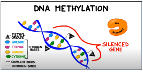
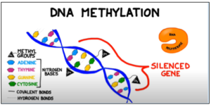
By contrast, epigenetic mechanisms are more easily reversed. One epigenetic mechanism is DNA methylation, where a methyl group (-CH3) is added to a cytosine nucleotide by the DNA methyltransferase enzyme. This extra functional group blocks transcription factors from binding to promoter regions nearby the methylated cytosine, in effect “silencing” the gene as it cannot be transcribed into mRNA. DNA methylation is important for differentiating cells into specific cell types by enabling cells to only express the most pertinent genes while still containing the entire genome (Moore et al., 2012). DNA methylation is reversible with the help of TET dioxygenase enzymes (Wu and Zhang, 2014). Geneticists have found DNA methylation to be a way to assess molecular aging in cells as a sort of “epigenetic clock”. By analyzing methylation patterns of the genome (the “methylome”), scientists can find the biological age as well as the rate of aging in an organism’s cells (Hannum et al., 2012).

Figure 1: Methyl groups attached to cytosine bases in a gene block the enzyme RNA polymerase from binding to the promoter region of a gene, preventing transcription. Adapted from BOGOBiology (2017)
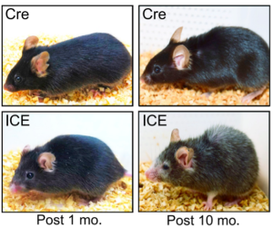
To investigate how changes to the epigenome affect aging in mice, Sinclair used a mouse system with induced changes to the epigenome (ICE). The genetically modified mice had a higher frequency of double-strand breaks (DSBs) in the DNA, which cause changes in the epigenome as cells are required to use their mechanisms of DNA repair more often . Sinclair’s method reduced the frequency of mutations by breaking the DNA strands in a way that left more whole unpaired nucleotides in the severed strand, making it more difficult for the cell to repair the DNA strand with a different sequence than before. While ICE mice had no significant difference in mutation frequencies versus control mice, DSBs in specific locations of the genome were observed as expected (Yang et al., 2023). Thus, any apparent changes in aging in the ICE mice of this study could be ascribed to the epigenetic DSB changes rather than mutations.

Figure 2: ICE treated mice after 10 months appear to have the physical hallmarks of aging, such as hair loss and reduced body mass, early compared to CRE control mice (Yang et. al. 2023).

The haggard appearance of the ICE 10 month old mice confirmed the early aging effects of the epigenetic changes (Figure 2). At the molecular level, analysis of DNA methylation at genome sites associated with age showed that ICE cells were approximately 1.5 times “older” than the control cells. With this artificially increased age came physiological consequences. ICE mice showed diminished short-term memory retention compared to Cre-control mice, as assessed by fear conditioning tests, and the ICE mice performed half as well in a Barnes Maze test as control, indicating decreased long-term memory. Additionally, ICE mice had decreased muscle mass and grip strength after 16 months. The authors attributed the cause of this accelerated aging to increased “faithful DNA repair” from the induced DSB breaks in the ICE mice, meaning that there were not significant mutations in the repair of DSBs.(Yang et al., 2023). During DSB repair, chromatin modifying factor proteins activate and move within a cell in a process known as “relocalization,” with repeated activation of this process known to cause epigenetic changes that silence genes normally expressed in young mice. Sinclair’s lab hypothesized that the relocalization of chromatin modifiers that occurs from repeated DSB repair associated with induced epigenetic changes lead to a gradual loss of cellular function associated with aging (Yang et al., 2023).

It may seem ironic that DNA strand break repair, a process meant to keep cells functioning when critical genes are damaged, is part of what ultimately causes the death of organisms. The fact that the mechanism implemented is epigenetic rather than genetic suggests that the effects of ageing may be reversible, like epigenetic mechanisms. In fact, Sinclair has shown that it is possible to partially undo the damaging effects of aging: after inducing OSK expression, which is a set of proteins known as “Yamanaka factors,” ICE mice exhibited some signs of rejuvenation in their eyes, kidneys, and muscles. Yamanaka factors like OSK are important in the fields of aging and regenerative medicine because they are keys to the synthesis of induced pluripotent stem cells, where somatic cells can become stem cells and potentially re-differentiate into other cell types (Takahashi and Yamanaka, 2006). The OSK treatment decreased the expression of age-associated markers in the kidney and muscle cells of the mice (Yang et al., 2023).

Sinclair’s experiments have shown the epigenome to be a key front in the investigation of aging, as the reversibility of changes to the epigenome can allow it to be a more accessible interface for scientists to interact with. The plasticity of the epigenome as demonstrated from the ability of Yamanaka factors to reverse the molecular indicators of aging mice show that there may be hope for science to bring this phenomenon to human epigenomes. Indeed, the news of reversing aging in mice made rounds in the media when Sinclair’s paper was published earlier this winter, and for good reason.

Works Cited

Al Aboud, N. M., Tupper, C., & Jialal, I. (2022). Genetics, Epigenetic Mechanism. In StatPearls. StatPearls Publishing. http://www.ncbi.nlm.nih.gov/books/NBK532999/

Bannister, A. J., & Kouzarides, T. (2011). Regulation of chromatin by histone modifications. Cell Research, 21(3), Article 3. https://doi.org/10.1038/cr.2011.22

BOGObiology (Director). (2017, October 11). Epigenetics: Nature vs. Nurture. https://www.youtube.com/watch?v=Q8BMP6HDIco

CDC. (2022, August 15). What is Epigenetics? | CDC. Centers for Disease Control and Prevention. https://www.cdc.gov/genomics/disease/epigenetics.htm

David Sinclair | The Sinclair Lab. (n.d.-a). Retrieved March 5, 2023, from https://sinclair.hms.harvard.edu/people/david-sinclair

Fernandez, A., O’Leary, C., O’Byrne, K. J., Burgess, J., Richard, D. J., & Suraweera, A. (2021). Epigenetic Mechanisms in DNA Double Strand Break Repair: A Clinical Review. Frontiers in Molecular Biosciences, 8, 685440. https://doi.org/10.3389/fmolb.2021.685440

Gilbert, S. F. (2000). Methylation Pattern and the Control of Transcription. Developmental Biology. 6th Edition. https://www.ncbi.nlm.nih.gov/books/NBK10038/

Hannum, G., Guinney, J., Zhao, L., Zhang, L., Hughes, G., Sadda, S., Klotzle, B., Bibikova, M., Fan, J.-B., Gao, Y., Deconde, R., Chen, M., Rajapakse, I., Friend, S., Ideker, T., & Zhang, K. (2013). Genome-wide Methylation Profiles Reveal Quantitative Views of Human Aging Rates. Molecular Cell, 49(2), 359–367. https://doi.org/10.1016/j.molcel.2012.10.016

Jin, B., Li, Y., & Robertson, K. D. (2011). DNA Methylation. Genes & Cancer, 2(6), 607–617. https://doi.org/10.1177/1947601910393957

Kulis, M., & Esteller, M. (2010). 2—DNA Methylation and Cancer. In Z. Herceg & T. Ushijima (Eds.), Advances in Genetics (Vol. 70, pp. 27–56). Academic Press. https://doi.org/10.1016/B978-0-12-380866-0.60002-2

Molecules discovered that extend life in yeast, human cells. (n.d.). EurekAlert! Retrieved March 5, 2023, from https://www.eurekalert.org/news-releases/664233.

Moore, L. D., Le, T., & Fan, G. (2013). DNA Methylation and Its Basic Function. Neuropsychopharmacology, 38(1), 23–38. https://doi.org/10.1038/npp.2012.112

Offord, Catherine. Two research teams reverse signs of aging in mice. (n.d.). Retrieved March 14, 2023, from https://www.science.org/content/article/two-research-teams-reverse-signs-aging-mice.

Takahashi, K., & Yamanaka, S. (2006). Induction of Pluripotent Stem Cells from Mouse Embryonic and Adult Fibroblast Cultures by Defined Factors. Cell, 126(4), 663–676. https://doi.org/10.1016/j.cell.2006.07.024

What is Epigenetics? The Answer to the Nature vs. Nurture Debate. (n.d.). Center on the Developing Child at Harvard University. Retrieved March 5, 2023, from https://developingchild.harvard.edu/resources/what-is-epigenetics-and-how-does-it-relate-to-child-development/

Wu, H., & Sun, Y. E. (2009). Reversing DNA Methylation: New Insights from Neuronal Activity–Induced Gadd45b in Adult Neurogenesis. Science Signaling, 2(64), pe17–pe17. https://doi.org/10.1126/scisignal.264pe17

Wu, H., & Zhang, Y. (2014a). Reversing DNA Methylation: Mechanisms, Genomics, and Biological Functions. Cell, 156(0), 45–68. https://doi.org/10.1016/j.cell.2013.12.019

Yang, J.-H., Hayano, M., Griffin, P. T., Amorim, J. A., Bonkowski, M. S., Apostolides, J. K., Salfati, E. L., Blanchette, M., Munding, E. M., Bhakta, M., Chew, Y. C., Guo, W., Yang, X., Maybury-Lewis, S., Tian, X., Ross, J. M., Coppotelli, G., Meer, M. V., Rogers-Hammond, R., … Sinclair, D. A. (2023). Loss of epigenetic information as a cause of mammalian aging. Cell, 186(2), 305-326.e27. https://doi.org/10.1016/j.cell.2022.12.027


Filed Under: Biology, Science

  • « Go to Previous Page
  • Page 1
  • Interim pages omitted …
  • Page 3
  • Page 4
  • Page 5
  • Page 6
  • Page 7
  • Go to Next Page »

Primary Sidebar

CATEGORY CLOUD

Biology Chemistry and Biochemistry Computer Science and Tech Environmental Science and EOS Honors Projects Math and Physics Psychology and Neuroscience Science

RECENT POSTS

  • Floating Systems: Jellyfish and Evolving Nervous Systems May 22, 2025
  • Biological ChatGPT: Rewriting Life With Evo 2 May 4, 2025
  • Unsupervised Thematic Clustering for Genre Classification in Literary Texts May 4, 2025

FOLLOW US

  • Facebook
  • Twitter

Footer

TAGS

AI AI ethics Alzheimer's Disease antibiotics artificial intelligence bacteria Bathymetry Beavers Biology brain Cancer Biology Cell Biology Chemistry and Biochemistry Chlorofluorocarbons climate change Computer Science and Tech CRISPR Cytoskeleton Depression dreams epigenetics Ethics Genes honors Luis Vidali Marine Biology Marine Mammals Marine noise Medicine memory Montreal Protocol Moss neurobiology neuroscience Nutrients Ozone hole Plants Psychology and Neuroscience REM seabirds sleep student superintelligence Technology therapy

Copyright © 2025 · students.bowdoin.edu