• Skip to primary navigation
  • Skip to main content
  • Skip to primary sidebar
  • Skip to footer

Bowdoin Science Journal

  • Home
  • About
    • Our Mission
    • Our Staff
  • Sections
    • Biology
    • Chemistry and Biochemistry
    • Math and Physics
    • Computer Science and Technology
    • Environmental Science and EOS
    • Honors Projects
    • Psychology and Neuroscience
  • Contact Us
  • Fun Links
  • Subscribe

Chemistry and Biochemistry

Ending the Biomedical Harvest: Synthetic Alternatives to Horseshoe Crab Blood for Bacterial Endotoxin Detection

December 3, 2023 by Gabe O'Brien

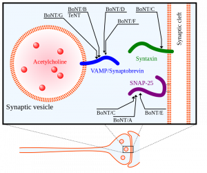
Did you know that horseshoe crabs have incredible immune systems? In fact, horseshoe crabs have the best immune systems out of all living invertebrates. Their secret? Blood. Horseshoe crab blood is very simple in composition, with only a single cell type in general circulation (the granular amebocyte) and only three proteins in the plasma of the blood (hemocyanin, C-reactive proteins, and a2-macroglobulin) (Armstrong et al., 2008). These proteins contribute to the horseshoe crab’s blood clotting system, protecting them from infection. Horseshoe crab blood has been found to be very sensitive to bacterial endotoxins found in illness-causing Gram-negative bacteria (Protecting Health). When horseshoe crab blood cells come into contact with bacterial endotoxin, they clot around it, preventing the bacterium from invading nearby cells (Natural History Museum 2020). 

With the rise of vaccine development, especially in the case of the Covid-19 pandemic, horseshoe crab blood plays an essential role in testing the safety of vaccines due to its endotoxin-detection properties. Additionally, large volumes of horseshoe crab blood can be collected easily, making it a convenient blood source (Armstrong et al., 2008). Despite all the beneficial applications of horseshoe crab blood, horseshoe crab bleeding leaves thousands of horseshoe crabs dead annually, causing their populations to be in decline (Maloney et al., 2018). A 2018 study has promoted a synthetic alternative to horseshoe crab blood, recombinant Factor C (rFC), and proven its efficacy in bacterial endotoxin detection. The use of rFC in vaccine development can eliminate the need for the use of actual horseshoe crab blood, sparing the horseshoe crab and promoting the conservation of this endangered species. 

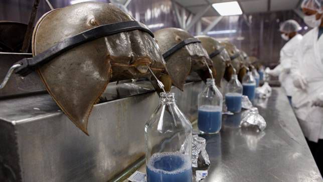
Typically, horseshoe crab blood is collected by the direct puncture of the heart under sterile conditions that minimize contamination by bacterial endotoxins (Figure 1). A large horseshoe crab can produce between 200 – 400 mL of blood, and the blood clotting system can be studied microscopically. The limitation of contamination by bacterial endotoxins is extremely important in the blood collection process, because cell clotting will compromise the effectiveness of the blood for its intended use of developing vaccines. Only undamaged horseshoe crabs are selected for blood collection, and the animal is bled by the insertion of a needle into the heart through the outer hinge joint of the horseshoe crab (Figure 2). The animal is then squeezed gently so that as much blood as possible can be deposited into the collection tube (Armstrong et al., 2008).

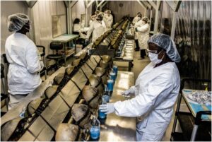
Figure 1: Horseshoe crab bleeding on a larger scale, with precautions taken to ensure sanitary, endotoxin-free conditions for blood collection.

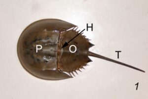
Figure 2: The three major components of the body of a horseshoe crab, including the prosoma (P), the opisthosoma (O), and the telson (T). The hinge (H) is where the prosoma meets the opisthosoma, and that is where the needle is inserted for blood collection. 

Following collection, horseshoe crab blood is ready for use in endotoxin detection (Armstrong et al., 2008). For example, in vaccine development, a Limulus amebocyte lysate (LAL) test detects the levels of clotting in horseshoe crab blood when it comes into contact with different vaccines. Horseshoe crab blood is very precise with detecting even small traces of endotoxin, making it an effective tool to identify small quantities of endotoxin present in potential vaccines (Protecting Health).  

Although horseshoe crab blood is effective in its ability to detect endotoxins, recombinant Factor C (rFC) can do the same job in a way that is more ecologically sustainable. Initially, rFC was discovered by scientists at the National University of Singapore, allowing them to visualize endotoxin detection using animal-free technology. Every year over 500,000 horseshoe crabs are captured and as much as ⅓ of their blood is drained, contributing to high mortality rates. On top of this, around 13% of the horseshoe crabs bled are later sold for bait, resulting in nearly 130,000 horseshoe crab victims to the biomedical industry. A 2018 study confirmed that the biomedical industry could reduce their use of horseshoe crab blood by nearly 90% if they were to employ rFC as a synthetic alternative for endotoxin detection processes. The 2018 study reviews multiple studies that show how rFC is just as effective as actual horseshoe crab blood in endotoxin detection, as rFC has been able to demonstrate the same high rate and sensitivity as horseshoe crab blood in detecting small amounts of endotoxin in a wide range of chemical structures. When endotoxin binds to a synthetic rFC molecule, it causes the rFC to fluoresce directly proportional to the concentration of endotoxin in a substance. rFC has even been able to demonstrate a higher rate of specificity for endotoxin detection (compared to horseshoe crab blood) in some studies (Maloney et al., 2018).

The most important next step of this research is to get synthetic rFC into the hands of the biomedical industry. Exposure to endotoxin can cause serious illness, making endotoxin detection for vaccines an essential part of the vaccine development process.  Even though there is ample evidence that rFC is equivalent to or better than horseshoe crab blood at detecting bacterial endotoxin, there are still limitations to the usage of rFC, as it is difficult for the biomedical industry to adopt new technologies quickly. Endotoxin detection testing is very highly regulated, so many pharmaceutical manufacturers may be hesitant to employ new detection technologies, as they may want to stick to traditional methods instead (Maloney et al., 2018). Despite these limitations, in order to progress towards horseshoe crab conservation, rFC should be produced and employed on a large scale so that the biomedical industry will no longer be solely reliant on the exploitation of horseshoe crabs for bacterial endotoxin detection.

Literature Cited

1. Armstrong, P., Conrad, M. Blood Collection from the American Horseshoe Crab, Limulus Polyphemus. J. Vis. Exp. (20), e958, doi:10.3791/958 (2008). 

2. “Horseshoe Crab Blood: The Miracle Vaccine Ingredient That’s Saved Millions of Lives.” Www.nhm.ac.uk, www.nhm.ac.uk/discover/horseshoe-crab-blood-miracle-vaccine-ingredient.html#:~:text=Horseshoe%20crab%20blood%20is%20bright.  

3. Maloney T, Phelan R, Simmons N. Saving the horseshoe crab: A synthetic alternative to horseshoe crab blood for endotoxin detection. PLoS Biol. 2018 Oct 12;16(10):e2006607. doi: 10.1371/journal.pbio.2006607. PMID: 30312293; PMCID: PMC6200278.  

4. “Protecting Health.” Www.horseshoecrab.org, www.horseshoecrab.org/med/health.html. Accessed 12 Nov. 2023.  


Filed Under: Biology, Chemistry and Biochemistry, Environmental Science and EOS Tagged With: Biology, blood, clinical testing, conservation, horseshoe crabs, Medicine, vaccine development

PedPRM Unveils Promising Treatment for Insomnia in Children with Autism Spectrum Disorder

December 3, 2023 by Fabiola Barocio Prieto '27

Getting enough sleep is widely considered crucial to our well-being. However, for some individuals with Autism, getting enough quality sleep is not as easy as it sounds. Autism Spectrum Disorder (ASD) is a developmental disorder that widely affects the U.S. population, as 1 in 36 children aged eight has been diagnosed with autism (CDC, 2023). The kind and severity of the symptoms that individuals with ASD may exhibit can vary along the ASD spectrum.  Sleeping, in particular, is a common challenge for those with ASD, as many suffer from problems related to the REM (Rapid Eye Movement) sleep phase, a phase critical for memory consolidation (Devnani & Hedge, 2015). This can quickly become a vicious cycle for some, as lack of sleep can increase the severity of other Autism-related symptoms but also affect both the individual and the quality of life of their family or caretakers. 

To investigate possible treatments, a recent study by Malow et al. focuses on the pharmacological approach of using melatonin to treat sleep disorders. Melatonin is a hormone produced by the body’s pineal gland to regulate circadian rhythm, allowing the body to relax according to the appropriate light-darkness cycles of the day. This study uses a small, long-release tablet (PedPRM) used to mimic endogenous melatonin secretion. It focused mainly on a younger population, from children to adolescents, who met two criteria. The first was that they had confirmed diagnoses of either Autism or Smith-Magenis syndrome and had also experienced sleep abnormalities. Smith-Magenis syndrome is also a developmental disorder that involves symptoms similar to those of ASD, affecting behavior, cognition, and sleep. They also had to have not previously seen improvements when using sleep hygiene treatments to be included in the study, such as establishing a strict bedtime routine and taking steps to provide a calm and comfortable sleep environment (CHOC, 2023).

With the study sample set, the study was conducted over 108 weeks and divided into four phases (Graph I). Throughout the four phases, participants’ caregivers would document sleep quality and total sleep time in a sleep and nap diary to record the efficacy of treatment. The study participants started the first stage with a 2-week period in which they were given placebos. If participants showed improvement while receiving a placebo, they would be removed from the study to reduce the possibility of external factors affecting the results (Scott et al., 2021). After clearing Phase 1, participants entered the second phase, which consisted of a double-blinded 13-week period in which they were randomly placed into either a placebo or treatment group (PedPRM). After this, Phase 3 comprised a longer 91-week open-label period in which both groups were combined. For the final phase of the study, participants were placed again in a 2-week single-blind placebo period to ensure that the drug had been completely removed from the participants with no adverse effects after stopping treatment.

Upon concluding the experimental period, the results to be considered for this study could be divided into three groups: participants’ sleep quality, caregivers’ well-being, and the participants’ growth development. With this data, the researchers found a significant

 decrease in sleep disturbance (Graph II-A) and an increase in caregiver satisfaction (II-B) and quality of life (II-C). These were most pronounced during the first half of the treatment. In the latter half of treatment, sleep disturbance continued to decrease but at a slower pace than the initial treatment phase. Fortunately, there were no reported deaths, and most adverse reactions included daytime fatigue and mood swings. However, the severity and extent of these were not detailed in the results of the study and offer the potential to be analyzed further.  

The study shows compelling initial evidence that PedPRM is an effective treatment for sleep disorders in individuals with ASD. However, as noted by the researchers, it also shows that constant active treatment is required as most sleep quality improvements are removed upon halting treatment. Since medications for children are generally more strictly controlled, PedPRM consistently demonstrates a possibility for effective pediatric treatment, even if for long-term medication. In particular, these findings are essential as it has been found that rapid-release melatonin is not helpful with maintaining sleep a couple of hours after administration, and it had long been considered a challenge to find small, swallowable prolonged-release tablets for children (Fliesler, 2022). As sleep interruption is something that mainly affects those with neurodevelopmental disorders, this is a significant step towards adequate treatment. However, it is essential to note that this pharmacological alternative should only be considered if behavioral interventions and sleep hygiene modifications have been attempted but have been found unsuccessful. 


Sources 

CDC. (2022, December 9). Centers for Disease Control and Prevention. https://www.cdc.gov/ncbddd/autism/facts.html 

CDC Newsroom. (2016, January 1). CDC. https://www.cdc.gov/media/releases/2023/p0323-autism.html 

CHOC – Children’s Hospital of Orange County. (2023, March 2). Autism and Sleep Hygiene – Children’s Hospital of Orange County. Children’s Hospital of Orange County. https://www.choc.org/programs-services/autism-neurodevelopmental/co-occurring-conditions-program/autism-and-sleep/ 

Devnani, P., & Hegde, A. U. (2015). Autism and sleep disorders. Journal of Pediatric Neurosciences, 10(4), 304. https://doi.org/10.4103/1817-1745.174438 

Fliesler, N. (2022, June 13). Melatonin for kids: Is it effective? Is it safe? – Boston Children’s Answers. Boston Children’s Answers. https://answers.childrenshospital.org/melatonin-for-children/#:~:text=There%20is%20some%20evidence%20to,the%20ability%20to%20swallow%20capsules. 

Furfaro, H. (2023, March 10). Sleep problems in autism explained. Spectrum | Autism Research News. https://www.spectrumnews.org/news/sleep-problems-autism-explained/ 

Lemoine, P., Garfinkel, D., Laudon, M., Nir, T., & Zisapel, N. (2011). Prolonged-release melatonin for insomnia – an open-label long-term study of efficacy, safety, and withdrawal. Therapeutics and Clinical Risk Management, 301. https://doi.org/10.2147/tcrm.s23036 

Malow, B. A., Findling, R. L., Schröder, C., Maras, A., Breddy, J., Nir, T., Zisapel, N., & Gringras, P. (2021b). Sleep, Growth, and puberty after 2 years of Prolonged-Release Melatonin in children with Autism Spectrum Disorder. Journal of the American Academy of Child and Adolescent Psychiatry, 60(2), 252-261.e3. https://doi.org/10.1016/j.jaac.2019.12.007 

Scott, A., Sharpe, L., Quinn, V. F., & Colagiuri, B. (2022). Association of single-blind placebo run-in periods with the placebo response in randomized clinical trials of antidepressants. JAMA Psychiatry, 79(1), 42. https://doi.org/10.1001/jamapsychiatry.2021.3204 

Signs & Symptoms | Autism Spectrum Disorder (ASD) | NCBDDD | CDC. (2023, January 11). Centers for Disease Control and Prevention. https://www.cdc.gov/ncbddd/autism/signs.html

Filed Under: Biology, Chemistry and Biochemistry, Psychology and Neuroscience, Science Tagged With: Autism, Medicine, sleep

mRNA: Unpacking the Research that led to the COVID-19 Vaccines and the 2023 Nobel Prize in Medicine

November 19, 2023 by Maren Cooper '27

        By 2023, 81.4% of the United States population have received at least one dose of the COVID-19 mRNA vaccine, and just about everyone has heard of these lifesaving shots (CDC, 2023). But how many of us actually know how they work? In general, vaccinations expose the body to a weakened strain of a virus that initiates an immune response so that the virus can be recognized, and quickly neutralized, in the future (Jain, 2021). This works because of the structure of our immune system, which is split up into innate immunity and adaptive immunity. Innate immunity is the immediate response that our bodies put up when exposed to a pathogen; it sends out macrophages to gobble up infected cells, creates an inflammatory response to kill off the infection, and uses a host of molecules and pathways to destroy the virus. Adaptive immunity involves the presence of B and T cells, which arise in a viral infection and create immunological memory through antibodies and memory cells so that a future infection of the same virus is immediately recognized and shut down. Vaccines work to stimulate this adaptive immunity, while minimizing innate immunity.

        mRNA vaccines use this same basic principle, but instead of injecting a weakened strain of the actual virus into someone, they instead inject a piece of mRNA, or messenger RNA, which codes for a viral protein that stimulates this immune response (Jain, 2021).

Figure 1: The methodology of mRNA vaccines. Adapted from Jain, et. al., 2021

mRNA vaccines are much easier to make than traditional vaccines because they don’t require growing and inactivating a virus, and they don’t carry the risk that live vaccines have of infecting the subject, but up until recently, they weren’t even remotely possible because in vitro mRNA would create a huge inflammatory response when entering the body (Karikó, 2005). However, Hungarian biochemist Katalin Karikó and her colleague Drew Weissman were undeterred, and their research to create mRNA without an inflammatory response paved the way for mRNA vaccines and won them the Nobel Prize in Medicine in 2023 (Karikó, 2005).

        Karikó and Weissman’s research focused on in vitro mRNA’s stimulation of Toll-like receptors (TLRs), which recognize pathogens and activate signaling pathways leading to an inflammatory response (Karikó, 2005). They discovered that mammalian mRNA, which doesn’t stimulate an immune response, contains many base pair modifications, whereas bacterial RNA, which is known to stimulate a response, has no modifications (Karikó, 2005). In vitro mRNA, grown in a lab rather than from mammalian cell cultures, also has no, or very few, modifications. This led the researchers to question whether base pair modifications were how cells distinguished between foreign and non-foreign RNA, and thus which pieces of RNA stimulate an immune response (Karikó, 2005). They discovered that in vitro RNA activates three main TLRs known for activating an inflammatory response, but that nucleoside modifications limit that activation. Most importantly, they discovered that the suppressed immune response is proportional to the number of nucleotide modifications, and that these modifications also increase the stability of in vitro mRNA in the body, another original roadblock in the usage of mRNA for vaccines. (Karikó, 2005).

Figure 2: The use of base modifications to suppress inflammatory response of RNA. Adapted from Nobelprize.org

        Combined with later research also by Karikó and Weissman discovering that modified base-pairs also create increased protein production and other research designing more stable lipid carriers to deposit the mRNA into human cells, mRNA became a promising new vaccine technology, eventually allowing the COVID-19 vaccines to be produced in record times, shortening the pandemic and granting all of us our lives back. But the road to Karikó’s research being recognized, and especially the road to the Nobel Prize, was not predetermined. Karikó’s research was long overlooked, in part because of her identity as a Hungarian immigrant. She was “demoted four times” at UPenn, being told that her research didn’t measure up to their standards, but she continued to persevere, making her both a scientific success story, and a personal one (Shrikant, 2023). The COVID-19 vaccines created by Pfizer and Moderna were the first mRNA vaccines to be released, but future research is focusing on creating a universal flu vaccine and possibly even an HIV vaccine, all using mRNA technologies, which could drastically improve the landscape of viral infections, both in the US and across the globe.  


Literature Cited

Karikó, K., Buckstein, M., Ni, H., & Weissman, D. (2005). Suppression of RNA Recognition by Toll-Like Receptors: The Impact of Nucleoside Modification and the Evolutionary Origin of RNA. Immunity, 23(2), 165–175. https://doi.org/10.1016/j.immuni.2005.06.008

 Karikó, K., Muramatsu, H., Welsh, F. A., Ludwig, J., Kato, H., Akira, S., & Weissman, D. (2008). Incorporation of Pseudouridine into mRNA yields Superior Nonimmunogenic Vector with Increased Translational Capacity and Biological Stability. Molecular Therapy, 16(11), 1833–1840. https://doi.org/10.1038/mt.2008.200

 Jain, S., Venkataraman, A., Wechsler, M. E., & Peppas, N. A. (2021). Messenger RNA-Based Vaccines: Past, Present, and Future Directions in the Context of the COVID-19 Pandemic. Advanced Drug Delivery Reviews, 179, 114000. https://doi.org/10.1016/j.addr.2021.114000

 The Nobel Prize in Physiology or Medicine 2023. (n.d.). NobelPrize.Org. Retrieved October 22, 2023, from https://www.nobelprize.org/prizes/medicine/2023/press-release/

 CDC. (2020, March 28). Covid Data Tracker. Centers for Disease Control and Prevention. https://covid.cdc.gov/covid-data-tracker

Shrikant, A. (2023, October 6). Nobel Prize Winner Katalin Karikó was “demoted 4 times” at her old job. How she persisted: “You have to focus on what’s next.” CNBC. https://www.cnbc.com/2023/10/06/nobel-prize-winner-katalin-karik-on-being-demoted-perseverance-.html 


Filed Under: Biology, Chemistry and Biochemistry, Science

Biomimicry Within Bowdoin: The Ongoing Development of Peptoids

April 4, 2023 by Anika Sen

To solve complex human health issues, scientists have more recently turned to biomimicry. Biomimicry, also known as biomimetics, is a field that develops synthetic materials, systems or machines that are derived from the principles of natural biological processes (Nature). Concepts within biomimetics are currently being used to design regenerative medicine and newer drugs for diseases such as cancer. In fact, within Bowdoin, Professor Benjamin Gorske, is undertaking a comprehensive research on developing methods to create drugs that attempt to inhibit signaling pathways within cancer, or interrupt the signal transduction pathways involved in the development of plaques in Alzheimer disease. 

Professor Gorske explains that these aforementioned diseases can be addressed by “controlling the signaling proteins”. By interrupting the signaling process, the underlying issue of these diseases – which are often so diverse and hard to target – doesn’t need to be treated. However, a difficulty that comes with targeting signaling proteins is that they bind to other molecules over a vast space, and are implicated in many other signaling pathways. Therefore, these proteins cannot simply be targeted by small molecular drugs, as they would be impossible to effectively block the proteins and thus are called “undruggable molecules”. Instead, Professor Gorske turns to attempting to create a biological molecule that can mimic the other molecules that bind to the target signaling protein – which are normally much bigger than current drug molecules. 

One of the targets that Professor Gorske looks into in his lab are the signaling proteins within the Hippo pathway. The Hippo pathway normally controls the size of organs by regulating cell proliferation (division of cells) and apoptosis (programmed cell death) (Cell Signaling Technology). Ultimately, this pathway controls the expression of certain genes that are involved in the proliferation process. This pathway is involved in cancer when it is disregulated, as it continuously sends a signal to the nucleus to express these genes encoding for cell proliferation, thus leading to the uncontrollable growth of cancer. The Hippo pathway involves a lot of signaling proteins that are solely involved in this signaling pathway. Most of these signaling proteins contain a WW domain – a distinct functional unit of the protein that mediates the interactions these proteins have with other molecules (EMBL-EBI). All the signaling proteins with this specific domain in this pathway generally bind to proteins with polyproline type 2 helices (PPII). Professor Gorske’s lab aims to design a molecule to contain this PPII structural component to effectively target the signaling proteins (with the WW domain) in the Hippo pathway.

Fig 1: Diagram of the Hippo Pathway. The signaling protein YAP (yes-associated protein/transcription coactivator) is an example of a molecule with the WW domain.

However, we can’t utilize naturally formed proteins with these PPII helices as the proteases in our body would find the related drugs as foreign and destroy them. Thus these helices need to be made artificially, forming molecules called peptoids. Peptoids are a class of molecules that mimic the structure and function of helical peptides (Chongsiriwatana et al., 2008). They are very biostable, relatively easy to make, and most importantly proteases don’t destroy these molecules. Thus Professor Gorske has chosen to make specific peptoids that perhaps could target these signaling proteins in his lab.

Fig 2: Comparison of the general molecular structure of a naturally-derived peptide versus an artificially-produced peptoid.

However, an issue that arose with creating these peptoids is that the peptoids often folded to form polyproline type 1 helices (PPI) rather than PPII helices. This is due to the differences in orientation of the connected amides (subunits of the peptoids). This leads to peptoids with PP1 helices being much more compressed than peptoids with PPII helices. While they do work as good lung surfactants and other antimicrobials, they are not suitable to target signaling proteins. 

Fig 3: Comparison of the 3D structure of polyproline 1 helix (PPI) and polyproline 2 helix (PPII), and the orientation of the respective amide subunits.

Through experimenting, Professor Gorske has found out in his lab that the best way to encourage amides to connect in the desired PPII orientation is through adding side chains on the amide subunits, and the side chains are additionally thionated – the carbon is double bonded to sulfur instead of oxygen. Currently, he is investigating whether adding more than one thionated residue can lead to the whole peptoid to adopt the desired PPII helical structure. 

This method of creating peptoids that Professor Gorske is working to devise can be implicated in so many uses within medicine. As peptoid drugs can mimic biological molecules, they can more precisely target the required proteins, to help inhibit or promote signaling pathways back to normal. Professor Gorske’s research is therefore crucial to the ongoing development of medicine and healthcare. 

References

“Biomimetics Articles from across Nature Portfolio.” Nature News, Nature Publishing Group, https://www.nature.com/subjects/biomimetics. 

Chen, Yu-An, et al. “WW Domain-Containing Proteins Yap and Taz in the Hippo Pathway as Key Regulators in Stemness Maintenance, Tissue Homeostasis, and Tumorigenesis.” Frontiers in Oncology, vol. 9, 2019, https://doi.org/10.3389/fonc.2019.00060. 

Chongsiriwatana, Nathaniel P., et al. “Peptoids That Mimic the Structure, Function, and Mechanism of Helical Antimicrobial Peptides.” Proceedings of the National Academy of Sciences, vol. 105, no. 8, 2008, pp. 2794–2799., https://doi.org/10.1073/pnas.0708254105. 

Embl-Ebi. “What Are Protein Domains?” What Are Protein Domains? | Protein Classification, https://www.ebi.ac.uk/training/online/courses/protein-classification-intro-ebi-resources/protein-classification/what-are-protein-domains/. 

“Hippo Signaling.” Cell Signaling Technology, 2010, https://www.cellsignal.com/pathways/hippo-signaling-pathway.


Filed Under: Chemistry and Biochemistry, Science

The Battle of the Medications: The Connection Between Antidepressants and Antibiotic Resistance in Bacteria

April 2, 2023 by Sam Koegler

         The consumption of antidepressant medications has skyrocketed in recent decades, reaching more than 337 million prescriptions written in 2016 in the United States alone (Wang et. al. 2019). For many individuals, these drugs are critical to maintaining everyday health as they treat many life-threatening psychiatric disorders. While their exact mechanisms differ, these medications travel in the bloodstream to the brain where they are able to influence the release of chemicals known as neurotransmitters that generate emotional states. However, while their intended target is the brain, these drugs continue to circulate throughout the body, thereby interacting with other organs and structures (Wang et. al. 2019).

         In their 2019 study, researchers led by Iva Lukic used data indicating the presence of antidepressants in the digestive tract to investigate the effect of these medications on the gut microbiome. After treating mice with different types of antidepressants, the team noticed a change in the types of bacteria present within the gut when compared to controls (Lukic et. al. 2019). This discovery that antidepressants could impact the types of bacteria present within the body ultimately led researcher Jianhua Guo to question the additional effects that these medications could have on bacteria. As antibiotics have also been shown to affect the composition of the gut microbiome, Guo began by investigating if the antidepressant fluoxetine could help Escherichia coli cells survive in the presence of various antibiotics. After finding that exposure to this medication did increase E. coli’s resistance to antibiotic treatments, Guo decided to expand his hypothesis to examine the overall connection of antidepressant usage with antibiotic resistance in bacteria.

        Collaborating with researchers Zue Wang and Zhigang Yue, Guo’s lab began by choosing five major types of antidepressant medications: sertraline, escitalopram, bupropion, duloxetine, and agomelatine. These medications differ in the ways that they prevent the reuptake of serotonin and norepinephrine in the brain, thereby allowing the researchers to examine the effects of various types of antidepressants that may be prescribed to patients. Then, E. coli bacteria were added to media containing varying concentrations of these five antidepressants. Once these cells were treated with antidepressants, the researchers began to test the cells’ resistance against antibiotics. In order to accurately reflect antibiotic use in the real world, the tested antibiotics covered the six main categories of antibiotic medications available on the market. The antidepressant-treated bacteria were then swabbed onto plates containing one of the tested antibiotics to observe cell growth. Based on the growth present on these plates, the researchers were able to estimate the incidence rate of bacterial resistance of E. coli bacteria treated with different antidepressants.

         Through this experiment, the lab observed that E. coli cells grown in sertraline and duloxetine, two antidepressants that inhibit the reuptake of serotonin, exhibited the greatest number of resistant cells across all the tested antibiotics (fig. 1). They also noted that E. coli cells exhibiting resistance to one antibiotic often demonstrate some level of resistance to other antibiotics as well. After detecting a correlation between antibiotic-resistance development and exposure to antidepressants, the lab tested the concentration dependence of this effect. While lowering the concentration of antidepressants seemed to decrease the amount of resistant E. coli cells, resistant cells continued to appear on the plates over time, suggesting that lowering antidepressant dosages only prolongs the process of developing antibiotic resistance.

Figure 1: These graphs showcase the change in the number of antibiotic-resistant E. coli cells after exposure to antidepressants over sixty days. The title of each graph indicates the tested antibiotic while the colored trend lines on the graph represent one of the five, tested antidepressants. On the y-axis of each graph, the fold change measurement is used to describe the change in the number of resistant cells that develop over time. As demonstrated by the purple and yellow trend lines, duloxetine and sertraline are associated with the greatest development of resistant cells to each of the four represented antibiotics. (Adapted from Wang et. al. 2019)

         After analyzing this data, the researchers were confronted with a question: what about anti-depressants led to the development of antibiotic resistance in bacteria? To examine this question, the lab used flow cytometry to examine what was happening within the bacterial cells. This lab technique uses a fluorescent dye that binds to specific intercellular target molecules, thereby allowing these components to be visualized. After applying this dye to resistant cells grown on the antibiotic agar plates, the researchers noticed the presence of specific oxygen compounds known as reactive oxygen species (ROS). Unstable ROS bind to other molecules within a cell, disrupting normal functioning and causing stress. Elevated cellular stress levels have been shown to induce the transcription of specific genes in bacteria that produce proteins to help return the cell to normal functioning (Wang et. al. 2019).

         ROS molecules have been shown to induce the production of efflux pumps in bacteria, leading the lab to investigate if these structures were involved in the antibiotic resistance of E. coli cells. Efflux pumps are structures in the cell membrane of a protein that pump harmful substances out of the cell. The lab mapped the genome to look for activated genes associated with the production of this protein. According to the computer model, more DNA regions in resistance bacteria coding for efflux pumps were active than in the Wild Type. The researchers then concluded that efflux pumps were being produced in response to antidepressant exposure. These additional efflux pumps removed antibiotic molecules in resistant E. coli, thereby allowing them to survive in the presence of lethal drugs.

         The antibiotic resistance uncovered in this study was significant and persistent. Even one day of exposure to antidepressants like sertraline and duloxetine led to the presence of resistant cells. Furthermore, the team demonstrated that these antibiotic-resistant capabilities often do not disappear over time; rather, they are inherited between generations of bacteria, leading to the proliferation of dangerous cells unsusceptible to available treatments. The next logical step towards validating the connection between antidepressants and antibiotic resistance would include studying the gut microbiomes of patients taking anti-depressants to look for antibiotic-resistant bacteria.

         This study reveals a novel issue that must be attended to.  In 2019, 1.27 million deaths worldwide could be directly attributed to antibiotic-resistant microbes, a number expected to grow to 10 million by the year 2050 (O’Neill 2023). These “superbugs” present a dangerously growing reality. If the correlation between antidepressant use and antibiotic resistance is left uninvestigated, superbugs will likely continue to develop even as antibiotic use is regulated and monitored to battle them. Only by taking this connection seriously will researchers be able to fully grapple with and battle the growing antibiotic resistance trends, thereby preventing common infections from becoming death sentences. 

 Sources:

CDC. (2022, July 15). The biggest antibiotic-resistant threats in the U.S. Centers for Disease Control and Prevention. https://www.cdc.gov/drugresistance/biggest-threats.html

Drew, L. (2023). How antidepressants help bacteria resist antibiotics. Nature. https://doi.org/10.1038/d41586-023-00186-y

Jin, M., Lu, J., Chen, Z., Nguyen, S. H., Mao, L., Li, J., Yuan, Z., & Guo, J. (2018). Antidepressant fluoxetine induces multiple antibiotics resistance in Escherichia coli via ROS-mediated mutagenesis. Environment International, 120, 421–430. https://doi.org/10.1016/j.envint.2018.07.046 

Lukić, I., Getselter, D., Ziv, O., Oron, O., Reuveni, E., Koren, O., & Elliott, E. (2019). Antidepressants affect gut microbiota and Ruminococcus flavefaciens is able to abolish their effects on depressive-like behavior. Translational Psychiatry, 9(1), 1–16. https://doi.org/10.1038/s41398-019-0466-x

O’Neill, J. (Ed.). (2016). Tackling Drug-Resistant Infections Globally: Final Report and Recommendations. The Review on Antimicrobial Resistance. https://amr-review.org/sites/default/files/160518_Final%20paper_with%20cover.pdf

Thompson, T. (2022). The staggering death toll of drug-resistant bacteria. Nature. https://doi.org/10.1038/d41586-022-00228-x

Wang, Y., Yu, Z., Ding, P., Lu, J., Mao, L., Ngiam, L., Yuan, Z., Engelstädter, J., Schembri, M. A., & Guo, J. (2023). Antidepressants can induce mutation and enhance persistence toward multiple antibiotics. Proceedings of the National Academy of Sciences, 120(5), e2208344120. https://doi.org/10.1073/pnas.2208344120

Filed Under: Biology, Chemistry and Biochemistry, Science Tagged With: antibiotics, antidepressants, bacteria

Ferroptosis-Related LncRNAs Found in Colon Cancer

April 2, 2023 by Emma Cheung

Colon cancer is the malignant growth of tumor cells in the large intestine. It is the third most common cancer and has the fourth highest death rate out of all types of cancer. Current treatments are limited, as they can be painful for the patient by killing healthy cells alongside cancer cells, and there is no guarantee that the treatments will completely eliminate all cancer cells.

Ferroptosis is a type of programmed cell death caused by high intracellular iron levels, which in turn activates cell death pathways. It differs from traditional cell death, apoptosis, since it is triggered by high iron intracellular concentrations. Rather than affecting the cell’s genetic material or plasma membrane, ferroptosis causes cell death through shrinking mitochondria and increasing mitochondrial membrane density. 

Long noncoding RNAs (lncRNAs) are a type of RNA that does not code for protein synthesis. While they don’t code for protein, lncRNAs have other functions, such as controlling gene regulation through unwinding chromatin for transcription and consequent translation and RNA processing. In regards to cancer, lncRNAs have been proven to contribute to proliferation, metastasis, and reproduction of malignant cells, and can therefore be indicators of the disease and its prognosis. Ferroptosis-related lncRNAs (FRLs), which influence the titular cellular process, in particular have been identified as possible indicators of cancer prognosis, yet not much is known.

The purpose of Wu et al (2022)’s study was to determine the molecular functions of FRLs in colon cancer. In this experiment, RNA sequencing data and genes related to ferroptosis were obtained from databases. In addition, human intestinal epithelial cells and various human colon cancer cell lines and colon cancer cell samples taken from patients at the Gastrointestinal Surgery Department of Xiangya 3rd Hospital were tested for cell composition via CIBERSORT, and had their RNA extracted for qRT-PCR and analysis. Malondialdehyde (MDA), Fe2+, reactive oxygen species (ROS), and IC50 levels of various drugs were also tested in these cells, as they all have a role in controlling ferroptosis and the consequent cell death. It was found that 26 different FRLs had some relationship to colon cancer, most of them being risk genes, genes specifically associated with the onset of cancer. Two lncRNAs, AP003555.1 and AC005841.1, had a significant relationship to colon cancer, as seen by the increased MDA, Fe2+, and ROS levels in cells with those two lncRNAs silenced and their knockout inhibiting cell proliferation.


Figure 1: Construction and validation of the ferroptosis-related lncRNA signature model in the training cohort, validation and overall groups. (A–C) The distribution of the risk scores and the distributions of overall survival status and risk score in the training, validation and overall groups. (D–F) The Kaplan–Meier curves for survival status and survival time in the training, validation and overall groups. (G–I) The receiver operating characteristic (ROC) curve shows the potential of the prognostic ferroptosis-related lncRNAs signature in predicting 1-, 2-, and 3-year overall survival (OS) in the training, validation and overall groups. (J–L) AUC of ROC curves comparing the prognostic accuracy of the risk score and other prognostic factors in the training, validation and overall groups.


Sources

Li, Jie, Feng Cao, He-liang Yin, Zi-jian Huang, Zhi-tao Lin, Ning Mao, Bei Sun & Gang Wang (2020), Ferroptosis: past, present and future, Cell Death and Disease, Volume 11, Issue 2, Page 88


Mármol, Inés, Cristina Sánchez-de-Diego, Alberto Pradilla Dieste, Elena Cerrada, and María Jesús Rodriguez Yoldi (2017) Colorectal Carcinoma: A General Overview and Future Perspectives in Colorectal Cancer, International Journal of Molecular Sciences, Volume 18, Issue 1, Pages 197. 


Qian, Yuchen, Lei Shi, and Zhong Luo (2020) Long Non-coding RNAs in Cancer: Implications for Diagnosis, Prognosis, and Therapy, Frontiers in Medicine


Wu, Zhiwei, Zhixing Lu1, Liang Li, Min Ma, Fei Long, Runliu Wu, Lihua Huang, Jing Chou, Kaiyan Yang, Yi Zhang, Xiaorong Li, Gui Hu, Yi Zhang, and Changwei Lin (2022) Identification and Validation of Ferroptosis-Related LncRNA Signatures as a Novel Prognostic Model for Colon Cancer, Sec. Cancer Immunity and Immunotherapy, Volume 12


Yao, Run-Wen, Yang Wang & Ling-Ling Chen (2019) Cellular functions of long noncoding RNAs, Nature Cell Biology, Volume 21, Issue 5, Pages 542-551


Yu, Haitao, Pengyi Guo, Xiaozai Xie, Yi Wang, and Gang Chen (2017) Ferroptosis, a new form of cell death, and its relationships with tumourous diseases, Journal of Cellular and Molecular Medicine, Volume 21, Issue 4, Pages 648–657


Zhang, Kaiming, Liqin Ping, Tian Du, Gehao Liang, Yun Huang, Zhiling Li, Rong Deng, and Jun Tang (2021) A Ferroptosis-Related lncRNAs Signature Predicts Prognosis and Immune Microenvironment for Breast Cancer, Frontiers in Molecular Bioscience

Filed Under: Biology, Chemistry and Biochemistry

Targeting the MYC Proto-Oncogene, BHLH Transcription Factor (MYC) interaction network in B-cell lymphoma via histone deacetylase 6 inhibition

November 11, 2022 by Emma Cheung

According to the World Health Organization (WHO), in 2020, cancer was responsible for the deaths of almost ten million people worldwide. Such statistics place cancer as a leading cause of death worldwide, second to heart disease. Cancer is when a series of mutations occurs in a cell, resulting in uncontrollable cellular division that eventually leads to interference in the function of vital organs. One of the more common types of cancer is lymphoma, the malignant growth of tumor cells of the lymphatic system. Current treatments for lymphoma include radiation therapy and chemotherapy, but these treatments can have drawbacks: they can be painful for the patient by killing healthy cells alongside cancer cells, and there is no guarantee that the treatments will completely eliminate all cancer cells. With a treatment that specifically targets the malignant cells, we can better treat lymphoma as well as other types of cancers.

MYC is a gene that when expressed in moderation, is responsible for maintaining cellular functions such as the cell cycle, apoptosis (programmed cell death), and protein production. It does so through “recruiting” enzymes such as histone acetyltransferases p300/CBP or the histone deacetylases (HDACs) to regulate expression of other genes. However, dysregulation of MYC expression can cause these cell functions to lose control as HDACs will have no means of regulation, genes to aid in the increase in cellular processes and pathways that would lead to the cell to become cancerous. MYC has also been found to be overexpressed in other types of cancers, such as uterine leiomyosarcoma.

The purpose of this project was to determine the effect of HDAC6 inhibitor Marbostat-100 (M-100) on oncogenic MYC expression levels in mice with MYC-induced aggressive B-cell lymphoma. In this experiment, mice with B-cell lymphoma as well as human B cell lymphoma cells were treated with various concentrations of M-100. It was found that all experimental concentrations of M-100 caused HDAC inhibition and reduction of MYC expression and protein levels, consequently inducing apoptosis in the murine and human cancer cells and statistically significantly increasing the mice’s survival rates. Therefore, MYC inhibition could be a possible therapeutic treatment for cancers like B-cell lymphoma.

Sources

https://www.nature.com/articles/s41388-022-02450-3

https://www.who.int/news-room/fact-sheets/detail/cancer

https://www.cancer.gov/about-cancer/treatment/types

Filed Under: Biology, Chemistry and Biochemistry

Immortality: a biological possibility

November 6, 2022 by Anika Sen

Immortality is biologically possible. It has been biologically possible ever since the discovery of the ‘immortal’ jellyfish Turritopsis. dohrnii in the Mediterranean Sea in 1883. This cnidarian is an exception to the normal cycle of life and death; they have an extra stage in their life cycle known as ‘rejuvenation’ where the mature medusa can metamorphose back into its juvenile form – as polyps. This is usually in response to damage or natural deterioration with age. If they are not eaten or killed by predators, they can rejuvenate and live forever. Therefore there could actually be an existing Turritopsis. dohrnii jellyfish that has lived since the time when dinosaurs roamed the Earth, as this species of jellyfish have been floating in the oceans since 66 million years ago. 

Fig 1: The immortal jellyfish: Turritopsis. dohrnii (American Museum of Natural History, 2015)

The diagram on the left of Figure 2 shows what the life cycle of a jellyfish normally is. The fertilized egg first grows into a small larva, known as a planula (Pascual-Torner, 2021). The planula the proceeds to ground itself into a solid surface and form a polyp where it develops a digestive system and reproduces asexually to form a colony (Pascual-Torner, 2021). A section of the polyp within the colony develops a new set of nerves and muscle, which can swim, grow and feed independently; eventually growing into a medusa, a full grown adult jellyfish which can reproduce sexually (Pascual-Torner, 2021). If not by being eaten or injured by a predator, old age is usually what kills these jellyfish (Pascual-Torner, 2021). 

Fig 2: The life cycles of Turritopsis. rubra (‘normal’ lifecycle) and Turritopsis. dohrnii  (Pascual-Torner, 2021)

Aging is generally governed by cellular senescence – formally defined as a state of cell cycle arrest where proliferating cells stop responding towards growth-promoting stimuli (Osterloff). This is usually in response to stresses such as telomere dysfunction and persistent DNA damage (Cell Signaling Technology). The number of senescent cells normally increases with age, negatively impacting other biological processes and impairs any potential for pluripotency – ability of a cell to differentiate into any type of specialized cell – and the possibility of regeneration. But this cnidarian species is able to challenge this trait and reverse their life cycle even after reaching sexual maturity, through a process called ontogeny reversal.

Comparative genomic studies have been conducted in hopes to find the genes involved in ontogeny reversal. In the study conducted by Pascual-Torner et al. (2021), the genes involved in aging and DNA repair were compared between Turritopsis. dohrnii and Turritopsis. rubra, which can’t rejuvenate at mature stages. Some of their findings suggested that Turritopsis. dohrnii may have “more efficient replicative mechanisms and repair systems” (Pascual-Torner et al., 2021). This includes the amplification of the genes POLD1 and POLA2, which encode for the enzyme DNA polymerase. This enzyme is involved in DNA replication, therefore its respective genes being amplified suggest enhanced replication in this cnidarian species. Furthermore, duplications to certain DNA repair genes such as XRCC5, GEN1, RAD51C, and MSH2 suggest more efficient DNA repair mechanisms (Pascual-Torner et al., 2021). A more efficient DNA repair mechanism reduces DNA damage and the triggers for cellular senescence, resulting in the slowing down of aging. In addition, there are also many other genomic differences noted by the study that also contribute to reducing the stressors for senescence that are not mentioned in this article. 

However, the ability Turritopsis. dohrnii has for ontogeny reversal implies that this jellyfish species additionally possesses some sort of cell reprogramming mechanism. To promote dedifferentiation, where cells grow in reverse from a differentiated stage to a less differentiated stage, there should be pathways that target the enzyme PRC2 (polycomb repression complex 2) and pluripotency related genes (Pascual-Torner et al., 2021). PRC2 catalyzes methylation of a specific set of histones to silence specific genes that enhance and maintain pluripotency in embryonic stem cells (Pascual-Torner et al., 2021). According to the same study, the silencing of PCR2 targets and activation of pluripotency targets was observed in Turritopsis. dohrnii (Pascual-Torner et al., 2021). Through these mechanisms, pluripotency is enhanced, leading the jellyfish to be able to form undifferentiated cells and thereby reversing back into its cyst stage, which is similar to its planula stage, as shown in Figure 2. 

It is mind-blowing that a mere floating sea creature is able to reverse its lifecycle and biologically be immortal. There is still a lot unknown about its process to being able to be immortal; scientists have only just started to uncover the basics from their genomic analysis. Maybe as scientists go deeper into uncovering Turritopsis. dohrnii’s strange immortality, we can start to think about whether we can transfer this ability to other creatures, even humans – or if we even want to? Now that immortality is actually a reality, do we want it to be a biological possibility for other creatures, for us? 

References

“Cellular Senescence.” Cell Signaling Technology, https://www.cellsignal.com/science-resources/overview-of-cellular-senescence. 

Matsumoto, Yui, and Maria Pia Miglietta. “Cellular Reprogramming and Immortality: Expression Profiling Reveals Putative Genes Involved in Turritopsis Dohrnii’s Life Cycle Reversal.” Genome Biology and Evolution, edited by Dennis Lavrov, vol. 13, no. 7, July 2021, p. evab136. DOI.org (Crossref), https://doi.org/10.1093/gbe/evab136.

Osterloff, Emily. “Immortal Jellyfish: The Secret to Cheating Death.” Natural History Museum, https://www.nhm.ac.uk/discover/immortal-jellyfish-secret-to-cheating-death.html. 

Pascual-Torner, Maria, et al. “Comparative Genomics of Mortal and Immortal Cnidarians Unveils Novel Keys behind Rejuvenation.” Proceedings of the National Academy of Sciences, vol. 119, no. 36, Sept. 2022, p. e2118763119. DOI.org (Crossref), https://doi.org/10.1073/pnas.2118763119.

“The ‘Immortal’ Jellyfish That Resets When Damaged: AMNH.” American Museum of Natural History, 2015, https://www.amnh.org/explore/news-blogs/on-exhibit-posts/the-immortal-jellyfish.


Filed Under: Biology, Chemistry and Biochemistry, Science

Small but Mighty: The Role of Micro-RNAs and Nanotechnology in Revolutionizing Cancer Treatment

November 6, 2022 by Sam Koegler

When you think of cancer, your mind may automatically jump to the terrifying realities of this disease: hair loss, pale skin, constant shivering, and nausea. All of these hallmarks of a cancer patient result from a current popular treatment regimen: chemotherapy. While these medications are effective for many cancer patients, they can be devastatingly hard to endure and are not always an option for every patient. Past cancer history and certain genetic mutations in tumor cells can lead to drug resistance that takes chemotherapy off the table as a treatment option. In an attempt to provide cancer patients with new medication options outside of chemotherapeutics, researchers have turned to an unassuming molecule: micro-RNA. 

Micro-RNAs (miRNAs) are short, non-coding sections of RNA that function in gene regulation cascades. Through binding to a certain region of DNA, these small biomolecules can suppress the translation of that gene into a protein. While this suppression process is a normal function of human gene regulation and protein production, dysregulation of miRNAs can lead to different levels of protein expression throughout a cell, disrupting regular maintenance processes. This dysregulation is often observed in cancer cells, as miRNAs can function as significant influencers of many hallmarks of cancer, such as cell proliferation and immortality (Ferdows, Bijan Emiliano, et al). This ability to influence cancer growth and metastasis makes miRNAs promising targets for treatments that slow the progression of tumors.

Due to the degradation of foreign RNAs in the human body, delivering miRNA treatments to target cells has proven difficult. In order to lessen this challenge, scientists have turned to nanotechnology to increase the efficacy of miRNA-targeted treatments. Nanotechnology encompasses many different organic and synthetic casings that prevent molecules from being degraded by the human body’s natural defense systems. Lipid-based nanoparticles often take the forefront of nanotechnology research. These particles are easily assimilated into the body because of their biological similarity to the lipid-based cell membrane. Cationic lipids are able to bind with the negatively charged phosphate groups in miRNA particles, creating a protective layer around these miRNA particles that can easily bind to target cells (Ferdows, Bijan Emiliano, et al). The biocompatibility found with lipid-based nanoparticles is expanded upon in extracellular vesicles, a type of molecule secreted by cells to facilitate intercellular communication. These lipid pockets are a promising target for miRNA delivery because they share many of the same biological and chemical qualities as their mother cell. Although organic nanoparticles are proving to be effective drug-delivery machines, researchers have also begun to examine the potential of using inorganic compounds to protect miRNAs from degradation. One such compound is gold-iron oxide nanoparticles (GIONS). In addition to the negatively charged GION surface that allows it to bind and transport miRNA, this nanoparticle class can also aid in the diagnosis of tumors. These particles appear on CT and MRI scans, and their appearance can help physicians determine where a tumor is located and how treatment should progress (Ferdows, Bijan Emiliano, et al).

While nanoparticle-based miRNA treatments have yet to hit mainstream cancer treatment plans, current research projects show that these medications have a promising future. In mice with transplanted lung tumors taken from human patients, cationic lipid particles have been used to deliver miRNA particles to study the effect of these treatments on patients with late-diagnosis lung cancer. Researchers used these lipid-based nanoparticles to deliver miRNA-660 to the MIR660, a gene responsible for enabling the activation of the crucial p53 tumor suppressor that results in the killing of cancer cells. In 8 weeks, the mice were shown to have 50% reduced tumor growth compared to controls, a promising result for applying this treatment in human lung cancer patients (Moro, Massimo, et al). 

GION-coated tumor-derived extracellular vesicles (TEVs) have also shown promising results as nanoparticles used in miRNA-based treatments for cancers such as breast cancer. Researchers have bound these particles to a type of naturally occurring miRNAs called anti-miRNA-21. This molecule suppresses oncomiR-21, a type of miRNA associated with assisting cancer development and growth by inhibiting apoptosis, a type of self-programmed cell death that occurs when a cell is functioning abnormally. OncomiR-21 deactivation enables a variety of proteins to regain function, allowing the cellular pathway that signals cell death to resume. When anti-miRNA-21 bound GION-TEVs were administered to breast cancer cells along with low levels of the chemotherapeutic doxorubicin, researchers found that the cells were killed almost three times quicker as compared to cells treated with doxorubicin alone (Bose RJC et al). This experimental result shows significant promise that nanoparticle-based miRNA treatments could be used in combination with chemotherapy in the future to help strengthen tumor cell apoptosis and reduce drug resistance that results from high doses of the same chemotherapeutic (Bose RJC et al). 

While these treatments show extremely significant promise, more research is needed to determine their efficacy in human patients. Specifically, inorganic nanoparticles such as GIONs require additional research to ensure their delivery is minimally toxic to human patients (Ferdows, Bijan Emiliano, et al). Fortunately, the results of current experiments demonstrate that nanoparticle-based miRNA cancer drugs could have a significant role in the future treatment of cancer patients. These treatments are minimally invasive and have the potential to allow physicians and researchers to target miRNAs to patient-specific genetic markers in tumor cells. This individualization could give patients with uniquely mutated tumors a chance for a longer lifespan or remission. In addition, the use of these treatments could reduce reliance on chemotherapy, thereby lessening drug resistance found in cancer patients with tumor recurrences (Ferdows, Bijan Emiliano, et al). Through further funding and research, nanoparticle-based miRNA cancer treatments could become the next big wave of cancer drugs to hit hospitals across the world, giving patients new hope for recovery and life after cancer. 


References

Bose RJC, Uday Kumar S, Zeng Y, Afjei R, Robinson E, Lau K, Bermudez A, Habte F, Pitteri SJ, Sinclair R, Willmann JK, Massoud TF, Gambhir SS, Paulmurugan R. Tumor Cell-Derived Extracellular Vesicle-Coated Nanocarriers: An Efficient Theranostic Platform for the Cancer-Specific Delivery of Anti-miR-21 and Imaging Agents. ACS Nano. 2018 Nov 27;12(11):10817-10832. doi: 10.1021/acsnano.8b02587. Epub 2018 Oct 22. PMID: 30346694; PMCID: PMC6684278.

Ferdows, Bijan Emiliano, et al. “RNA Cancer Nanomedicine: Nanotechnology-Mediated RNA Therapy.” Nanoscale, vol. 14, no. 12, 2022, pp. 4448–55. DOI.org (Crossref), https://doi.org/10.1039/D1NR06991H.

Moro, Massimo, et al. “Coated Cationic Lipid-Nanoparticles Entrapping MiR-660 Inhibit Tumor Growth in Patient-Derived Xenografts Lung Cancer Models.” Journal of Controlled Release, vol. 308, Aug. 2019, pp. 44–56. DOI.org (Crossref), https://doi.org/10.1016/j.jconrel.2019.07.006.

Filed Under: Biology, Chemistry and Biochemistry

Toxin Therapy

March 1, 2021 by Joanna Lin '22

While the growth of mold on fruits and vegetables forgotten in the fridge is not an atypical occurrence, lethal spores slowly sprouting in improperly preserved or fermented foods lead to more than a smelly fridge. The Clostridium botulinum bacterium produces deadly botulinum toxins (BoNT) that destroy proteins critical for the release of acetylcholine, the neurotransmitter primarily responsible for muscular function, into the neuromuscular synapse. The simple bacterium may be microscopic, but its ability to inhibit signals in the muscular network are potent and can induce irreversible paralysis. 

Clostridium botulinum produces lethal toxins that disrupt muscular contraction.
Photo credits: Dr. Phil Luton/Science Photo Library/Corbis

Exocytosis, the release of neurotransmitters into the synapse via vesicle-membrane fusion, primarily requires the complete assembly of three proteins: SNAP-25, syntaxin, and synaptobrevin. The bridging of these proteins between the vesicle and the plasma membrane are crucial for neurotransmitter release. Once the vesicles bind to the plasma membrane, neurotransmitters are released into the synapse and the action potential signals from the presynaptic neurons are sent to the postsynaptic muscle fibers. When these signals are blocked, however, muscle contractions are inhibited — initiating paralysis. 


Several types of botulinum toxins target critical proteins for exocytosis and inhibit the release of acetylcholine.

The structure of BoNT allows it to penetrate neurons and cleave the proteins that transfer the signals for movement. The toxins have 2 subunits, a light and heavy chain, which work together to penetrate the neuron and wreak havoc. The heavy chain dictates which neurons are affected by the toxins by strongly binding to the external membrane. They facilitate the entry of the light chain into the cytoplasm of synaptic terminals, which then disrupts exocytosis by snipping the critical proteins for vesicle-membrane fusion. The structure of the light chain determines which proteins are cleaved. The toxins ultimately causes a paralytic effect by inhibiting membrane fusion of vesicles and acetylcholine release at neuromuscular junctions.

The extreme potency and lethality of botulinum toxins makes them potentially fatal bioweapons. Small amounts of BoNT can be deadly, where “a single gram of crystalline toxin, evenly dispersed and inhaled, can kill more than one million people.” The lethal dose for humans orally is estimated to be 30 ng and by inhalation 0.80 to 0.90 µg. An estimate of only 39.2 g of pure BoNT could eradicate humankind. While the inhibition of neurotransmitter release is irreversible, the paralytic effects are felt in full force by four to seven days after exposure. The long latency of effects can delay alarm and medical treatment. While some paralytic effects may be mediated by the growth of new nerve terminals and synaptic connections, these recovery processes can take up to months.

The lethality of these toxins have been harnessed for a range of purposes, from cosmetic procedures to treatments for movement disorders. BoNT is colloquially well-known as Botox, the drug commonly used to smooth facial wrinkles and enhance a youthful appearance. Beyond the surface, Botox has also been FDA-approved to treat chronic migraines, excessive sweating, and several other medical conditions. Other applications are under investigation, but the botulinum toxins have been found to reduce tremors, tics, muscle spasms, and other movement disorders that derive from debilitating neurological diseases.

The potential uses of these toxins may enhance the quality of life for many people. While the use of deadly botulinum toxins for medical treatments may seem unorthodox, these compounds have proven to be incredibly versatile in their application.

Filed Under: Chemistry and Biochemistry, Psychology and Neuroscience Tagged With: BoNT, C. botulinum, Clostridium botulinum, neurobiology

  • « Go to Previous Page
  • Page 1
  • Page 2

Primary Sidebar

CATEGORY CLOUD

Biology Chemistry and Biochemistry Computer Science and Tech Environmental Science and EOS Honors Projects Math and Physics Psychology and Neuroscience Science

RECENT POSTS

  • Floating Systems: Jellyfish and Evolving Nervous Systems May 22, 2025
  • Biological ChatGPT: Rewriting Life With Evo 2 May 4, 2025
  • Unsupervised Thematic Clustering for Genre Classification in Literary Texts May 4, 2025

FOLLOW US

  • Facebook
  • Twitter

Footer

TAGS

AI AI ethics Alzheimer's Disease antibiotics artificial intelligence bacteria Bathymetry Beavers Biology brain Cancer Biology Cell Biology Chemistry and Biochemistry Chlorofluorocarbons climate change Computer Science and Tech CRISPR Cytoskeleton Depression dreams epigenetics Ethics Genes honors Luis Vidali Marine Biology Marine Mammals Marine noise Medicine memory Montreal Protocol Moss neurobiology neuroscience Nutrients Ozone hole Plants Psychology and Neuroscience REM seabirds sleep student superintelligence Technology therapy

Copyright © 2025 · students.bowdoin.edu