• Skip to primary navigation
  • Skip to main content
  • Skip to primary sidebar
  • Skip to footer

Bowdoin Science Journal

  • Home
  • About
    • Our Mission
    • Our Staff
  • Sections
    • Biology
    • Chemistry and Biochemistry
    • Math and Physics
    • Computer Science and Technology
    • Environmental Science and EOS
    • Honors Projects
    • Psychology and Neuroscience
  • Contact Us
  • Fun Links
  • Subscribe

Chemistry and Biochemistry

Engineered Nanoparticles Enable Selective Gene Therapy in Brain Tumors

May 8, 2024 by Sophie Nigrovic '24

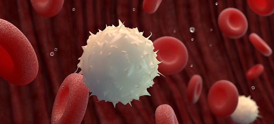
Inborn protective mechanisms present challenges for therapies targeting cancers of the brain. Engineered nanoparticles permit the selective delivery of CRISPR-Cas9 to glioblastoma tumors.

Glioblastoma accounts for almost half of all cancerous tumors originating in the brain.1 Even with maximum safe treatment, the median survival period for patients is less than 1.5 years.2 However, survival varies widely by age, from a 0.9% 5-year survival rate for patients over 75 years old to an 18.2% 5-year survival rate for patient 0-19 years old.3 Nevertheless, the overall 5-year survival rate remains low at 5%2 and novel therapies are urgently needed for glioblastoma treatment. Zou et al. present a highly specific CRISPR-Cas9-based gene therapy for glioblastoma.4

Gene therapies offer an attractive treatment for cancers. The goal of gene therapy is to mutate or remove deleterious DNA sequences such that they are unable to be transcribed and translated into functioning proteins. In the most prevalent technique, CRISPR-Cas9, single guide RNA (sgRNA) identifies and binds to the target DNA sequence. It then recruits Cas9 proteins to excise parts of the target DNA sequence. Mutations are generated as the cell tries to repair the damaged DNA.5

While a powerful tool, researchers have struggled to effectively deliver CRISPR-Cas9 to their cellular target. Delivery is particularly complex in brain tumors such as glioblastoma. Traditionally medications are trafficked through the body and delivered to their target through the bloodstream. However, the brain is a more complex and protected system. Primarily, selective delivery of therapeutics to tumor cells is necessary to protect neuronal function. Moreover, the brain is separated from the blood stream by a thin layer of cells termed the blood-brain barrier (BBB). The BBB allows for selective permeation of compounds into the brain, shielding neurons from toxins while permitting the passage of essential nutrients.6 Previous studies have sought to transport CRISPR-Cas9 therapies across the BBB using viruses as delivery capsules7 or circumvent the BBB altogether via intercranial injection of therapeutics.8 Yet these methods carry risk, either an immune response to the viral vector or complications from the invasive injection.

Zou et al. sought to resolve the issues of specific cell targeting and BBB permeability in CRISPR-Cas9 delivery by encapsulating the gene editing complex within a nanoparticle. The researchers chose to target Polo-like kinase 1 (PLK1) using CRISPR-Cas9 gene therapy. PLK1 is an attractive for selective gene therapy due to its higher overexpression by glioblastoma cells and by the proliferative glioblastoma subtype in particular.9 Moreover, inhibition of PLK1 has been shown to reduce tumor growth and induce cell death.9

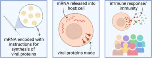
The researchers encapsulated Cas9 and sgPLK1 in a neutrally charged nanoparticle for delivery. The small size of nanoparticles, which are measured in nanometers, allow for easy transport through the bloodstream and uptake by cells. Taking advantage of the high expression of lipoprotein receptor-related protein-1 (LRP-1) on both BBB endothelial cells and glioblastoma tumor cells, they decorated the nanocapsule surface with LRP-1 ligand angiopep-2 peptide to facilitate selective uptake by BBB and glioblastoma cells (Figure 1). Zou and her colleagues bound the nanoparticle together with disulfide bonds as an added layer of selectivity for glioblastoma cell delivery. In the high glutathione environment of a glioblastoma cell, the nanoparticle dissolves, releasing its contents. However, glutathione concentrations are lower in BBB endothelial cells and healthy neurons, reducing the dissolution of the nanoparticles and leaving healthy DNA alone.


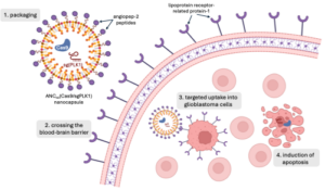
Figure 1. Nanoparticles enable permeation of the blood brain barrier (BBB) and selective delivery of the CRISPR/Cas9 system to glioblastoma (GBM) cells. Angiopep-2 peptides, which decorate the nanoparticle’s surface, bind with lipoprotein receptor-related protein-1 (LRP-1) overexpressed on BBB and GBM cells. Following uptake into GBM cells, the nanoparticles dissolve in the high glutathione environment, releasing the Cas9 nuclease and single guide RNA (sgRNA) targeting Polo-like kinase 1 (PLK-1) genes. Zou et al. demonstrated the selective mutation of PLK-1 induced apoptosis in GBM cells with minimal off-target effects.


Through Cas9/sgPLK1 delivery by nanoparticle, Zou et al. demonstrated a 53% reduction in expression of the targeted gene in vitro. PLK1 gene editing was cell selective, with negligible genetic mutation in the healthy surrounding brain tissue. While nanoparticles with and without disulfide cross-linking were capable of gene editing, the disulfide cross-linked nanoparticle induced almost 400% more mutations. Glioblastoma cells treated with disulfide cross-linked nanoparticles were also over 3 times more likely to undergo apoptosis cell death. Mice grafted with patient glioblastoma tumors treated with Cas9/sgPLK1 nanocapsules experienced an almost 3-fold extension in life expectancy, suggesting this treatment as a viable anti-glioblastoma therapy.5

Yet in order to be effectively applied as a cancer therapy, the efficiency of this nanoparticle delivery system must be increased. In mouse glioblastoma models, Zou et al. only achieved a maximum accumulation of 12% and effected a 38% knockdown of PLK1.5 Despite their low magnitude, these values are far greater than similar gene therapy treatments currently studied, suggesting nanoparticles present an innovation in the delivery of CRISPR-Cas9 to difficult to access tumors.

In addition to the improved efficacy over existing systems, the work of Zou et al. opens the door for less invasive administration of treatment. In contrast to previous gene therapies administered directly to the tumor through intercranial injection, the BBB penetration and tumor-specific accumulation of the nanoparticles may permit systemic administration. Zou et al. injected the nanoparticles intravenously, but treatment may even be given as a pill taken orally, eliminating any surgical intervention. Moreover, due to their modular design, the engineered nanoparticles may be adapted as targeted treatments for other tumors. Exchanging the angiopep-2 peptides for another ligand would facilitate uptake by cells expressing the corresponding receptor. The load carried within the nanoparticle could be altered to contain a different sgRNA targeting a new gene or another therapeutic entirely as a complementary treatment. Nanoparticle delivery systems like that studied by Zou et al. contains many layers of selectivity, offering hope for effective delivery of treatment to previously inaccessible tumors.


Works Cited:

  1. Wirsching, H.-G. & Weller, M. Glioblastoma. in Malignant Brain Tumors : State-of-the-Art Treatment (eds. Moliterno Gunel, J., Piepmeier, J. M. & Baehring, J. M.) 265–288 (Springer International Publishing, Cham, 2017). doi:10.1007/978-3-319-49864-5_18.
  2. Delgado-López, P. D. & Corrales-García, E. M. Survival in glioblastoma: a review on the impact of treatment modalities. Clin Transl Oncol 18, 1062–1071 (2016).
  3. Ostrom, Q. T. et al. CBTRUS Statistical Report: Primary Brain and Central Nervous System Tumors Diagnosed in the United States in 2007–2011. Neuro Oncol 16, iv1–iv63 (2014).
  4. Lino, C. A., Harper, J. C., Carney, J. P. & Timlin, J. A. Delivering CRISPR: a review of the challenges and approaches. Drug Delivery 25, 1234–1257 (2018).
  5. Zou, Y. et al. Blood-brain barrier–penetrating single CRISPR-Cas9 nanocapsules for effective and safe glioblastoma gene therapy. Sci Adv 8, eabm8011.
  6. Dotiwala, A. K., McCausland, C. & Samra, N. S. Anatomy, Head and Neck: Blood Brain Barrier. in StatPearls (StatPearls Publishing, Treasure Island (FL), 2024).
  7. Song, R. et al. Selection of rAAV vectors that cross the human blood-brain barrier and target the central nervous system using a transwell model. Molecular Therapy Methods & Clinical Development 27, 73–88 (2022).
  8. Lee, B. et al. Nanoparticle delivery of CRISPR into the brain rescues a mouse model of fragile X syndrome from exaggerated repetitive behaviours. Nat Biomed Eng 2, 497–507 (2018).
  9. Lee, C. et al. Polo-Like Kinase 1 Inhibition Kills Glioblastoma Multiforme Brain Tumor Cells in Part Through Loss of SOX2 and Delays Tumor Progression in Mice. Stem Cells 30, 1064–1075 (2012).

Filed Under: Biology, Chemistry and Biochemistry, Psychology and Neuroscience, Science Tagged With: blood-brain barrier, CRISPR, glioblastoma, nanoparticles

The Melting Arctic’s Impact on the Gulf of Maine

December 6, 2023 by Zoe Peterson '25

Recent observation and nutrient analysis in the Gulf of Maine has found that within the past 50 years nutrient sources have become more limited, impacting the entire ecosystem. The Gulf of Maine receives nutrient-rich waters from the continental slope that enters through the Northeast Channel, north of Georges Bank (figure 1). These continental slope waters originate off southern Newfoundland and travel into the Gulf of Maine passing by Labrador and the Scotian Shelf, all the while accumulating and retaining its high concentration of nutrients. Nutrients from this water source, such as nitrate and silicate that exist in excess within continental slope water, make the Gulf of Maine a highly productive area. Nitrate is of particular interest as it is often the limiting nutrient. In other words, nitrate is often scarce in an ecosystem and therefore is the nutrient that puts a cap on the accumulation of biomass such as phytoplankton. However, recent observation and nutrient analysis in the Gulf of Maine has found that within the past 50 years nutrient sources have become more limited, impacting the entire ecosystem.  

              Figure 1. Map of Gulf of Maine

Since the 1970s, studies have shown a notable decrease in the abundance of nitrate in the Gulf of Maine. Along with this change, the deep waters in the Gulf of Maine have become cooler and less salty. In 2010, Townsend et al suggested  that these changes all originate from the accelerating melting of ice in the Arctic. Since salt does not freeze, when water freezes in the Arctic, the ice it forms is made of freshwater. As this freshwater melts at a faster rate than the Earth has previously seen, it changes the salinity of the water, making it fresher and therefore less dense. Deep ocean circulation is based on density and so with this change in density, comes a change in the way water circulates the planet. 

Given the changes in densities of water in the Arctic, a new source of water from the bottom of the Atlantic ocean carrying far less nutrients now supplies the Gulf of Maine.  With the changes in deep ocean circulation patterns, now water entering the Gulf of Maine passes closer to the bottom of the ocean. As this water passes the ocean floor, microbes in the sediment remove nitrogen from the water (for use as a nutrient), a process called denitrification. While this benefits ecosystems at the bottom of the deep ocean, by the time the water reaches the Gulf of Maine, much of the nitrate in the water has already been used. 

The Gulf of Maine will become less productive as ecosystems are supplied with low concentrations of nitrate for long periods of time. Phytoplankton, the first step of the food web, absorb these nutrients and use them for growth. Once phytoplankton are less abundant, animals that rely on them for food will begin to struggle. As nitrogen deficiency continues up the food chain, it will eventually reach the larger fish upon which we in Maine rely on for our food. The gradual loss of nitrogen rich waters to the Gulf of Maine is not only a sad reminder of climate change’s far reaching consequences, but also presents a growing issue for the fishing industry in Maine which relies on the productivity of the water. 


Work Cited

Townsend, D. W., Pettigrew, N. R., Thomas, M. A., Neary, M. G., McGillicuddy, D. J., & O’Donnell, J. (2015). Water masses and nutrient sources to the Gulf of Maine. Journal of Marine Research, 73(3), 93–122. doi:10.1357/002224015815848811

Filed Under: Chemistry and Biochemistry, Environmental Science and EOS, Science Tagged With: Gulf of Maine, Ice melt, Nutrients

Fine-tuning of Chemotherapeutic Drug Reactions through Ruthenium Organic Complexes

December 4, 2023 by Ahmad Harb '25

The development of cancer treatment reagents aims at optimizing the reactivity of the reagent with the cellular DNA while reducing the reactivity with other bodily sites. This is in order to maximize cytotoxicity to cancer cells while reducing the side effects associated with chemotherapy (Wang, 2005).

Organometallic complexes, organic compounds with one or more metallic central atoms, have been used to control the release of compounds involved in key biological reactions (Renfrew, 2014). In the context of cancer chemotherapy, cisplatin complexes have been successfully developed to react with guanosine 5’ monophosphate, or GMP, as a potential binding site in the cell’s DNA, while avoiding the reactions associated with side effects (Dasari, 2014 & Reedjik, 2003).

The development of chemotherapeutic reagents requires the fine-tuning of the ligand substitution reactions that the organometallic complex can undergo. Ligand substitution reactions are reactions where one or more of the substituents bonded to the metal atom in the organometallic complex are replaced by a compound from the surrounding environment. An example is shown in the figure below.

Fig 1. Example of an organometallic complex undergoing a ligand substitution reaction. When dissolved in pyridine, the chromium complex, [Cr(TPP)(Br)(H2O)], reacts with the solvent, and two of its ligands (Br and OH2) are replaced by pyridine compounds (Py) (Okada, 2012). 

In a 2005 study, a group of chemists tested multiple ligand-substitution reactions of Ruthenium (Ru) complexes to test the possibility of the development of a competitor for cisplatin in chemotherapy (Wang, 2005). The Ruthenium complex studied have “stool”-like structures with an arene upper part and a tetrahedral Ruthenium compound which contains the leaving group. The researchers use X-ray crystallography and other characterization methods to identify the structure of every ruthenium complex involved in the study.

Fig 2. The structure of the Ruthenium organometallic complexes tested in the study. The basic structure is shown on the upper left. The “arene” can be any of the structures shown, while the leaving group, X, can be any of the structures shown as well as a halide or pseudohalide (Wang, 2005). 

Given the high concentration of chloride anions in the bloodstream and the intercellular fluid, the substitution of X by chloride is a main mechanism by which the reagent is lost before it can attack cancer cells (Wang, 2005). Previous research has shown that hydrolysis (substitution of X by a water molecule) is an essential activation step in the reagent’s reaction with GMP (Chen, 2003). The researchers thus investigated how the choice of the arene and the leaving group within the ruthenium complex can affect the reaction rates such that the reagent is cytotoxic but is inactive before reaching its target site. 

The reaction rate with chloride was established by dissolving the complexes in a 104mM NaCl solution, mimicking the high-chloride media within the body, and monitoring the formation of the substitution reaction product using the same characterization methods involved in the identification of the complexes.

The hydrolysis rate was established by allowing the aqueous solutions of the complexes to equilibrate for 24-48 hours at 37°C, mimicking body temperature. The formation of the hydrolysis product was monitored using the same characterization methods. The reaction equilibria were determined using high-performance liquid chromatography. 

The reaction with GMP is believed to be the final step in the reagent’s activityagainst cancer cells. The reaction rate was established by dissolving 0.5mM of each complex in a 0.5mM aqueous GMP solution. The product formation rate was determined using the same methods as the other reactions.

The researchers summarized their key resultsin the table below:

The data show that complex 13, for example, has a faster reaction rate with chloride than GMP, and will, therefore, be lost before attacking cancer cells if it were to be used as a chemotherapyreagent. Data shows that complexes 15 and 17, on the other hand, react faster with water and GMP than chloride, which makes them more suitable for chemotherapy. Complex 1 can undergo hydrolysis and react with GMP at a relatively high rate. A key finding in this research is that complex 21 can bind to GMP without undergoing hydrolysis, skipping a previously thought required first step. 

The overall cytotoxicity of each complex was compared to cisplatin by determining the concentration of the complex which caused at least 50% inhibition in the growth of ovarian cancer cells, IC50 values. As previous research predicted, high hydrolysis rates correlated with high cytotoxicity (Chen, 2003). Cisplatin has IC50 = 0.6µM; chemotherapy reagents are considered to have good cytotoxic activity if they have IC50 < 18µM. Some of the same complexes discussed above, with the exception of complex 17 and 21 had IC50 values competitive with cisplatin (IC50 < 6µM). Furthermore, the studied Ru complexes exhibited cytotoxicity towards cisplatin-resistant ovarian cancer cells (Wang, 2005).

Overall, the results show that the rates of the reactions involved in chemotherapy can be fine tuned by the choice of the ligand within the ruthenium complex. These results can be used in the future development of novel chemotherapy reagents.  

Work Cited:

Chen, H., Parkinson, J. A., Morris, R. E., & Sadler, P. J. (2003). Highly selective binding of organometallic ruthenium ethylenediamine complexes to nucleic acids: novel recognition mechanisms. Journal of the American Chemical Society, 125(1), 173-186.

Dasari, S., & Tchounwou, P. B. (2014). Cisplatin in cancer therapy: molecular mechanisms of action. European journal of pharmacology, 740, 364-378.

Okada, K., Sumida, A., Inagaki, R., & Inamo, M. (2012). Effect of the axial halogen ligand on the substitution reactions of chromium (III) porphyrin complex. Inorganica Chimica Acta, 392, 473-477.

Reedijk, J. (2003). New clues for platinum antitumor chemistry: kinetically controlled metal binding to DNA. Proceedings of the National Academy of Sciences, 100(7), 3611-3616.

Renfrew, A. K. (2014). Transition metal complexes with bioactive ligands: mechanisms for selective ligand release and applications for drug delivery. Metallomics, 6(8), 1324-1335.

Wang, F., Habtemariam, A., van der Geer, E. P., Fernández, R., Melchart, M., Deeth, R. J., … & Sadler, P. J. (2005). Controlling ligand substitution reactions of organometallic complexes: Tuning cancer cell cytotoxicity. Proceedings of the National Academy of Sciences, 102(51), 18269-18274.

Filed Under: Biology, Chemistry and Biochemistry Tagged With: Chemistry and Biochemistry, Medicine

Unpacking the Ethical Implications of Human Germline Editing

December 3, 2023 by Kennedy Kirkland '27

On December 30th, 2019, biophysicist Dr. He Jiankui was sentenced to three years in prison for forging ethical review documents and misleading doctors into unknowingly implanting gene-edited embryos into two women in China (Normile 2019). One of these women had a set of twins and the other had a single child. These children are now the first genetically modified humans in history to be resistant to HIV. They can pass this modification to the next generation, and their whereabouts are still unknown (Greely 2019). Jiankui used CRISPR/Cas9, a gene editing tool, to modify their germlines and edit the CCR5 gene, a contributor to broad immune responses (his focus was its importance to HIV viruses).

Based on the National Natural Science Foundation of China, there has been a total of 3.7 billion yuan (roughly $576 million) government spending on embryonic stem cell research from 1997 to 2019 (Lou 2021). Germline gene editing research is allowed, but establishing a pregnancy with genetically modified embryos has been outlawed by multiple regulations (Xinqing 2014). Similarly to China, the National Institutes of Health within the US estimates that human embryonic stem cell research has received $1.48 billion in government funding since 2009. There are no current laws or regulations that ban germline gene editing conducted through private funding within the US, but it would have to be approved by the FDA for marketing and clinical studies. No proposals have been submitted (​​Genetic Literacy Project 2019). Dr. Jiankui has since been disgraced by the scientific community around the world for his actions. Some scientists believe that germline gene editing is not only an unethical practice but a potentially dangerous one that could lead to a new era of eugenics with irreversible harm (Genetic Literacy Project 2019). Little is known about the intricate details of Dr. Jiankui’s experiment, but what has been revealed poses several hard-hitting ethical questions. Should we manipulate the next generation of humans? What are the ethical dilemmas in the advancement of this technology? To create an informed opinion on the matter, it is helpful to first understand the intricacies of Dr. Jiankui’s experiment, and how exactly he made the first genetically modified children.

Dr. Jiankui conducted human germline genome editing, which refers to the technique of modifying not only the genetic information of a subject but also what can be passed down to the next generation (Normile 2019). Germ cells can create a new generation (ex. sperm and eggs). So germ cells, and cells that produce germ cells, are known as the germline. These are different from somatic cells, which are body cells that continuously divide throughout a person’s lifetime and play a minimal role in gene inheritance (Greely 2019). Certain alleles or alterations to CCR5 within the human genome can provide resistance to HIV. Thus, Jiankui used CRISPR to alter CCR5 in HIV-susceptible patients to make them resistant to the disease.

CRISPR, which stands for clustered regularly interspaced short palindromic repeats, is a technique originally found in bacteria as a defense system to render inserted viral DNA ineffective. It also allows for the modification of DNA (Rapini 2023). The enzyme Cas-9 can cut pieces of DNA and is guided to its location based on an attached strand of RNA (called the guide RNA strand). By cutting a targeted sequence of DNA, it can render protein-coding genes inactive or disfunctional. After Cas-9 has cut the target DNA, researchers can choose to place a modified DNA sequence in the vacant space (Fig 1).

Figure 1. The editing technique of CRISPR involves the guide RNA strand, guide RNA sequence, Cas-9 enzyme, and target DNA (Roach 2015).

While little is known about Dr. Jiankui’s specific technique in using the CRISPR/Cas-9 system, we know that he modified the gene CCR5 (Greely 2019). CCR5 is a gene on chromosome 3 that encodes for a protein called C-C chemokine receptor type 5 (also shortened to CCR5) (Normile 2019). CCR5, along with another receptor called CD4, is utilized by the HIV virus to bind to bacteria-detecting/destruction cells called macrophages (white blood cells) and infect them. The HIV virus protein envelope binds to the primary receptor CD4 on the macrophage with the gp120 protein, which are exterior “protective” protein around the HIV cell. If the CCR5 coreceptor is present as well, it can successfully enter the macrophage, release viral RNA and enzymes, and infect the macrophage. This infection alters the function of the macrophage, causing it to assemble and release viruses. Eventually, the gene that codes for the gp120 receptor is altered by a mutation and can now bind to a different co-receptor called CXCR4 which is found on the CD4 plus T-cells. (Prakash 2019). The same process of virus construction occurs within T-cells, but as the viruses leave the T-cell it ruptures the plasma membrane. This kills the T-cells, causing a weakened immune response and eventually resulting in the onset of AIDS.

Figure 2. The mechanism by which the HIV viral envelope infects the macrophage creates mutated viral cells with different gp120 receptors and destroys T-cells. (Prakash 2019) 

It is believed that Dr. Jiankui used the CRISPR/Cas-9 system to delete 32 base pairs of the CCR5 gene, therefore making it produce non-functional copies of the CCR5 protein. This was done in the hope that HIV would be unable to infect the white blood cells of the babies born from the embryos (Normile 2019). If Jiankui only modified somatic cells using this technique, the children may or may not have gained protection against HIV and could not pass it down to the next generation.

In 2015 (before He’s experiment was released) Jennifer Doudna, one of two researchers who published their findings on CRISPR, convened with the U.C Berkeley’s Institute for Genome Innovation, which was composed of Nobel laureates and esteemed professors of bioethics. They concluded that conducting germline editing would be irresponsible until matters such as balancing the potential risks and benefits have been concluded and that there is a broad societal consensus about the appropriate use of proposed applications of this technology (Cohan, 2018). Jennifer Doudna and Emmanuelle Charpentier created this technology to find ways to edit out faulty genes and cure diseases at the source—within our genetic code (Doudna 2019). However, after the release of He’s experiment, Doudna publicly stated her evolving concerns relating to this application of her discovery. During her national press tour in 2019, she shared a frightening dream she had in which a colleague of her’s asked about CRISPR but later turned out to be Hitler (Than 2019). This likely stems from the horrifying history of Nazis and the eugenics movement, which stemmed from the idea of improving the genetic quality of humans and sterilizing and or eliminating those with unfavorable traits. She also voiced that her opinion on germline genome editing has been evolving from an outright ban to warranting it in certain circumstances. However, her underlying opinion remains that it would have to be under transparent and safe circumstances and concern a medical need that was unmet by any technology. In addition to Doudna, countless other scientists have come out in disagreement with He Jianku’s work including researchers from Stanford University, Harvard Medical School, and the NIH (Cohen 2018). Specifically, a journalist from Science magazine states that representatives from eight countries who attended the International Summit on Human Genome Editing in Hong Kong came to a consensus that his actions were irresponsible, violated international norms, lacked transparency, and did not have sufficient medical justification (Cohen 2018).

Jennifer Doudna stated that her goal was to edit out faulty genes, but that definition leaves a lot to interpretation. How can we as a society come together to define the parameters of a faulty gene? From treating HIV to Down Syndrome, these conditions hold more than just health implications, but social ones as well. If the scientific community deems it accessible to prevent Down syndrome using human germline genome editing, are they stating that humans that currently possess this condition are “wrong” and have “faulty genes” in need of correction? Whoever holds the power to manipulate this technology essentially has the power to decide what the next generation of humans will be capable of and what genes are deemed “faulty” within our society. Jianku’s reckless use of technology was majorly deemed wrong for its lack of transparency, safety, and following set regulations upheld by currency bioethical standards. However, in situations like parents wanting their children to be healthy, it is completely understandable that human germline editing sounds like an intriguing way to protect their children. For these reasons, it is imperative to familiarize ourselves with the development of this technology, and its continued push for regulations across the world to maintain ethical standards. Further, it is also important to ask ourselves hard-hitting questions such as: should one person, scientist, or government have the power to determine what the next generation will possess, look like, or become? We as members of society must continue to stay informed in a world with the capacity to genetically modify human beings.

References:

  1. Bhanu Prakash. “How the HIV Infection Cycle Works.” Proceum Pvt. Ltd. January 2019. https://www.youtube.com/watch?v=GyofqO1TRjU
  2. Brianna Rapini, Sarina Peterson, “Genetic Engineering.” September 2023. https://www.youtube.com/watch?v=CDw4WPng2iE
  3. Dennis Normile. “Chinese scientist who produced genetically altered babies sentenced to 3 years in jail.” Science. December 2019.https://www.science.org/content/article/chinese- scientist-who-produced-genetically-altered-babies-sentenced-3-years-jail
  4. Deng Luo, Zihui Xu, Zhongjing Wang, Wenzhuo Ran. “China’s Stem Cell Research and Knowledge Levels of Medical Practitioners and Students.” Stem Cells International. 2021. https://doi.org/10.1155/2021/6667743.
  5. Doudna J. 2019. Faculty Research Page. Department of Molecular & Cell Biology. http://mcb.berkeley.edu/faculty/BMB/doudnaj.html.
  6. Dylan Roach, Tanya Lewis. “CRISPR, the gene-editing tech that’s making headlines, explained in one graphic.” Insider. December 2015. https://www.businessinsider.com /crispr-gene-editing-explained-2015-12
  7. Genetic Literacy Project. 2019 July 23. United States: Germline / Embryonic. Global Gene Editing Regulation Tracker. https://crispr-gene-editing-regs-tracker.geneticliteracy project.org/united-states-embryonic-germline-gene-editing/.
  8. Henry T Greely. “CRISPR’d babies: human germline genome editing in the ‘He Jiankui affair.’” Journal of Law and the Biosciences. Volume 6, Issue 1. October 2019, Pages 111–183, https://doi.org/10.1093/jlb/lsz010
  9. Indra Mani. “CRISPR-Cas9 for treating hereditary diseases.” Progress in molecular biology and translational science. Volume 181. February 2017. https://pubmed.ncbi.nlm.nih.gov/34127193/
  10. John Cohen. “After last week’s shock, scientists scramble to prevent more gene-edited babies.” Science. December 2018.https://www.science.org/content/article/after-last- weeks-shock-scientists-scramble-prevet-more-gene-edited-babies
    Join David Ignatius. “Walter Isaacson & Jennifer Doudna join Washington Post Live to discuss CRISPR.” Washington Post. March 2021. https://www.youtube.com/ watch?v=cKHuuALENZk
  11. Than K. 2019 Nov 12. AI and gene-editing pioneers to discuss ethics. Stanford News. [accessed 2023 Dec 3]. https://news.stanford.edu/2019/11/12/ai-gene-editing-pioneers- discuss-ethics/#:~:text=Doudna%20decried%20the%20act%20but.
  12. Yi Zheng et al. “Structure of CC Chemokine Receptor 5 with a Potent Chemokine Antagonist Reveals Mechanisms of Chemokine Recognition and Molecular Mimicry by HIV.” Immunity. Volume 46, Issue 6. June 2017. https://www.cell.com/immunity /pdf/S1074-7613(17)30218-2.pdf
  13. ‌Zhang Xinqing, Zhang Wenxia, Zhao Yandong. “The Chinese Ethical Review System and its Compliance Mechanisms.” TRUST. September 2014. https://trust-project.eu/ wp-content/uploads/2016/03/Chinese-Ethics-Review-System.pdf

Filed Under: Biology, Chemistry and Biochemistry, Science Tagged With: Bioethics, Genes, HIV

Inverse Vaccines: A New Way to Treat Autoimmune Disorders

December 3, 2023 by Divya Bhargava '26

Williams, 2023

Of the 8.1 billion people in the world, 1 in 10 have an autoimmune disorder. There are 8.1 billion people in the world, and 810 million of these people, or 1 in 10, have an autoimmune disorder (“1 in 10 people”, 2023). Autoimmune disorders are a category of conditions in which the body attacks itself. Although management systems for most of these types of conditions have been developed, autoimmune diseases still cannot be cured (“Autoimmune disorders”). In fact, even with management of their symptoms, up to 50% of patients with autoimmune disorders still experience impairment in their health-related quality of life (Pryce and Fontana, 2017). The development of so-called “inverse vaccines” may provide the much needed mechanism to help find a cure for this class of conditions by teaching the body not to attack itself. 

A traditional vaccine works because it helps the body learn to recognize parts of foreign pathogens and builds up the body’s immune response against these pathogens so that the response is stronger and happens more quickly after recognition. In people with autoimmune diseases, the body also forms an immune response against self molecules because it mistakenly identifies them as foreign antigens, or pathogenic molecules (usually proteins or sugars) that induce an immune response. The idea behind “inverse vaccines” is that instead of building up the immune system’s response to foreign antigens, they could suppress the response to self-antigens by helping to teach the body to recognize these misidentified molecules as self. 

A study led by Andrew Tremain at the University of Chicago’s Pritzker School of Medicine is one of the groups who are involved in this novel inverse vaccine research. The inverse vaccines they developed contain modified copies of the self-antigens that are targeted by the immune system which are attached to long chains of sugars called polysaccharides (Tremain et al., 2023). These polysaccharide chains guide the self-antigens to the liver, which plays an important role in the establishment of tolerance to these molecules. Once these modified self-antigens arrive at the liver, specialized immune cells pick them up and then inhibit the action of T cells against them through T cell uptake (Leslie, 2023). T cells are a type of immune cell that carry out part of the typical response against molecules identified as foreign or invaders through either cytotoxic or signaling based immune responses. The inhibition of the T cell response against misidentified self-antigens reduces or prevents the autoimmune response that causes the body to attack itself, thereby acting as a kind of “inverse vaccine”. 

Once Tremain et al. developed their inverse vaccine, they conducted testing to determine its efficacy and viability as a method for increasing tolerance to self-antigens in those with autoimmune disorders. First, they wanted to ascertain whether inverse vaccines could truly provide inhibition of an immune response. To do this, they injected an egg white protein into mice as an experimental foreign antigen to trigger a strong immune response. Then an inverse vaccine against the egg white protein was injected to suppress the response to the original dose of the protein. In their analysis, they found that the T cells that would’ve responded to the egg white protein were not present. These results suggested that the inverse vaccine blocked the typical immune response, demonstrating viability as a treatment method against stimulated immune responses (Tremain et al. 2023).

However, Tremain and his colleagues still had to demonstrate the efficacy of inverse vaccines in inhibiting an autoimmune response rather than one caused by a foreign antigen. To do this, they induced an autoimmune disease called experimental autoimmune encephalomyelitis (EAE) in mice. In EAE, the immune system attacks myelin, the substance responsible for forming insulation around nerve cell axons. EAE is a particularly informative experimental model in mice because it mimics multiple sclerosis (MS), a human autoimmune disorder. Once they induced EAE, Tremain et al. injected an inverse vaccine made up of a polysaccharide carrying part of a myelin protein. Physiological analysis after injection suggested that this treatment had stopped mice from developing EAE. Furthermore, injection of a different inverse vaccine targeting an alternate form of EAE showed prevention of symptom relapse. This means that Tremain et al. were able to demonstrate inhibition of autoimmune responses against two types of EAE in mice, representing two different types of MS, providing a heartening outlook on this research (Tremain et al., 2023). 

In summary, inverse vaccines had the ability to turn off immune responses to particular antigens in mice. These results are a promising sign for the ability of inverse vaccines to combat autoimmune diseases. Additionally, initial clinical trials testing the safety and efficacy of inverse vaccine strategy to increase tolerance to self-antigens in humans have had positive results so far for immune disorders such as multiple sclerosis and celiac disease where the misidentified self-antigens are known (Leslie, 2023). However research into tolerance-increasing strategies tends to stall both because we do not know which self-antigens are attacked in several autoimmune disorders, and because the mechanisms that produce tolerance after antigens are brought to the liver are not well understood (“Immune Tolerance”). This means that even if autoimmune vaccines prove to be a viable form of treatment for autoimmune disorders, they will only be able to treat diseases with known self-antigens until further research into the antigen and tolerance mechanisms is conducted. Nevertheless, inverse vaccine research is incredibly promising and has the potential to help hundreds of millions of people worldwide.

Works Cited

Pryce, CR., and Fontana, A. (2017). Depression in autoimmune diseases. Current Topics in Behavioral Neurosciences, 31. https://doi.org/10.1007/7854_2016_7 

1 in 10 people suffer from autoimmune diseases. (2023). Neuroscience News.  https://neurosciencenews.com/population-autoimmune-disease-23198/ 

Autoimmune disorders. (n.d.). Retrieved November 5, 2023, from  http://www.betterhealth.vic.gov.au/health/conditionsandtreatments/autoimmune-disorders

Tremain, A.C., Wallace, R.P., Lorentz, K.M. et al. Synthetically glycosylated antigens for the antigen-specific suppression of established immune responses. Nat. Biomed. Eng 7, 1142–1155 (2023). https://doi.org/10.1038/s41551-023-01086-2 

Leslie, M. (2023). ‘Inverse vaccine’ could help tame autoimmune diseases, Science. https://www.science.org/content/article/inverse-vaccine-could-help-tame-autoimmune-diseases 

Immune Tolerance in Autoimmune Disease, Immune Tolerance Network. (n.d). Retrieved November 5, 2023, from https://www.immunetolerance.org/researchers/clinical-trials/autoimmune-disease 

Understanding Multiple Sclerosis, Oregon Health and Science University. Retrieved November 5, 2023, from  https://www.ohsu.edu/brain-institute/understanding-multiple-sclerosis 

Williams, S. (2023). “Inverse vaccine” shows potential to treat multiple sclerosis and other autoimmune diseases. Pritzker School of Molecular Engineering,  The University of Chicago. https://pme.uchicago.edu/news/inverse-vaccine-shows-potential-treat-multiple-sclerosis-and-other-autoimmune-diseases

Filed Under: Biology, Chemistry and Biochemistry, Science Tagged With: autoimmune disease, autoimmune disorder, inverse vaccine, multiple sclerosis, Vaccine

Plant Talk: Eavesdropping on Underground Plant Communication

December 3, 2023 by Riley Simon '26

Have you ever looked at a tiny sapling, a winding vine, or a massive oak tree and felt like they have some sort of personality? With the rustle of some leaves or the snap of a twig it might seem like these plants are talking to each other. As it turns out, these fantasies aren’t too far from the truth. Vascular plants (which consist of most plants other than mosses and algae) can actually communicate. These plants can exchange messages through their root systems with the help of mycorrhizal fungi. These fungi exist in a mutualistic relationship with plants and, along with acting as a living walkie talkie, they provide many survival benefits to the plants they live with. 

To be clear, vascular plants aren’t chatting in some sort of plant language in the same way that we talk to each other. Instead, they communicate through the transfer of infochemicals. “Infochemical” is an umbrella term for substances released by one plant and detected by another (Chen 2018). Infochemicals can take the form of plant hormones or nutrients and are passed between plants through the soil. The problem with this system is that transport through the soil is incredibly inefficient. When infochemicals move from plant to plant, they can quickly be absorbed by organic material or degrade in the soil such that they do not reach the intended “listener” plant. This is where mycorrhizal fungi come into play.

Mycorrhizal fungi (MF) are distinguished from other fungi by the symbiotic relationship that they have with plant roots. MF attach to plant roots and perform beneficial services for the plant in return for the carbon necessary for MF’s survival. MF networks add large amounts of surface area to plant root systems, allowing for the more efficient uptake of nutrients to the plants such as nitrogen, phosphorus, and carbon. The MF relationship increases efficiency of water collection, enhances photosynthesis, and improves resistance to pathogens. (Barto 2012). 

When it comes to plant communication, MF act as “superhighways” for the infochemicals to travel from plant to plant. Instead of having to travel through the soil, infochemicals can be safely transported between plants through common mycorrhizal networks (CMNs). CMNs are made up of interconnected networks of fungal branches, which span the distance between plant roots (Chen 2018). These networks are not exclusive to one species of plant because MF are not host specific and therefore can associate with multiple species at the same time. This allows for messages, in the form of infochemicals, to be passed efficiently between plants of varying species. This method is exponentially more efficient than infochemical transport through the soil, allowing plants to communicate much easier.

You might be wondering, what do these plants have to talk about? It turns out, they have a whole lot to discuss. The world can be a dangerous place and plants use these CMN superhighways as an emergency warning system to let neighboring plants know about potential threats. A plant that experiences a disturbance, such as infection by a pathogen or herbivore attack, can send signals to surrounding plants to let them know of the potential danger. The plants receiving the message can then increase their defense to better prepare for the threat. This exact phenomenon has been observed in neighboring plants where one plant is infected with a pathogen, and then surrounding uninfected plants respond to infochemical signals by activating defense proteins (Chen 2018).

Beyond plant defense, there is still a lot to learn about how plants are communicating and what kinds of things they are “talking” about. There are still questions to be answered such as how plant relatedness impacts infochemical transfer or how far these networks can span underground. If we continue to eavesdrop on this “plant talk” then we can start to understand the interconnected nature of plant communities even better.

Works Cited

Barto, E. K., Weidenhamer, J. D., Cipollini, D., & Rillig, M. C. (2012). Fungal superhighways: do common mycorrhizal networks enhance below ground communication?. Trends in plant science, 17(11), 633–637. https://doi.org/10.1016/j.tplants.2012.06.007

Chen, M., Arato, M., Borghi, L., Nouri, E., & Reinhardt, D. (2018). Beneficial Services of Arbuscular Mycorrhizal Fungi – From Ecology to Application. Frontiers in plant science, 9, 1270. https://doi.org/10.3389/fpls.2018.01270

Bonazzi, D. (2021). The secret underground life of trees. Weizmann Compass. Retrieved December 3, 2023, from https://www.weizmann.ac.il/WeizmannCompass/sections/features/the-secret-underground-life-of-trees.


Filed Under: Biology, Chemistry and Biochemistry Tagged With: Biology, Communication, Fungi, Nutrients, Trees

Ending the Biomedical Harvest: Synthetic Alternatives to Horseshoe Crab Blood for Bacterial Endotoxin Detection

December 3, 2023 by Gabe O'Brien

Did you know that horseshoe crabs have incredible immune systems? In fact, horseshoe crabs have the best immune systems out of all living invertebrates. Their secret? Blood. Horseshoe crab blood is very simple in composition, with only a single cell type in general circulation (the granular amebocyte) and only three proteins in the plasma of the blood (hemocyanin, C-reactive proteins, and a2-macroglobulin) (Armstrong et al., 2008). These proteins contribute to the horseshoe crab’s blood clotting system, protecting them from infection. Horseshoe crab blood has been found to be very sensitive to bacterial endotoxins found in illness-causing Gram-negative bacteria (Protecting Health). When horseshoe crab blood cells come into contact with bacterial endotoxin, they clot around it, preventing the bacterium from invading nearby cells (Natural History Museum 2020). 

With the rise of vaccine development, especially in the case of the Covid-19 pandemic, horseshoe crab blood plays an essential role in testing the safety of vaccines due to its endotoxin-detection properties. Additionally, large volumes of horseshoe crab blood can be collected easily, making it a convenient blood source (Armstrong et al., 2008). Despite all the beneficial applications of horseshoe crab blood, horseshoe crab bleeding leaves thousands of horseshoe crabs dead annually, causing their populations to be in decline (Maloney et al., 2018). A 2018 study has promoted a synthetic alternative to horseshoe crab blood, recombinant Factor C (rFC), and proven its efficacy in bacterial endotoxin detection. The use of rFC in vaccine development can eliminate the need for the use of actual horseshoe crab blood, sparing the horseshoe crab and promoting the conservation of this endangered species. 

Typically, horseshoe crab blood is collected by the direct puncture of the heart under sterile conditions that minimize contamination by bacterial endotoxins (Figure 1). A large horseshoe crab can produce between 200 – 400 mL of blood, and the blood clotting system can be studied microscopically. The limitation of contamination by bacterial endotoxins is extremely important in the blood collection process, because cell clotting will compromise the effectiveness of the blood for its intended use of developing vaccines. Only undamaged horseshoe crabs are selected for blood collection, and the animal is bled by the insertion of a needle into the heart through the outer hinge joint of the horseshoe crab (Figure 2). The animal is then squeezed gently so that as much blood as possible can be deposited into the collection tube (Armstrong et al., 2008).

Figure 1: Horseshoe crab bleeding on a larger scale, with precautions taken to ensure sanitary, endotoxin-free conditions for blood collection.

Figure 2: The three major components of the body of a horseshoe crab, including the prosoma (P), the opisthosoma (O), and the telson (T). The hinge (H) is where the prosoma meets the opisthosoma, and that is where the needle is inserted for blood collection. 

Following collection, horseshoe crab blood is ready for use in endotoxin detection (Armstrong et al., 2008). For example, in vaccine development, a Limulus amebocyte lysate (LAL) test detects the levels of clotting in horseshoe crab blood when it comes into contact with different vaccines. Horseshoe crab blood is very precise with detecting even small traces of endotoxin, making it an effective tool to identify small quantities of endotoxin present in potential vaccines (Protecting Health).  

Although horseshoe crab blood is effective in its ability to detect endotoxins, recombinant Factor C (rFC) can do the same job in a way that is more ecologically sustainable. Initially, rFC was discovered by scientists at the National University of Singapore, allowing them to visualize endotoxin detection using animal-free technology. Every year over 500,000 horseshoe crabs are captured and as much as ⅓ of their blood is drained, contributing to high mortality rates. On top of this, around 13% of the horseshoe crabs bled are later sold for bait, resulting in nearly 130,000 horseshoe crab victims to the biomedical industry. A 2018 study confirmed that the biomedical industry could reduce their use of horseshoe crab blood by nearly 90% if they were to employ rFC as a synthetic alternative for endotoxin detection processes. The 2018 study reviews multiple studies that show how rFC is just as effective as actual horseshoe crab blood in endotoxin detection, as rFC has been able to demonstrate the same high rate and sensitivity as horseshoe crab blood in detecting small amounts of endotoxin in a wide range of chemical structures. When endotoxin binds to a synthetic rFC molecule, it causes the rFC to fluoresce directly proportional to the concentration of endotoxin in a substance. rFC has even been able to demonstrate a higher rate of specificity for endotoxin detection (compared to horseshoe crab blood) in some studies (Maloney et al., 2018).

The most important next step of this research is to get synthetic rFC into the hands of the biomedical industry. Exposure to endotoxin can cause serious illness, making endotoxin detection for vaccines an essential part of the vaccine development process.  Even though there is ample evidence that rFC is equivalent to or better than horseshoe crab blood at detecting bacterial endotoxin, there are still limitations to the usage of rFC, as it is difficult for the biomedical industry to adopt new technologies quickly. Endotoxin detection testing is very highly regulated, so many pharmaceutical manufacturers may be hesitant to employ new detection technologies, as they may want to stick to traditional methods instead (Maloney et al., 2018). Despite these limitations, in order to progress towards horseshoe crab conservation, rFC should be produced and employed on a large scale so that the biomedical industry will no longer be solely reliant on the exploitation of horseshoe crabs for bacterial endotoxin detection.

Literature Cited

1. Armstrong, P., Conrad, M. Blood Collection from the American Horseshoe Crab, Limulus Polyphemus. J. Vis. Exp. (20), e958, doi:10.3791/958 (2008). 

2. “Horseshoe Crab Blood: The Miracle Vaccine Ingredient That’s Saved Millions of Lives.” Www.nhm.ac.uk, www.nhm.ac.uk/discover/horseshoe-crab-blood-miracle-vaccine-ingredient.html#:~:text=Horseshoe%20crab%20blood%20is%20bright.  

3. Maloney T, Phelan R, Simmons N. Saving the horseshoe crab: A synthetic alternative to horseshoe crab blood for endotoxin detection. PLoS Biol. 2018 Oct 12;16(10):e2006607. doi: 10.1371/journal.pbio.2006607. PMID: 30312293; PMCID: PMC6200278.  

4. “Protecting Health.” Www.horseshoecrab.org, www.horseshoecrab.org/med/health.html. Accessed 12 Nov. 2023.  


Filed Under: Biology, Chemistry and Biochemistry, Environmental Science and EOS Tagged With: Biology, blood, clinical testing, conservation, horseshoe crabs, Medicine, vaccine development

PedPRM Unveils Promising Treatment for Insomnia in Children with Autism Spectrum Disorder

December 3, 2023 by Fabiola Barocio Prieto '27

Getting enough sleep is widely considered crucial to our well-being. However, for some individuals with Autism, getting enough quality sleep is not as easy as it sounds. Autism Spectrum Disorder (ASD) is a developmental disorder that widely affects the U.S. population, as 1 in 36 children aged eight has been diagnosed with autism (CDC, 2023). The kind and severity of the symptoms that individuals with ASD may exhibit can vary along the ASD spectrum.  Sleeping, in particular, is a common challenge for those with ASD, as many suffer from problems related to the REM (Rapid Eye Movement) sleep phase, a phase critical for memory consolidation (Devnani & Hedge, 2015). This can quickly become a vicious cycle for some, as lack of sleep can increase the severity of other Autism-related symptoms but also affect both the individual and the quality of life of their family or caretakers. 

To investigate possible treatments, a recent study by Malow et al. focuses on the pharmacological approach of using melatonin to treat sleep disorders. Melatonin is a hormone produced by the body’s pineal gland to regulate circadian rhythm, allowing the body to relax according to the appropriate light-darkness cycles of the day. This study uses a small, long-release tablet (PedPRM) used to mimic endogenous melatonin secretion. It focused mainly on a younger population, from children to adolescents, who met two criteria. The first was that they had confirmed diagnoses of either Autism or Smith-Magenis syndrome and had also experienced sleep abnormalities. Smith-Magenis syndrome is also a developmental disorder that involves symptoms similar to those of ASD, affecting behavior, cognition, and sleep. They also had to have not previously seen improvements when using sleep hygiene treatments to be included in the study, such as establishing a strict bedtime routine and taking steps to provide a calm and comfortable sleep environment (CHOC, 2023).

With the study sample set, the study was conducted over 108 weeks and divided into four phases (Graph I). Throughout the four phases, participants’ caregivers would document sleep quality and total sleep time in a sleep and nap diary to record the efficacy of treatment. The study participants started the first stage with a 2-week period in which they were given placebos. If participants showed improvement while receiving a placebo, they would be removed from the study to reduce the possibility of external factors affecting the results (Scott et al., 2021). After clearing Phase 1, participants entered the second phase, which consisted of a double-blinded 13-week period in which they were randomly placed into either a placebo or treatment group (PedPRM). After this, Phase 3 comprised a longer 91-week open-label period in which both groups were combined. For the final phase of the study, participants were placed again in a 2-week single-blind placebo period to ensure that the drug had been completely removed from the participants with no adverse effects after stopping treatment.

Upon concluding the experimental period, the results to be considered for this study could be divided into three groups: participants’ sleep quality, caregivers’ well-being, and the participants’ growth development. With this data, the researchers found a significant

 decrease in sleep disturbance (Graph II-A) and an increase in caregiver satisfaction (II-B) and quality of life (II-C). These were most pronounced during the first half of the treatment. In the latter half of treatment, sleep disturbance continued to decrease but at a slower pace than the initial treatment phase. Fortunately, there were no reported deaths, and most adverse reactions included daytime fatigue and mood swings. However, the severity and extent of these were not detailed in the results of the study and offer the potential to be analyzed further.  

The study shows compelling initial evidence that PedPRM is an effective treatment for sleep disorders in individuals with ASD. However, as noted by the researchers, it also shows that constant active treatment is required as most sleep quality improvements are removed upon halting treatment. Since medications for children are generally more strictly controlled, PedPRM consistently demonstrates a possibility for effective pediatric treatment, even if for long-term medication. In particular, these findings are essential as it has been found that rapid-release melatonin is not helpful with maintaining sleep a couple of hours after administration, and it had long been considered a challenge to find small, swallowable prolonged-release tablets for children (Fliesler, 2022). As sleep interruption is something that mainly affects those with neurodevelopmental disorders, this is a significant step towards adequate treatment. However, it is essential to note that this pharmacological alternative should only be considered if behavioral interventions and sleep hygiene modifications have been attempted but have been found unsuccessful. 


Sources 

CDC. (2022, December 9). Centers for Disease Control and Prevention. https://www.cdc.gov/ncbddd/autism/facts.html 

CDC Newsroom. (2016, January 1). CDC. https://www.cdc.gov/media/releases/2023/p0323-autism.html 

CHOC – Children’s Hospital of Orange County. (2023, March 2). Autism and Sleep Hygiene – Children’s Hospital of Orange County. Children’s Hospital of Orange County. https://www.choc.org/programs-services/autism-neurodevelopmental/co-occurring-conditions-program/autism-and-sleep/ 

Devnani, P., & Hegde, A. U. (2015). Autism and sleep disorders. Journal of Pediatric Neurosciences, 10(4), 304. https://doi.org/10.4103/1817-1745.174438 

Fliesler, N. (2022, June 13). Melatonin for kids: Is it effective? Is it safe? – Boston Children’s Answers. Boston Children’s Answers. https://answers.childrenshospital.org/melatonin-for-children/#:~:text=There%20is%20some%20evidence%20to,the%20ability%20to%20swallow%20capsules. 

Furfaro, H. (2023, March 10). Sleep problems in autism explained. Spectrum | Autism Research News. https://www.spectrumnews.org/news/sleep-problems-autism-explained/ 

Lemoine, P., Garfinkel, D., Laudon, M., Nir, T., & Zisapel, N. (2011). Prolonged-release melatonin for insomnia &ndash; an open-label long-term study of efficacy, safety, and withdrawal. Therapeutics and Clinical Risk Management, 301. https://doi.org/10.2147/tcrm.s23036 

Malow, B. A., Findling, R. L., Schröder, C., Maras, A., Breddy, J., Nir, T., Zisapel, N., & Gringras, P. (2021b). Sleep, Growth, and puberty after 2 years of Prolonged-Release Melatonin in children with Autism Spectrum Disorder. Journal of the American Academy of Child and Adolescent Psychiatry, 60(2), 252-261.e3. https://doi.org/10.1016/j.jaac.2019.12.007 

Scott, A., Sharpe, L., Quinn, V. F., & Colagiuri, B. (2022). Association of single-blind placebo run-in periods with the placebo response in randomized clinical trials of antidepressants. JAMA Psychiatry, 79(1), 42. https://doi.org/10.1001/jamapsychiatry.2021.3204 

Signs & Symptoms | Autism Spectrum Disorder (ASD) | NCBDDD | CDC. (2023, January 11). Centers for Disease Control and Prevention. https://www.cdc.gov/ncbddd/autism/signs.html

Filed Under: Biology, Chemistry and Biochemistry, Psychology and Neuroscience, Science Tagged With: Autism, Medicine, sleep

mRNA: Unpacking the Research that led to the COVID-19 Vaccines and the 2023 Nobel Prize in Medicine

November 19, 2023 by Maren Cooper '27

        By 2023, 81.4% of the United States population have received at least one dose of the COVID-19 mRNA vaccine, and just about everyone has heard of these lifesaving shots (CDC, 2023). But how many of us actually know how they work? In general, vaccinations expose the body to a weakened strain of a virus that initiates an immune response so that the virus can be recognized, and quickly neutralized, in the future (Jain, 2021). This works because of the structure of our immune system, which is split up into innate immunity and adaptive immunity. Innate immunity is the immediate response that our bodies put up when exposed to a pathogen; it sends out macrophages to gobble up infected cells, creates an inflammatory response to kill off the infection, and uses a host of molecules and pathways to destroy the virus. Adaptive immunity involves the presence of B and T cells, which arise in a viral infection and create immunological memory through antibodies and memory cells so that a future infection of the same virus is immediately recognized and shut down. Vaccines work to stimulate this adaptive immunity, while minimizing innate immunity.

        mRNA vaccines use this same basic principle, but instead of injecting a weakened strain of the actual virus into someone, they instead inject a piece of mRNA, or messenger RNA, which codes for a viral protein that stimulates this immune response (Jain, 2021).

Figure 1: The methodology of mRNA vaccines. Adapted from Jain, et. al., 2021

mRNA vaccines are much easier to make than traditional vaccines because they don’t require growing and inactivating a virus, and they don’t carry the risk that live vaccines have of infecting the subject, but up until recently, they weren’t even remotely possible because in vitro mRNA would create a huge inflammatory response when entering the body (Karikó, 2005). However, Hungarian biochemist Katalin Karikó and her colleague Drew Weissman were undeterred, and their research to create mRNA without an inflammatory response paved the way for mRNA vaccines and won them the Nobel Prize in Medicine in 2023 (Karikó, 2005).

        Karikó and Weissman’s research focused on in vitro mRNA’s stimulation of Toll-like receptors (TLRs), which recognize pathogens and activate signaling pathways leading to an inflammatory response (Karikó, 2005). They discovered that mammalian mRNA, which doesn’t stimulate an immune response, contains many base pair modifications, whereas bacterial RNA, which is known to stimulate a response, has no modifications (Karikó, 2005). In vitro mRNA, grown in a lab rather than from mammalian cell cultures, also has no, or very few, modifications. This led the researchers to question whether base pair modifications were how cells distinguished between foreign and non-foreign RNA, and thus which pieces of RNA stimulate an immune response (Karikó, 2005). They discovered that in vitro RNA activates three main TLRs known for activating an inflammatory response, but that nucleoside modifications limit that activation. Most importantly, they discovered that the suppressed immune response is proportional to the number of nucleotide modifications, and that these modifications also increase the stability of in vitro mRNA in the body, another original roadblock in the usage of mRNA for vaccines. (Karikó, 2005).

Figure 2: The use of base modifications to suppress inflammatory response of RNA. Adapted from Nobelprize.org

        Combined with later research also by Karikó and Weissman discovering that modified base-pairs also create increased protein production and other research designing more stable lipid carriers to deposit the mRNA into human cells, mRNA became a promising new vaccine technology, eventually allowing the COVID-19 vaccines to be produced in record times, shortening the pandemic and granting all of us our lives back. But the road to Karikó’s research being recognized, and especially the road to the Nobel Prize, was not predetermined. Karikó’s research was long overlooked, in part because of her identity as a Hungarian immigrant. She was “demoted four times” at UPenn, being told that her research didn’t measure up to their standards, but she continued to persevere, making her both a scientific success story, and a personal one (Shrikant, 2023). The COVID-19 vaccines created by Pfizer and Moderna were the first mRNA vaccines to be released, but future research is focusing on creating a universal flu vaccine and possibly even an HIV vaccine, all using mRNA technologies, which could drastically improve the landscape of viral infections, both in the US and across the globe.  


Literature Cited

Karikó, K., Buckstein, M., Ni, H., & Weissman, D. (2005). Suppression of RNA Recognition by Toll-Like Receptors: The Impact of Nucleoside Modification and the Evolutionary Origin of RNA. Immunity, 23(2), 165–175. https://doi.org/10.1016/j.immuni.2005.06.008

 Karikó, K., Muramatsu, H., Welsh, F. A., Ludwig, J., Kato, H., Akira, S., & Weissman, D. (2008). Incorporation of Pseudouridine into mRNA yields Superior Nonimmunogenic Vector with Increased Translational Capacity and Biological Stability. Molecular Therapy, 16(11), 1833–1840. https://doi.org/10.1038/mt.2008.200

 Jain, S., Venkataraman, A., Wechsler, M. E., & Peppas, N. A. (2021). Messenger RNA-Based Vaccines: Past, Present, and Future Directions in the Context of the COVID-19 Pandemic. Advanced Drug Delivery Reviews, 179, 114000. https://doi.org/10.1016/j.addr.2021.114000

 The Nobel Prize in Physiology or Medicine 2023. (n.d.). NobelPrize.Org. Retrieved October 22, 2023, from https://www.nobelprize.org/prizes/medicine/2023/press-release/

 CDC. (2020, March 28). Covid Data Tracker. Centers for Disease Control and Prevention. https://covid.cdc.gov/covid-data-tracker

Shrikant, A. (2023, October 6). Nobel Prize Winner Katalin Karikó was “demoted 4 times” at her old job. How she persisted: “You have to focus on what’s next.” CNBC. https://www.cnbc.com/2023/10/06/nobel-prize-winner-katalin-karik-on-being-demoted-perseverance-.html 


Filed Under: Biology, Chemistry and Biochemistry, Science

Biomimicry Within Bowdoin: The Ongoing Development of Peptoids

April 4, 2023 by Anika Sen

To solve complex human health issues, scientists have more recently turned to biomimicry. Biomimicry, also known as biomimetics, is a field that develops synthetic materials, systems or machines that are derived from the principles of natural biological processes (Nature). Concepts within biomimetics are currently being used to design regenerative medicine and newer drugs for diseases such as cancer. In fact, within Bowdoin, Professor Benjamin Gorske, is undertaking a comprehensive research on developing methods to create drugs that attempt to inhibit signaling pathways within cancer, or interrupt the signal transduction pathways involved in the development of plaques in Alzheimer disease. 

Professor Gorske explains that these aforementioned diseases can be addressed by “controlling the signaling proteins”. By interrupting the signaling process, the underlying issue of these diseases – which are often so diverse and hard to target – doesn’t need to be treated. However, a difficulty that comes with targeting signaling proteins is that they bind to other molecules over a vast space, and are implicated in many other signaling pathways. Therefore, these proteins cannot simply be targeted by small molecular drugs, as they would be impossible to effectively block the proteins and thus are called “undruggable molecules”. Instead, Professor Gorske turns to attempting to create a biological molecule that can mimic the other molecules that bind to the target signaling protein – which are normally much bigger than current drug molecules. 

One of the targets that Professor Gorske looks into in his lab are the signaling proteins within the Hippo pathway. The Hippo pathway normally controls the size of organs by regulating cell proliferation (division of cells) and apoptosis (programmed cell death) (Cell Signaling Technology). Ultimately, this pathway controls the expression of certain genes that are involved in the proliferation process. This pathway is involved in cancer when it is disregulated, as it continuously sends a signal to the nucleus to express these genes encoding for cell proliferation, thus leading to the uncontrollable growth of cancer. The Hippo pathway involves a lot of signaling proteins that are solely involved in this signaling pathway. Most of these signaling proteins contain a WW domain – a distinct functional unit of the protein that mediates the interactions these proteins have with other molecules (EMBL-EBI). All the signaling proteins with this specific domain in this pathway generally bind to proteins with polyproline type 2 helices (PPII). Professor Gorske’s lab aims to design a molecule to contain this PPII structural component to effectively target the signaling proteins (with the WW domain) in the Hippo pathway.

Fig 1: Diagram of the Hippo Pathway. The signaling protein YAP (yes-associated protein/transcription coactivator) is an example of a molecule with the WW domain.

However, we can’t utilize naturally formed proteins with these PPII helices as the proteases in our body would find the related drugs as foreign and destroy them. Thus these helices need to be made artificially, forming molecules called peptoids. Peptoids are a class of molecules that mimic the structure and function of helical peptides (Chongsiriwatana et al., 2008). They are very biostable, relatively easy to make, and most importantly proteases don’t destroy these molecules. Thus Professor Gorske has chosen to make specific peptoids that perhaps could target these signaling proteins in his lab.

Fig 2: Comparison of the general molecular structure of a naturally-derived peptide versus an artificially-produced peptoid.

However, an issue that arose with creating these peptoids is that the peptoids often folded to form polyproline type 1 helices (PPI) rather than PPII helices. This is due to the differences in orientation of the connected amides (subunits of the peptoids). This leads to peptoids with PP1 helices being much more compressed than peptoids with PPII helices. While they do work as good lung surfactants and other antimicrobials, they are not suitable to target signaling proteins. 

Fig 3: Comparison of the 3D structure of polyproline 1 helix (PPI) and polyproline 2 helix (PPII), and the orientation of the respective amide subunits.

Through experimenting, Professor Gorske has found out in his lab that the best way to encourage amides to connect in the desired PPII orientation is through adding side chains on the amide subunits, and the side chains are additionally thionated – the carbon is double bonded to sulfur instead of oxygen. Currently, he is investigating whether adding more than one thionated residue can lead to the whole peptoid to adopt the desired PPII helical structure. 

This method of creating peptoids that Professor Gorske is working to devise can be implicated in so many uses within medicine. As peptoid drugs can mimic biological molecules, they can more precisely target the required proteins, to help inhibit or promote signaling pathways back to normal. Professor Gorske’s research is therefore crucial to the ongoing development of medicine and healthcare. 

References

“Biomimetics Articles from across Nature Portfolio.” Nature News, Nature Publishing Group, https://www.nature.com/subjects/biomimetics. 

Chen, Yu-An, et al. “WW Domain-Containing Proteins Yap and Taz in the Hippo Pathway as Key Regulators in Stemness Maintenance, Tissue Homeostasis, and Tumorigenesis.” Frontiers in Oncology, vol. 9, 2019, https://doi.org/10.3389/fonc.2019.00060. 

Chongsiriwatana, Nathaniel P., et al. “Peptoids That Mimic the Structure, Function, and Mechanism of Helical Antimicrobial Peptides.” Proceedings of the National Academy of Sciences, vol. 105, no. 8, 2008, pp. 2794–2799., https://doi.org/10.1073/pnas.0708254105. 

Embl-Ebi. “What Are Protein Domains?” What Are Protein Domains? | Protein Classification, https://www.ebi.ac.uk/training/online/courses/protein-classification-intro-ebi-resources/protein-classification/what-are-protein-domains/. 

“Hippo Signaling.” Cell Signaling Technology, 2010, https://www.cellsignal.com/pathways/hippo-signaling-pathway.


Filed Under: Chemistry and Biochemistry, Science

  • « Go to Previous Page
  • Page 1
  • Page 2
  • Page 3
  • Go to Next Page »

Primary Sidebar

CATEGORY CLOUD

Biology Chemistry and Biochemistry Computer Science and Tech Environmental Science and EOS Honors Projects Math and Physics Psychology and Neuroscience Science

RECENT POSTS

  • From Pack to Patient: Improvising Stability in the Backcountry May 3, 2026
  • Between Concentration and Decentralization: The Changing Landscape of AI Power May 3, 2026
  • An Eight-Week Shot at Equality: Advancing Male Hormonal Contraceptives May 3, 2026

FOLLOW US

  • Facebook
  • Twitter

Footer

TAGS

AI AI ethics Alzheimer's Disease antibiotics artificial intelligence bacteria Bathymetry Biology brain Cancer Biology Cell Biology Chemistry and Biochemistry Chlorofluorocarbons climate change Computer Science and Tech CRISPR Cytoskeleton Depression Dermatology dreams emergency medicine epigenetics Ethics Genes Gut microbiota honors Marine Biology Marine Mammals Marine noise Medicine memory Montreal Protocol neurobiology neuron neuroscience Nutrients Ozone hole Psychology and Neuroscience REM seabirds sleep student Technology therapy Women's health

Copyright © 2026 · students.bowdoin.edu