• Skip to primary navigation
  • Skip to main content
  • Skip to primary sidebar
  • Skip to footer

Bowdoin Science Journal

  • Home
  • About
    • Our Mission
    • Our Staff
  • Sections
    • Biology
    • Chemistry and Biochemistry
    • Math and Physics
    • Computer Science and Technology
    • Environmental Science and EOS
    • Honors Projects
    • Psychology and Neuroscience
  • Contact Us
  • Fun Links
  • Subscribe

Biology

Floating Systems: Jellyfish and Evolving Nervous Systems

May 22, 2025 by Camilla White

Jellyfish are just one species within the phylum cnidaria. A phylum is a broad level of taxonomic classification that includes many different species, with cnidaria additionally including coral and anemones. Cnidaria provides comparative neuroscience information due to the simple behaviors that the species within the phylum exhibit. Despite their shared phylum that creates nerve cells with similar properties, the species have dramatically different nervous systems, allowing for unique perspectives on the diversity, origins, and evolution of neural systems within species (Cunningham et al., 2024). Comparative neuroscience information is the study of nervous systems across a variety of animal species. Through this research, the evolutionary changes in the brain’s structure can be examined, allowing scientists to see how differences in nervous systems shape certain behaviors (Miller et al., 2019). Neuroscience researchers can use an all-optical interrogation, in which they study and manipulate neural systems using light, upon these species, allowing them to image and photograph the neuronal networks in the creature for further examination. 

Fig 1. Photo of Clytia hemisphaerica (Clytia Hemisphaerica Medusa – 13673149 ❘ Science Photo Library, n.d.)

Jellyfish are major contributors to ocean ecosystems. Their reproductive, foraging, and defensive behaviors all uniquely impact the ecosystem at large. What is notable about jellyfish, however, is that these behaviors are shaped out of decentralized, regenerative nervous systems. Rather than the creature being controlled by the neurons in its brain, the jellyfish’s neurons are spread throughout the body (“Thinking without a Centralized Brain,” n.d.). This allows the various parts of its body to have a role in controlling and processing information. Additionally, the nervous system itself has the ability to repair and restore itself, allowing damaged nerves to be replaced by new ones (Gaskill, 2018). 

Jellyfish are the most complicated species of Cnidaria, due to various behaviors that demonstrate their higher level functioning compared to other species in the phylum. They have the ability to move in 3-dimensions, capture and consume other creatures, and the ability to escape from predators and other potential threats. Notably, jellyfish also exhibit courtship behaviors and sleep states, despite lacking a central brain. These behaviors are  due to their sensory structures, made up by two nervous systems: one which controls their swimming and another that controls all other behaviors. The jellyfish’s nervous systems can respond to each other, despite the lack of a central controller (Cunningham et al., 2024). 

A recent scientific investigation conducted at Caltech by Anderson et al. sought to explore how the jellyfish can be used to conduct neuroscience research. Clytia hemisphaerica is a species of jellyfish that has recently been adopted into a genetic neuroscience model. It has previously been used as a model to study evolution, embryology, regeneration, and other fields. This species of jellyfish is a particularly useful model for neuroscience research because its genome is already sequenced and assembled from the birth of the creature, with whole-animal single-celled Ribonucleic Acid (RNA) sequences formed within the species. Rather than using multiple animals to sequence the RNA, Clytia hemisphaerica has the capability to provide the necessary amounts of cells needed to be examined. Using multiplexed single-cell RNA sequencing, in which individual animals were indexed and pooled from control and perturbation conditions into a single sequencing run (Chari et al., 2021). Clytia hemisphaerica is the only jellyfish whose RNA sequences are being used to rapidly develop genomic tools. These tools can be tested and utilized by researchers, allowing them to explore brain function and neurological disorders through this model (Cunningham et al., 2024).

The last common ancestor of Clytia hemisphaerica was a hydrozoan jellyfish, which are able to perform specific behaviors even if certain body parts are detached from the body. Hydrozoan jellyfish, notably, have the ability to cycle back and forth between various stages of their life–allowing them to live for large expanses of time. When the body parts exist in an intact organism, they also have the ability to perform more complex behaviors. These include different feeding behaviors and mechanisms. The swimming behavior of the Clytia hemisphaerica also reveals key information about the neuromechanics behind different behaviors of jellyfish. As an example, although jellyfish spend a majority of their lives swimming, there are periods where they may start and stop. These periods are only exhibited when there is food passing or defensive behaviors are exhibited. When these behaviors are examined, neuroscientists can ponder and develop further conclusions about multi-sensory integration, motor control, and the mechanisms that underlie behavioral states (Cunningham et al., 2024). Figure 1

Fig 2. The Evolution, Life Cycle, and Genetic Tools of Clytia hemisphaerica (Cunningham et al., 2024)

Through using neural population imaging, in which researchers have the ability to monitor large groups of neurons through calcium and voltage imaging, on the whole-organism scale through the Clytia hemisphaerica, emergent properties of function networks can be uncovered (Zhu et al., 2022). Without this model, scientists would have to use traditional single-cell unit recordings, requiring using fine tools just to see the individual activity of a single neutron, or anatomical studies, which would not provide the same amount of potential discoveries that new techniques with Clytia hemisphaerica provide. Through using this species as a model, researchers can uncover more knowledge and data about nervous system evolution and function, particularly for neural regeneration.

Neural regeneration is particularly important in the treatment of injury and disease in the nervous system. It aids in cognitive recovery following neurodegeneration, helping rebuild neurons and nervous tissue (Steward et al., 2013). Through neural regeneration, the nervous system may regain its functions, allowing for betterment of quality of life. By continuing to examine species capable of neural regeneration, we may learn to apply this to the human nervous system, allowing us to move forward in curing traumatic brain injuries and degeneration of the brain and its abilities (Neuroregeneration – an Overview | Sciencedirect Topics, n.d.).


References:

Chari, T., Weissbourd, B., Gehring, J., Ferraioli, A., Leclère, L., Herl, M., Gao, F., Chevalier, S., Copley, R. R., Houliston, E., Anderson, D. J., & Pachter, L. (2021). Whole animal multiplexed single-cell rna-seq reveals plasticity of clytia medusa cell types. bioRxiv. https://doi.org/10.1101/2021.01.22.427844

Cunningham, K., Anderson, D. J., & Weissbourd, B. (2024). Jellyfish for the study of nervous system evolution and function. Current Opinion in Neurobiology, 88, 102903. https://doi.org/10.1016/j.conb.2024.102903

Gaskill, M. (2018, November 20). No brain? For jellyfish, no problem | blog | nature | pbs. Nature. https://www.pbs.org/wnet/nature/blog/no-brain-for-jellyfish-no-problem/

Miller, C. T., Hale, M. E., Okano, H., Okabe, S., & Mitra, P. (2019). Comparative principles for next-generation neuroscience. Frontiers in Behavioral Neuroscience, 13. https://doi.org/10.3389/fnbeh.2019.00012

Neuroregeneration—An overview | sciencedirect topics. (n.d.). Retrieved April 27, 2025, from https://www.sciencedirect.com/topics/neuroscience/neuroregeneration

Steward, M. M., Sridhar, A., & Meyer, J. S. (2013). Neural regeneration. Current Topics in Microbiology and Immunology, 367, 163–191. https://doi.org/10.1007/82_2012_302

Thinking without a centralized brain: The intelligence of the octopus. (n.d.). WHYY. Retrieved April 27, 2025, from https://whyy.org/segments/thinking-without-a-centralized-brain-the-intelligence-of-the-octopus/

Zhu, F., Grier, H. A., Tandon, R., Cai, C., Agarwal, A., Giovannucci, A., Kaufman, M. T., & Pandarinath, C. (2022). A deep learning framework for inference of single-trial neural population dynamics from calcium imaging with sub-frame temporal resolution. Nature Neuroscience, 25(12), 1724–1734. https://doi.org/10.1038/s41593-022-01189-0

Filed Under: Biology, Chemistry and Biochemistry Tagged With: Biology, Jellyfish, Marine Biology

POTS vs Atomoxetine: The Unseen Interaction

May 4, 2025 by Martina Tognato Guaqueta

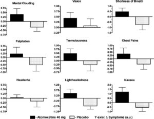
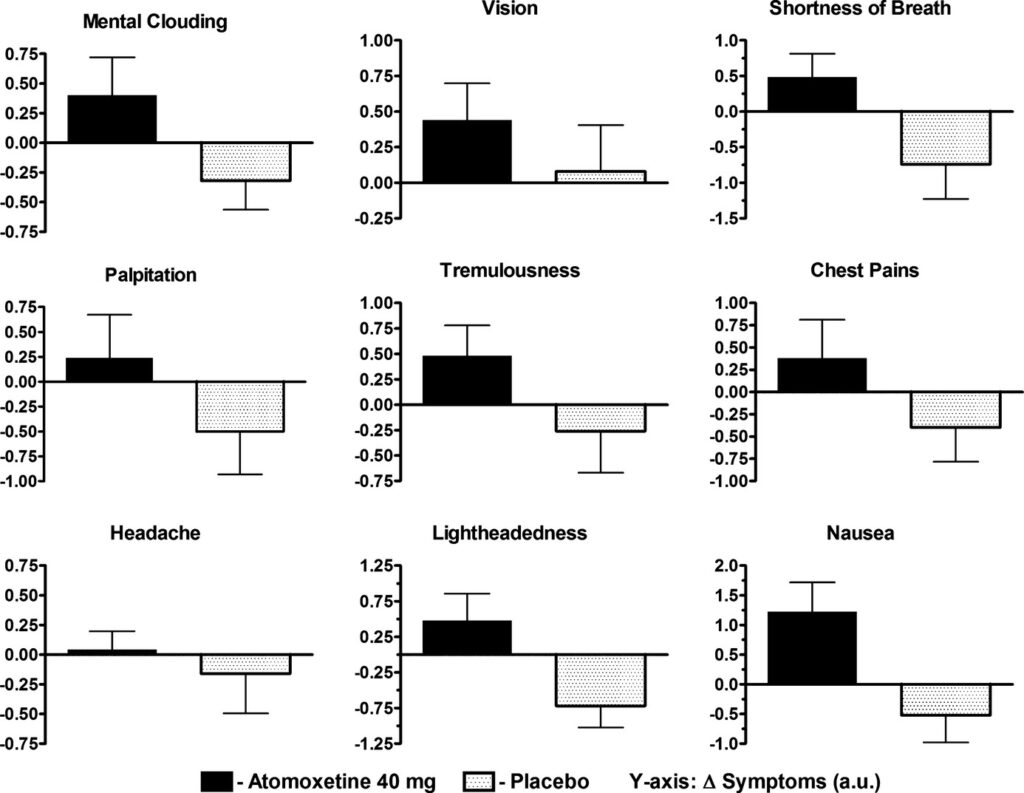
Graph describing the effects of the medication on POTS symptoms

Postural Orthostatic Tachycardia Syndrome (POTS) is a malfunction in the body’s autonomic nervous system. Rather than the blood vessels below their heart compensating by constricting, when a person with POTS goes from a lying to a standing position, a large amount of blood pools in the legs and abdomen. Normally, the blood vessels in the lower extremities constrict to maintain appropriate blood pressure throughout the whole body and help return the blood to the heart and head. The autonomic system (the part of the nervous system that is in charge of the involuntary aspects of the body) responds to low blood pressure by releasing norepinephrine and adrenaline, which cause vasoconstriction and a rise in heart rate. In POTS patients, vessels do not respond to the hormones and remain vasodilated. This combination of high heart rate and insufficient blood flow to the brain causes characteristic dizziness, fainting, and fatigue. POTS can be aggravated by a variety of things, including strenuous exercise, caffeine, hot environments, and certain medications (POTS, n.d.). 

One such class of medications is norepinephrine reuptake inhibitors (NOIs). Used to treat ADHD, major depressive disorder, and narcolepsy, NOIs block the uptake of norepinephrine in the synapses (De Crescenzo et al., 2018). This type of medication allows norepinephrine to stay in the blood longer, elevating mood and energy levels and enhancing focus. A common side effect is an elevated heart rate, which aggravates POTS. 

Green et al. conducted the first study examining the acute effects of atomoxetine on POTS patients. The study was composed of 27 patients and a variety of tests. A baseline was created to manage the patients’ diets. This entailed removing methylxanthines from their diet, which includes caffeine among other compounds, and moderating sodium and potassium intake. Additionally, all long-term medications were suspended for at least 5 half-life periods to ensure no hormonal effects would be present. All of these measures were taken to minimize the exacerbation of POTS symptoms (Green et al., 2013). 

All patients received the atomoxetine and the placebo (on different days). During this time, a posture study was done. Measurements of heart rate (HR), systolic blood pressure (SBP), diastolic blood pressure (DBP), mean arterial pressure (MAP), and plasma catecholamines were taken during a lying position and a standing position. This targets the effects of POTS, highlighting the possible impact of the atomoxetine. 

The posture study was paired with the medication study. During the medication study, patients were asked to fill out a symptom feedback form before the experiment, and every hour up to 4 hours after drug administration. This is because peak atomoxetine concentration occurs 1-2 hrs after ingestion. The Vanderbilt Orthostatic Symptom Score (VOSS) was used on the symptom feedback form, where patients are asked to rank the following on a scale from 1-10: mental clouding, brain fog, shortness of breath, palpitations, tremors, headache, tightness in the chest, blurred vision, and nausea. The lowest (1) is no symptom burden, and 10 is the worst. 

Researchers found that when patients took atomoxetine, their symptom burden increased. This presented a statistically significant increase in heart rate and a general upward trend in blood pressure throughout the 4 hours. In the case of the placebo, there was a decrease in symptom burden as the 4-hour period progressed. 

Atomoxetine is a non-stimulant medication used to treat ADHD; unfortunately, the stimulant alternatives are found to have similar effects on POTS patients. Due to a susceptibility to heart rate changes, ADHD medication negatively interacts with the condition and must be administered with exceeding caution. This interaction is important for prescribing professionals to be aware of. As this is a relatively under-researched intersection, consideration of mechanisms and close patient-doctor communication is necessary when considering medication. 

Figure 1: Results of VOSS with and without atomoxetine (Green et al., 2013)

Graph describing the effects of the medication on POTS symptoms

References

De Crescenzo, F., Ziganshina, L. E., Yudina, E. V., Kaplan, Y. C., Ciabattini, M., Wei, Y., & Hoyle, C. H. (2018). Noradrenaline reuptake inhibitors (NRIs) for attention deficit hyperactivity disorder (ADHD) in adults. The Cochrane Database of Systematic Reviews, 2018(6), CD013044. https://doi.org/10.1002/14651858.CD013044

Green, E. A., Raj, V., Shibao, C. A., Biaggioni, I., Black, B. K., Dupont, W. D., Robertson, D., & Raj, S. R. (2013). Effects of norepinephrine reuptake inhibition on postural tachycardia syndrome. Journal of the American Heart Association, 2(5), e000395. https://doi.org/10.1161/JAHA.113.000395

POTS: Causes, Symptoms, Diagnosis & Treatment. (n.d.). Cleveland Clinic. Retrieved April 8, 2025, from https://my.clevelandclinic.org/health/diseases/16560-postural-orthostatic-tachycardia-syndrome-pots


Filed Under: Biology Tagged With: ADHD, Biology, Medicine, POTS

TMJ Arthroscopy: How well does it work in Ehlers Danlos patients?

May 4, 2025 by Martina Tognato Guaqueta

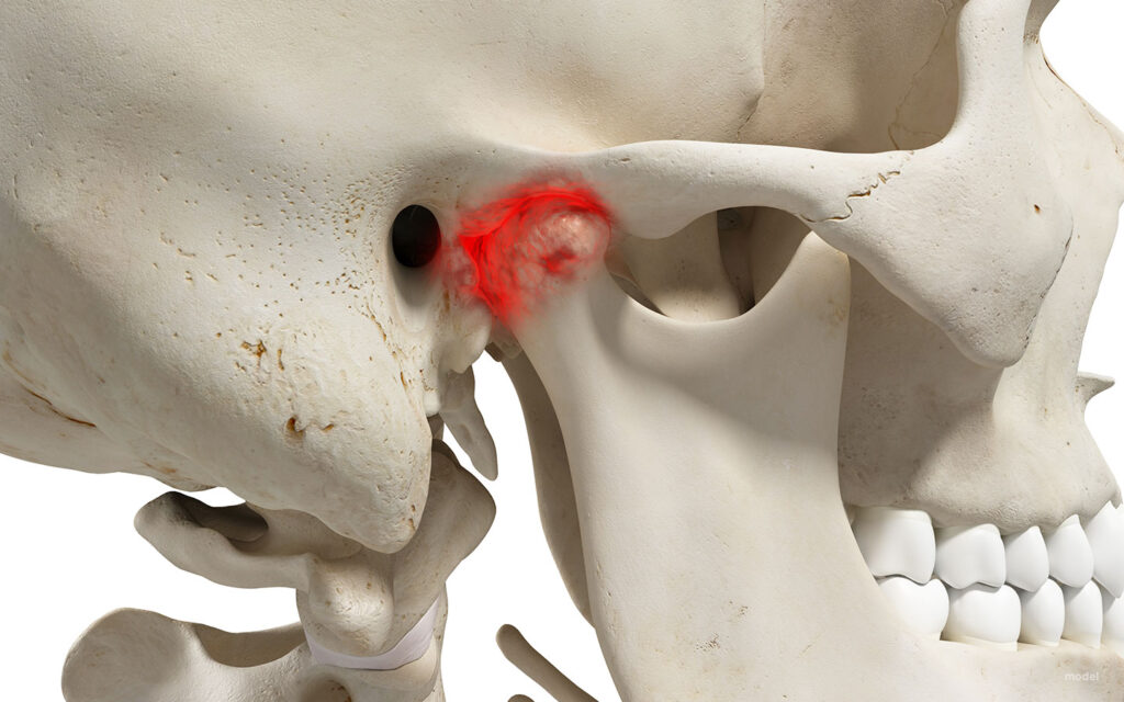
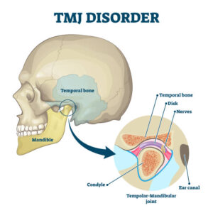
The temporomandibular joint (TMJ) is responsible for the mandible’s chewing, talking, and all movement. As a condylar joint, this joint allows motion in two planes: side to side and up and down. A disc cushions the bone to facilitate this motion and serves as a lubricant. 


Figure 1: TMJ diagram (TMJ Disorders – Symptoms and Causes, n.d.).

 Temporomandibular disorders (TMD) often arise from damage or irritation relating to the disc. To determine the condition of the joint, physicians use the Wilkes scale, which allows for the assessment of internal derangement/damage of the TMJ. TMD is often a symptom of a larger condition, it can arise from connective tissue disorders, injury, teeth grinding, etc.


Figure 2: Wilkes Scale descriptive table (Table 1 . Wilkes Classification of TMJ Internal Derangement, n.d.)


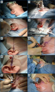
A patient’s placement on this scale indicates the type of treatment they could be a candidate for.  This could range from over-the-counter medications to physical therapy, to Botox injections, and a variety of surgeries. A surgery often seen is a TMJ arthroscopy.  The arthroscopic element refers to the minimally invasive approach assisted by a camera called an arthroscope. Done under general anesthesia, a surgeon will enter the joint space through a small incision. The image of the joint is delivered to a screen through an arthroscope. During the procedure, the surgeon may reposition the disc, flush the joint, and remove scar tissue to alleviate pain (Arthroscopy for Temporomandibular Disorders (TMDs) | Kaiser Permanente, n.d.). This, however, is not the only option, and can only be decided upon looking at the full picture of a patient’s history—for example, a connective tissue disorder. 

Ehlers-Danlos (EDS) is a group of connective tissue disorders that can affect many systems, including the joints and, in turn, the TMJ.  Within the group, each particular variation has a different set of symptoms. For example, vascular EDS causes the blood vessels to rupture, whereas hypermobile EDS (hEDS) causes frequent joint dislocations. Some types of EDS have a clear genetic link, however, the origins of hEDS have yet to be understood. Due to the overly flexible nature of their joints, hEDS patients often encounter issues with their TMJ.  (Ehlers-Danlos Syndrome – Symptoms and Causes, n.d.).

Jerjes et al. conducted a retrospective case study that looked at the outcomes of 18 hEDS patients who underwent arthroscopic surgery to treat their TMDs. All patients were female and between the ages of 23-60 years old. Due to the EDS, TMJ dislocation was a common history within the sample. In turn, the dislocations were linked to the damage and pain, which were visualized during the arthroscopies. This demonstrated a link between the state on the TMJ and hEDS status. Moreover, 12 out of the 18 experienced TMD bilaterally, meaning there was pain on both sides (Jerjes et al., 2010). 

Furthermore, it seemed as though the intervention was most commonly done at/by the Wilkes Stage III (9 patients at Stage III and 5 at Stage II ). As seen in Figure 2, Stage III is characterized by moderate disc deformity and frequent pain/dislocations.  As the Wilkes Scale goes on, the condition becomes more painful and harder to treat. However, although minimally invasive, arthroscopy is considered a more extreme treatment for TMJ. Over the years, the procedure has developed and improved, reaching an 80-90% success rate (Insel et al., 2020). Despite this, the National Institute of Dental and Craniofacial Research brochure still has a negative outlook on the surgical route as a treatment for TMDs (Jerjes et al., 2010). 

Notwithstanding the controversy, Jerjes et al. demonstrate positive post-operative results. The pain subsided quickly (within 1 week post-operative) for 15 out of 18 patients and 5-6 weeks for the remaining 3. Delayed healing occurred in 4 out of 18 patients. Slow healing is a characteristic of hEDS and could be a potential reason in this case for the post-surgical complication. Most importantly, the mouth opening measured 6 months postoperatively increased from an average of approximately 23-28 mm,  with all final postoperative results being positive. 

Overall, the study aims to illustrate the success of TMJ arthroscopy in hEDS patients and suggests it as the first consideration for invasive procedures. Due to hEDS patients often having a difficult time with wound healing, a minimally invasive procedure is favored. Additionally, these patients have previously sought out other forms of treatment before surgery. This study does not suggest that surgery should be an initial consideration, but it should not be forgotten as an option. 


Figure 3: Outside view of an TMJ arthroscopy 

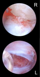
Figure 4: Internal photo is TMJ arthroscopy


The hEDS population is small; therefore, studies that center on them and how treatments interact with their condition are few and far between. Moreover, providers for TMJ-related surgeries are scarce. In turn, this middle ground (as noted by Jerjes et al. ) is a neglected area of research. Further development would focus on EDS patients more and seek to understand the interconnectedness of EDS and joint pain and how it can impact the choice of treatments. 


References

Arthroscopy for Temporomandibular Disorders (TMDs) | Kaiser Permanente. (n.d.). Retrieved April 8, 2025, from https://healthy.kaiserpermanente.org/health-wellness/health-encyclopedia/he.arthroscopy-for-temporomandibular-disorders-tmds.hw209368

Ehlers-Danlos syndrome—Symptoms and causes. (n.d.). Mayo Clinic. Retrieved April 8, 2025, from https://www.mayoclinic.org/diseases-conditions/ehlers-danlos-syndrome/symptoms-causes/syc-20362125

Insel, O., Glickman, A., Reeve, G., Kahan, B., Tran, T., & Israel, H. (2020). New criteria demonstrate successful outcomes following temporomandibular joint (TMJ) arthroscopy. Oral Surgery, Oral Medicine, Oral Pathology and Oral Radiology, 130(1), e20–e21. https://doi.org/10.1016/j.oooo.2019.12.022

Jerjes, W., Upile, T., Shah, P., Abbas, S., Vincent, A., & Hopper, C. (2010). TMJ arthroscopy in patients with Ehlers Danlos syndrome: Case series. Oral Surgery, Oral Medicine, Oral Pathology, Oral Radiology, and Endodontology, 110(2), e12–e20. https://doi.org/10.1016/j.tripleo.2010.03.024

Table 1. Wilkes classification of TMJ internal derangement. (n.d.). ResearchGate. Retrieved April 8, 2025, from https://www.researchgate.net/figure/Wilkes-classification-of-TMJ-internal-derangement_tbl1_7691660

TMJ disorders—Symptoms and causes. (n.d.). Mayo Clinic. Retrieved April 8, 2025, from https://www.mayoclinic.org/diseases-conditions/tmj/symptoms-causes/syc-20350941

Top Five Fun Facts About The TMJ. (2022, July 14). Ladner Village Physiotherapy | Delta BC. https://ladnervillagephysio.com/blog/top-five-fun-facts-about-the-tmj


Filed Under: Biology Tagged With: Biology, Medicine, surgery, TMJ

Philip Spyrou in the Spotlight

December 9, 2024 by Noah Zuijderwijk

“The lab and the art studio are fundamentally the same space; you have a material, a question you want to answer, and you experiment” – Philip Spyrou

Give a teen unfettered access to the internet and they might transform into a brain-rotten screenager. Luckily, in Philip Spyrou’s case, hours spent looking at Reddit feeds and YouTube videos did not translate into cognitive decline. In fact, quite the opposite was true; he used his internet privileges to teach himself how to cultivate life. As a high school sophomore, Philip experimented with hydroponics and tried to grow mushrooms using soil he made with whole grains and a pressure cooker. His resourceful and creative fascination with life led him to a chemistry and visual arts double major at Bowdoin College. He now studies the role of proteins in neuron function as a senior researcher in Professor Henderson’s chemistry lab.

When Philip showed me around the Henderson lab, he explained that proteins play a near-infinite number of crucial roles in biological processes. One such process is the formation of synapses in the brain. In simple terms, a synapsis is the coming together of two neurons to exchange information – a crucial mechanism for routine brain function. However, neurons need the ability to “crawl” around brain tissue before they can find other neurons and form synapses. SRGAP proteins enable neurons to develop finger-like protrusions from the cell membrane with which they can “crawl”. Philip studies how the membrane attracts these proteins.

Though one might think studying neurons requires a lab furnished with preserved brains in glass jars, Philip’s research (disappointingly) does not involve Frankensteinian techniques. In fact, Philip works with model cells called Giant Unilamellar Vesicles (GUVs), which are artificial membrane systems used to study cell functions. By modifying the GUV’s membrane, he observes how different membrane compositions attract SRGAP proteins. These observations can then be mapped onto neurons to understand how they develop the ability to “crawl” through brain tissue.
 

To study this neuron crawling mechanism, Philip has to think beyond the two dimensions of a textbook. After all, a protein’s three-dimensional structure is key to its function. In this regard, his time in the art studio has proven valuable to his work in the lab. Philip believes the lab and the studio aren’t all that different, and that working with clay and ceramics has trained him on how to gather materials, ask questions, and design experiments with a three-dimensional mindset.


“I like thinking visually, structurally, and three-dimensionally about the biological processes I study”


The three-dimensionality of Philip’s research unfolds at the molecular scale. It requires him to spend most of his time thinking about intangible processes. But, he says, it helps him to think of the applications of his research. For example, loss of proteins that enable neuron cells to crawl around the brain might be implicated in cognitive disabilities and memory loss. This is something he hopes to continue researching by earning a PhD with the goal of eventually becoming a full-time researcher.

As Philip continues on this path toward becoming a scientist, he finds it important to keep reminding himself of where his passion for science comes from. His love for understanding life originated in his backyard when he figured out how to grow plants and mushrooms. Though he does not foresee himself going back to researching those forms of life any time soon, he does want to keep tapping in to his fifteen-year-old self’s creative fascination for life.

Filed Under: Honors Projects Tagged With: biochemistry, Biology, brain, chemistry, honors, neuron, neuroscience, student

Uncovering Our Inner Overlord: How DEADbox ATPases Built Their Empire Off Regulating RNA Maturation

December 9, 2024 by Lia Scharnau

Do you remember the simple days? Recall your fond memories of learning about organelles in introductory biology. This is where we learned our favorite biology fact, that the mitochondria is the powerhouse of the cell. Sigh, those were the days. Well, recently the field of biology has discovered a new type of organelles in the cell; membraneless organelles! They are formed through liquid-liquid phase separation (LLPS). If you imagine the droplets formed when you combined oil and water, that’s a form of LLPS. Membraneless organelles rely on LLPS for rapid and reversible cell compartmentalization.

In 2019, researcher Maria Hondele and her team took particular interest in investigating membraneless organelles, focusing specifically on DEAD-box ATPases (DDX) and their role in regulating them. DEAD-box ATPases keep ribonucleoprotein complexes from misfolding or building up over time. The role of DDX-mediated phase separation in compartmentalizing RNA processing is a rare cellular organization conserved across prokaryotes and eukaryotes over time (Hondele 2022). Highly conserved proteins have withstood the test of evolution and have continued to be passed down through generations without significant mutation. Hondele looked specifically at RNA-dependent DEAD-box ATPases because they regulate the RNA movement in and out of the membraneless organelles.

This investigation focused on  Dhh1, which is a DEAD-box ATPase specific to Saccharomyces cerevisiae (yeast). A wide range of assays were run to systematically determine the conditions required for the in vitro formation of Dhh1 liquid droplets. Liquid droplets are formed through LLPs and are indicators of membraneless organelles. Hondele found that liquid droplet formation is a fickle process that requires specific amounts of RNA and ATP to be added to the system and the cell environment to be at a low pH and salt concentration (Hondele 2019). Additionally from a DNA standpoint, the DDX itself must have low-complexity domain tails which means the ends of the proteins do not consist of a large variety of amino acids (Hondele 2019). 

After the initial investigation of the DDX ATPase and how it runs controls Dhh1 droplet formation, Hondele, and her team investigated DDX ATPase’s role in the regulation of RNA. Through a series of experiments, they found that DDX ATPases have played an extensive role in RNA regulation. The DDX ATPases can actually control the RNA maturation steps so they become spatially and temporally separated in distinct membraneless organelles (Hondele 2019). This means that each membraneless organelle may specialize in one step of the RNA maturation process so that the RNA must move between different organelles throughout the process. Of course, the release and transfer of RNA is regulated by ATPase activity, confirming DDX ATPase’s role as the omnipotent overlord of RNA. The DDXs derive their power from the low-complexity domains. These domains give DDXs the intrinsic ability to set up distinct compartments and when teamed up with the ATPases, they can influence the partitioning of RNA molecules between compartments (Hondele 2019).

Hondele and her team managed to uncover a complex and extensive dictatorship that has been operating for years under our very noses and in our very cells. The well-established and conserved cellular network of DEAD-box ATPases allows the RNA processing steps to be regulated, leading to DEAD-box ATPase control over maturation state, RNP composition, and ultimately RNA fate.

Unfortunately, we are still in the investigation phase and are yet to decide on how best to manipulate this dictatorship to benefit us. Current intelligence indicates that the dysregulation of DDXs could have pathological consequences that could contribute to the development of aggregation diseases, such as Parkinson’s, Alzheimer’s, Amyotrophic lateral sclerosis, and Frontotemporal Dementia (Gomes 2018). Luckily liquid-liquid phase separation has provided a mechanistic link between normal cellular function and disease phenotypes. Over time, these liquid droplets become more static and aggregated, likely leading these protein aggregates to be an end-stage phenotype after aberrant phase separation has overwhelmed cellular machinery that ordinarily reverses these altered phases (Gomes 2018). Through further study and comprehension of how DDXs contribute to these diseases, new treatments could be developed.


Literature Cited:

Gomes, E,. Shorter, J. The molecular language of membraneless organelles. J. Biol Chem. 2018; 294(18):7115-7127. 10.1074/jbc.TM118.001192

Hondele, M.,  Sachdev, R., Heinrich, S., Wang, J., Vallotton, P., Fontoura, B.M.A., Weis, K. DEAD-box ATPases are global regulators of phase-separated organelles. Nature. 2019; 573(7772):144-148. 10.1038/s41586-019-1502-y.

Hondele, M., Weis, K. The Role of DEAD-Box ATPases in Gene Expression and the Regulation of RNA-Protein Condensates. Annu Rev Biochem. 2022;  91:197-219. 10.1146/annurev-biochem-032620-105429. 

Filed Under: Biology, Science Tagged With: Biology, Cell Biology, Proteins

Gracie Scheve in the Spotlight

December 9, 2024 by Noah Zuijderwijk

“To do research, you have to be stubborn. But also, don’t be too hard on yourself” – Gracie Scheve

Around 600 million years ago, marine invertebrates emerged as Earth’s first multicellular organisms. Today, Gracie Scheve is scheming to make a career out of researching their extraordinary life cycles. Her interest in invertebrate evolution and development has not come out of the blue though. Gracie’s family would drive down from their home in Cincinnati, Ohio to Florida every summer for vacation when she was little. There, she would load buckets onto her paddle board, paddle out to sea, and collect countless jellyfish. Back on shore, she would spend hours marveling at her catch. Now, years later, Gracie has carved out her niche in invertebrate biology as a senior researcher in the Rogalski lab at Bowdoin College. With Professor Rogalski, she investigates reproductive strategies of Daphnia, or the common water flea.

In late Spring, Gracie brought me along to her study lake. While she collected water samples, Gracie explained that Daphnia are cyclical parthenogens. In simple terms, they can reproduce both sexually and asexually. Typically, their wild population is exclusively female. This all-female population reproduces asexually into the next generation of clonal daughters with every reproduction cycle. In Gracie’s words “Daphnia are girl bosses”. However, things change when they encounter stress. For example, disease can trigger Daphnia to produce males with which the females will sexually reproduce. This, in turn, results in more genetic diversity, which increases the population’s stress tolerance and survival probability. Through her research, Gracie hopes to gain more clarity on what stresses alter reproductive behavior, and by what mechanism.

Over the summer of 2024, Gracie observed an unexpected pattern in the field; the only stress factor that seemed related to an uptick in sexual reproduction was a novel fungal parasite. Though this might mean Gracie and Professor Rogalski will get to name a new genus of fungus, for now, it is leading to more questions than answers. For example, how does the fungus affect Daphnia? And is it truly inducing sexual reproduction, or was Gracie’s observation merely coincidental? Gracie is currently experimenting with this fungus in the lab. She admitted that she might not find the answers before the end of her senior year. However, she is excited about the novelty of her research.
 

“I want to go into a field where there are questions I am interested in that haven’t been answered yet”


Along with asking new questions comes a level of uncertainty that makes Gracie’s research unpredictable. It means that over the past months, Gracie has experienced many unexpected turns, like when all Daphnia had disappeared in mid-June. She recognizes these surprises are a natural part of research and that it is a good thing she is learning how to handle them now – especially because she hopes to take her next step into an evolutionary biology PhD program. Her undergraduate research experiences have taught her not only to be flexible, but also that research requires an underappreciated range of soft skills. Whereas quantitative skills and book smarts seem to prevail, Gracie shared that having an open mind, being persistent, and being patient with oneself are some of the most important qualities of a researcher. Wherever Gracie will go next, she will take these lessons with her.

“Fieldwork is frustrating sometimes because you’re not in control. And when you do have control in the lab, results might not map onto the field at all. Regardless, you have to be patient with yourself and your research.”

Filed Under: Honors Projects Tagged With: Biology, Daphnia, honors, student

Gut Viruses Might Be the Key to Life Saving Early Pancreatic Cancer Diagnosis

December 8, 2024 by Noah Zuijderwijk

New study links the community of viruses in our gut to early pancreatic cancer development – a potentially lifesaving discovery.

With a mortality-to-incidence ratio of over 90%, pancreatic cancer (PC) is among the most deadly forms of cancer. Since its early stages often bear no distinct symptoms, the disease grows stealthily until it’s too late. What’s more? Scientists foresee a near 100% increase in PC deaths, from 466 000 in 2020 to over 800 000 by 2040. To avert this grim future, researchers strive to develop methods for earlier detection, and subsequent earlier treatment. A 2022 study at the University of Tokyo linked PC to changes in gut microbiome composition. This has directed gastroenterologists’ focus to the microbiome in the search for new diagnostic tools.

The gut microbiome is often understood as the community of bacteria living in symbiosis with our digestive tract. Bacteria, like E. coli, break down our food in exchange for a safe habitat. However, bacteria do not aid our digestion for brownie points. As evolving creatures, they constantly test the limits of our gut ecosystems. As far as we understand, that’s where viruses come in; they regulate the bacterial population in our guts. The microbiome, therefore, consists of not only bacteria but also viruses. All viruses together make up our body’s virome.

An imbalance of bacteria and viruses has previously been observed in PC patients and is believed to be a factor in PC development. For example, the bacterium Roseburia intestinalis is significantly less abundant in the guts of PC patients compared to healthy individuals. This particular bacterium produces a cancer-inhibiting metabolite called butyrate, a substance that limits cancer development by suppressing inflammation and reducing the expression of genes involved in tumor cell growth. Other bacteria produce cancer promoting-metabolites, like lipopolysaccharide (LPS). This substance activates our immune system in the presence of pathogens, but also stimulates inflammation, and therefore, promotes cancer growth in the process. The balance of bacteria producing these two kinds of metabolites depends on the virome’s composition. Therefore, if we could identify the gut viruses causing imbalances, we might be able to diagnose patients earlier.

Researchers at Xi’an Jiaotong University took on this hypothesis when they performed a study that compared PC patient viromes to those of healthy individuals. They sequenced the DNA of 183 fecal samples from a Spanish and a German cohort with 101 PC patients and 82 healthy individuals. After sequencing the samples, they filtered out human DNA by comparing the sequenced DNA to an established human reference genome. They then used viral references to compare viral DNA from the samples to known viruses. After statistical analyses confirmed significant difference between the PC patient group and the healthy group, they identified which viruses were present in the PC patients’ guts, and how those differed from the ones found in healthy individuals. As pancreatic cancer severity increased, virome diversity decreased in PC patients. Additionally, the viruses present in the affected individuals targeted different bacteria compared to the gut viruses found in healthy individuals, offering a potential explanation for the relationship between unbalanced microbiomes and cancer growth.

Using their results, the researchers created models to differentiate PC patients from healthy individuals. These models succeeded with 87.9% accuracy. Though these findings do not offer the ultimate solution to late PC diagnoses, access to virome information could be used as a diagnostic tool in addition to the tools currently available. Namely, a viral DNA sequencing-based tool could identify the specific viral biomarkers linked to pancreatic cancer. In the future, at risk groups for PC might, therefore, be asked to supply fecal samples for gut virus analysis during routine check-ups. In the case that PC-linked biomarkers show up, these at-risk groups could be provided early treatment, potentially saving their lives.

Sources:

Miyabayashi, K., Ijichi, H., & Fujishiro, M. (2022). The Role of the Microbiome in Pancreatic Cancer. Cancers, 14(18), 4479. https://doi.org/10.3390/cancers14184479  

Zhang, P., Shi, H., Guo, R., Li, L., Guo, X., Yang, H., Chang, D., Cheng, Y., Zhao, G., Li, S., Zhong, Q., Zhang, H., Zhao, P., Fu, C., Song, Y., Yang, L., Wang, Y., Zhang, Y., Jiang, J., & Wang, T. (2024). Metagenomic analysis reveals altered gut virome and diagnostic potential in pancreatic cancer. Journal of Medical Virology, 96(7). https://doi.org/10.1002/jmv.29809

Cover image by magicmine, https://stock.adobe.com/search?k=pancreas+cancer&asset_id=343535067

Filed Under: Biology Tagged With: Biology, cancer, Cancer Biology, gut viruses, Pancreatic cancer, virome, viruses

The Dark Side of Antibiotics

May 8, 2024 by Maya Lall '27

Antibiotics are medications that fight infections caused by bacteria. But they can also lead to mental health issues, such as anxiety and depression, later in life. 

The discovery of antibiotics was one of the greatest medical advances of the 20th century. Antibiotics have significantly reduced mortality from infectious diseases and increased average life expectancy. However, they have a variety of side effects, including cognitive impairment and emotional disorders in adulthood (Adedji 2016). 

Antibiotics destroy bacteria in the gut, which can disrupt brain function. Gut microbiota are an important component of the gut-brain axis–the two-way line of communication between the gastrointestinal tract and the central nervous system. Gut microbiota produce neurotransmitters that regulate mood, such as dopamine, norepinephrine, and serotonin, which travel through the vagus nerve to the brain (Karakan et al 2021). Previous studies have found that antibiotic-induced gut microbiota depletion causes dysfunction of the gut-brain axis, increasing anxiety and depression-related behaviors (Mosaferi et al 2021).

Aging plays an important role in the development of both gut microbiota and the brain. Microbiota first appear at birth and rapidly colonize the intestinal tract. The composition and diversity of gut microbiota resembles adult level by 2 years of age and remains stable throughout adulthood before decreasing in old age. The brain develops until the mid-to-late 20s and starts to decline in middle age. It has been shown that antibiotic-induced gut microbiota depletion has negative effects on the brain (Li et al 2022). However, no prior research has been done on this relationship during the different stages of life. This study aimed to determine the connection between gut microbiota and cognitive and emotional function during the different life stages. 

In this experiment, the researchers used mice as models for human subjects, randomly assigning 75 mice to five groups. One group served as the control (Veh group) and was given distilled water from birth to death. The other four groups were given an antibiotic cocktail at different life stages: birth to death (Abx group), birth to postnatal day 21 (Abx infant group), postnatal day 21 to 56 (Abx adolescence group), and postnatal day 57 to 84 (Abx adulthood group).  

At postnatal day 85, the researchers randomly selected thirteen mice from each group for testing. They measured the cognitive function and emotion of the mice by using four traditional behavioral tests: open-field test (OFT), passive avoidance test (PAT), morris water maze test (MWM), and tail suspension test (TST). The OFT measured anxiety level by observing how long the mice moved in an open field for 5 minutes. The PAT measured short-term memory, which was defined as the difference in latency–the time it took mice to reenter a room that delivered an electric shock–between day 1 and day 2 (Jahn-Eimermacher et al 2011). The MWM measured spatial memory, which was defined as incubation time, or how long it took mice to find a submerged platform in a pool after a 5-day training period. The TST measured depression state by observing the duration of quiescence (motionless state) of the mice while they were suspended upside down for 4 minutes.

Figure 1 | Results of OFT, PAT, MVM, and TST tests. Researchers conducted four traditional behavioral tests on mice given water, as well as mice given antibiotics at different stages of life: birth to death, infancy, adolescence, and adulthood. They found that exposure to antibiotics from birth to death and in infancy led to the most severe cognitive and emotional dysfunction, followed by exposure in adolescence and adulthood (Li et al 2022).

The results of this study suggested that life cycle stages influence the relationship between gut microbiota and cognitive and emotional function. For the OFT test, the total movement time of the Abx, Abx infant, and Abx adolescent groups was significantly lower than the Veh group, indicating they were more anxious than the Veh group (Figure 1). In other words, exposure to antibiotics from birth to death, in infancy, and in adolescence caused anxiety-related behaviors. For the PAT test, the difference in latency for every Abx group was significantly lower than the Veh group, meaning all Abx groups reentered the room with the electric shock more quickly after training. These results signaled that short-term memory loss was greater in the Abx groups than the Veh group; exposure to antibiotics at any stage of life caused short-term memory loss. The MWM test found that the incubation time after the 5-day training period was significantly higher for the Abx and Abx infant groups than the Veh group, so they experienced more spatial memory loss than the Veh group; exposure to antibiotics from birth to death and in infancy caused spatial memory loss. The TST test found that the duration of quiescence in the Abx and Abx infant groups was significantly higher than the Veh group, implying they were more depressed than the Veh group. In other words, exposure to antibiotics from birth to death and in infancy caused depression-related behaviors.

The researchers’ findings align with previous work showing that depletion of gut microbiota causes cognitive impairment and emotional problems (Lach et al 2020). Furthermore, the researchers demonstrated that life cycle stages are an important factor in the relationship between gut microbiota and cognitive and emotional function. In particular, their findings strengthened the idea that infancy is a crucial stage of development of gut microbiota and the brain (Hunter et al 2023). Gut microbiota lost in infancy recovers over time; however, this depletion has lasting cognitive effects. In this study, mice given antibiotics in infancy exhibited similar behaviors in adulthood as mice given antibiotics from birth to death: anxiety, depression, memory loss, and learning ability decline. Exposure to antibiotics in infancy and in the long term led to the most severe cognitive and emotional dysfunction, followed by exposure in adolescence and adulthood.

The researchers’ findings also have implications for the treatment of mental health illnesses. Previous studies have shown that probiotics replace depleted gut microbiota, alleviating symptoms of anxiety and depression. Antidepressants and anxiolytics–current medications for anxiety and depression–cause side effects such as nausea, weight gain, insomnia, constipation, dizziness, agitation, and restlessness. Probiotics are associated with milder side effects, including gas and bloating (Bistas et al 2023). Probiotics are unlikely to treat severe depression and anxiety, but they are promising treatments for people with milder conditions. The next step is to identify and manufacture effective probiotics, which would revolutionize the field of psychiatry and improve the lives of people around the world.


References

Adedji, W.A. 2016. THE TREASURE CALLED ANTIBIOTICS. Annals of Ibadan Postgraduate Medicine. 14(2):56-57. https://www.ncbi.nlm.nih.gov/pmc/articles/PMC5354621/.

Bistas KG, Tabet JP. 2023. The Benefits of Prebiotics and Probiotics on Mental Health. Cureus Journal of Medical Science. 15(8):e43217. doi:10.7759/cureus.43217.

Hunter S, Flaten E, Petersen C, Gervain J, Werker JF, Trainor LJ, Finlay BB. 2023. Babies, bugs and brains: How the early microbiome associates with infant brain and behavior development. PLOS One. 18(8):e0288689. doi:10.1371/journal.pone.0288689.

Jahn-Eimermacher A, Lasarzik I, Raber J. 2011. Statistical analysis of latency outcomes in behavioral experiments. Behavioural Brain Research. 221(1):271-275. doi:10.1016/j.bbr.2011.03.007.

Karakan T, Ozkul C, Akkol EK, Bilici S, Sobarzo-Sánchez E, Capasso R. 2021. Gut-Brain Microbiota Axis: Antibiotics and Functional Gastrointestinal Disorders. Nutrients. 13(2):389. doi:10.3390/nu13020389.

Lach G, Fülling C, Bastiaanssen TFS, Fouhy F, O’Donovan AN, Ventura-Silva AP, Stanton C, Dinan TG, Cryan JF. 2020. Translational Psychiatry. 10(1):382. doi:10.1038/s41398-020-01073-0.

Li J, Pu F, Peng C, Wang Y, Zhang Y, Wu S, Wang S, Shen X, Li Y, Cheng R, He F. 2022. Antibiotic cocktail-induced gut microbiota depletion in different stages could cause host cognitive impairment and emotional disorders in adulthood in different manners. Neurobiology of Disease. 170:105757. doi:10.1016/j.nbd.2022.105757.

Mosaferi B, Jand Y, Salari AA. 2021. Gut microbiota depletion from early adolescence alters anxiety and depression-related behaviors in male mice with Alzheimer-like disease. Scientific Reports. 11:22941. doi:10.1038/s41598-021-02231-0.

Filed Under: Biology, Psychology and Neuroscience Tagged With: antibiotics, Anxiety, Biology, Depression, Gut microbiota

Smoke Signals: The Unexpected Long Term Effects of Smoking on the Immune System

April 30, 2024 by Divya Bhargava

Image Source: “Smoking has a Lasting Impact on the Immune System, 2024”

When we get sick, our bodies’ immune systems work to fight off infections by invading pathogens, or organisms like bacteria and viruses that cause disease. However, many factors such as lack of sleep and poor nutrition weaken our immune system, meaning that we are less able to stay healthy. It has been known that smoking is another one of these factors that weaken our immune systems, but a recent study from a group at the Institut Pasteur in France looking at the effects of a variety of factors on the immune system showed that the extent to which smoking plays a role is much higher than many would think. But to understand the results of this study, it is important to first understand the mechanisms the immune system uses to fight infection. 

The immune system has many different moving components, including two distinct branches. The first is the faster, more general innate immune system which has a similar response to all infections. The second is the adaptive immune system which is slower, memory-based, and is involved in pathogen specific response. Although the innate immune system involves general molecules that interact with all cells and the adaptive immune system has specialized molecules that interact with pathogens based on memory of past infection, they share one important class of signaling molecules. These molecules are called cytokines and their role is to coordinate both of these types of immune response. Cytokines are small molecules that are released by immune cells to communicate with other parts of the body and each other. This signaling results in deployment of a response by other immune cells against invading pathogens. However, levels of cytokine production exist in a very fine balance. In order to get the desired immune response, you need the exact right level of cytokines present. If levels are too high or too low, they could cause abnormalities including overactive immune response and inflammation or impaired immune responses. 

To investigate the effects of a variety of different factors on the immune system and cytokine responses of healthy individuals, a project called the Milieu Intérieur put together a cohort of 1000 healthy participants and has been studying variability in the immune system between these individuals (“The Milieu Intérieur Project”). In an investigation of this data, the group from Institut Pasteur, Saint André et al, analyzed 136 variables measured in the Milieu Intérieur Project that could be causing differences in cytokine secretion and immune response (Luo and Stent 2024). These variables included everything from demographics, to diet, to health habits like smoking, to social and environmental characteristics (Saint-André et al. 2024).

After they performed their initial statistical analysis, Saint André et al measured production of 13 disease relevant cytokines as a quantitative measure of immune response in populations with different demographics, health habits, and other characteristics. In the lab, they exposed blood samples from their sample population to 12 different molecules meant to serve as stimulants for the immune system (these molecules included things like viral and bacterial proteins). After this exposure, the authors tested cytokine production in both innate and adaptive immune cells, and once they had that data, they took their results one step further. The group also used epigenetics, or the study of changes in gene expression rather than the DNA code that makes up the genome to investigate possible reasons for variability in immune responses associated with factors tested. Their epigenetic evaluation consisted of analyzing the extent to which one epigenetic process, DNA methylation, occurred at specific regulators of signaling and metabolism (Saint-André et al. 2024) to assess changes associated with smoking. 

As previously stated, one of the authors’ main findings from the initial statistical analysis was that smoking had a large effect on cytokine response. In fact it had the same effect as age, sex, and genetics, three things many would consider much more directly impactful to the immune system than smoking. In their in vitro simulations, they found that smoking had a temporary effect on the ability of the innate immune system to function properly. This result is a relatively intuitive one. If you do something that is considered bad for you, it makes sense that you would get sick more easily. 

However, more surprisingly, they also found that smoking leaves a lasting effect on memory based adaptive immune responses even after cessation of smoking, meaning that even after people quit smoking, their immune systems still are impacted. They found that in samples from individuals who smoked there were higher levels of cytokine expression, especially of an inflammatory cytokine called CXCL5 that is secreted in response to bacterial infection. Secretion of this cytokine is associated with the presence of an inflammatory protein called CEACAM6 in the blood. Consistent upregulation of levels of this protein has been found to have links with multiple cancers such as colon cancer (Wu et al. 2024). In Saint André et al’s epigenetic investigation of this association, they found that DNA methylation, which results in a downregulation of gene expression and in this case an increase in cytokine production, is linked to smoking’s lasting effect on the immune system (Greenberg and Bourc’his 2019). DNA methylation was decreased at many of the sites they tested which are involved in regulation of signaling genes and metabolism. Decreased DNA methylation was likely impacting levels of cytokines in response to detection of pathogens. In these populations, smoking caused lasting changes in gene expression which resulted in long term changes in addition to the expected short term effects on the immune system. 

This study demonstrates that smoking can have lasting negative impacts on your health which are not limited to just lung damage. It is also associated with pro-inflammatory cancer pathways and epigenetic markers that cause increased cytokine production. This overproduction of cytokines can confuse cells and also cause increased inflammation. Over time the extra inflammation can damage tissues and lead to developments of other conditions, like the cancers previously mentioned and complications associated with overproduction of cytokines (“What are Cytokines”). These recent findings emphasize that it is important to consider the possible implications of smoking and all things that we expose ourselves to, and to keep in mind that new data is still being discovered.

Works Cited

The Milieu Intérieur Project Institut Pasteur. Luo,Y. and Stent,S. (2024) Smoking’s lasting effect on the immune system. Nature, 626,  724–725.

Saint-André,V., Charbit,B., Biton,A., Rouilly,V., Possémé,C., Bertrand,A., Rotival,M., Bergstedt,J., Patin,E., Albert,M.L., et al. (2024) Smoking changes adaptive immunity with persistent effects. Nature, 626, 827–835.

Wu,G., Wang,D., Xiong,F., Wang,Q., Liu,W., Chen,J. and Chen,Y. (2024) The emerging roles of CEACAM6 in human cancer (Review). International Journal of Oncology, 64, 1–15.

Greenberg,M.V.C. and Bourc’his,D. (2019) The diverse roles of DNA methylation in mammalian development and disease. Nat Rev Mol Cell Biol, 20, 590–607.

What are Cytokines? Types and Function Cleveland Clinic.

Smoking has a lasting impact on the immune system, a new study finds (2024) Euronews.

Filed Under: Biology Tagged With: Biology, Genes, Medicine

Plant Talk: Eavesdropping on Underground Plant Communication

December 3, 2023 by Riley Simon '26

Have you ever looked at a tiny sapling, a winding vine, or a massive oak tree and felt like they have some sort of personality? With the rustle of some leaves or the snap of a twig it might seem like these plants are talking to each other. As it turns out, these fantasies aren’t too far from the truth. Vascular plants (which consist of most plants other than mosses and algae) can actually communicate. These plants can exchange messages through their root systems with the help of mycorrhizal fungi. These fungi exist in a mutualistic relationship with plants and, along with acting as a living walkie talkie, they provide many survival benefits to the plants they live with. 

To be clear, vascular plants aren’t chatting in some sort of plant language in the same way that we talk to each other. Instead, they communicate through the transfer of infochemicals. “Infochemical” is an umbrella term for substances released by one plant and detected by another (Chen 2018). Infochemicals can take the form of plant hormones or nutrients and are passed between plants through the soil. The problem with this system is that transport through the soil is incredibly inefficient. When infochemicals move from plant to plant, they can quickly be absorbed by organic material or degrade in the soil such that they do not reach the intended “listener” plant. This is where mycorrhizal fungi come into play.

Mycorrhizal fungi (MF) are distinguished from other fungi by the symbiotic relationship that they have with plant roots. MF attach to plant roots and perform beneficial services for the plant in return for the carbon necessary for MF’s survival. MF networks add large amounts of surface area to plant root systems, allowing for the more efficient uptake of nutrients to the plants such as nitrogen, phosphorus, and carbon. The MF relationship increases efficiency of water collection, enhances photosynthesis, and improves resistance to pathogens. (Barto 2012). 

When it comes to plant communication, MF act as “superhighways” for the infochemicals to travel from plant to plant. Instead of having to travel through the soil, infochemicals can be safely transported between plants through common mycorrhizal networks (CMNs). CMNs are made up of interconnected networks of fungal branches, which span the distance between plant roots (Chen 2018). These networks are not exclusive to one species of plant because MF are not host specific and therefore can associate with multiple species at the same time. This allows for messages, in the form of infochemicals, to be passed efficiently between plants of varying species. This method is exponentially more efficient than infochemical transport through the soil, allowing plants to communicate much easier.

You might be wondering, what do these plants have to talk about? It turns out, they have a whole lot to discuss. The world can be a dangerous place and plants use these CMN superhighways as an emergency warning system to let neighboring plants know about potential threats. A plant that experiences a disturbance, such as infection by a pathogen or herbivore attack, can send signals to surrounding plants to let them know of the potential danger. The plants receiving the message can then increase their defense to better prepare for the threat. This exact phenomenon has been observed in neighboring plants where one plant is infected with a pathogen, and then surrounding uninfected plants respond to infochemical signals by activating defense proteins (Chen 2018).

Beyond plant defense, there is still a lot to learn about how plants are communicating and what kinds of things they are “talking” about. There are still questions to be answered such as how plant relatedness impacts infochemical transfer or how far these networks can span underground. If we continue to eavesdrop on this “plant talk” then we can start to understand the interconnected nature of plant communities even better.

Works Cited

Barto, E. K., Weidenhamer, J. D., Cipollini, D., & Rillig, M. C. (2012). Fungal superhighways: do common mycorrhizal networks enhance below ground communication?. Trends in plant science, 17(11), 633–637. https://doi.org/10.1016/j.tplants.2012.06.007

Chen, M., Arato, M., Borghi, L., Nouri, E., & Reinhardt, D. (2018). Beneficial Services of Arbuscular Mycorrhizal Fungi – From Ecology to Application. Frontiers in plant science, 9, 1270. https://doi.org/10.3389/fpls.2018.01270

Bonazzi, D. (2021). The secret underground life of trees. Weizmann Compass. Retrieved December 3, 2023, from https://www.weizmann.ac.il/WeizmannCompass/sections/features/the-secret-underground-life-of-trees.


Filed Under: Biology, Chemistry and Biochemistry Tagged With: Biology, Communication, Fungi, Nutrients, Trees

  • Page 1
  • Page 2
  • Go to Next Page »

Primary Sidebar

CATEGORY CLOUD

Biology Chemistry and Biochemistry Computer Science and Tech Environmental Science and EOS Honors Projects Math and Physics Psychology and Neuroscience Science

RECENT POSTS

  • Floating Systems: Jellyfish and Evolving Nervous Systems May 22, 2025
  • Biological ChatGPT: Rewriting Life With Evo 2 May 4, 2025
  • Unsupervised Thematic Clustering for Genre Classification in Literary Texts May 4, 2025

FOLLOW US

  • Facebook
  • Twitter

Footer

TAGS

AI AI ethics Alzheimer's Disease antibiotics artificial intelligence bacteria Bathymetry Beavers Biology brain Cancer Biology Cell Biology Chemistry and Biochemistry Chlorofluorocarbons climate change Computer Science and Tech CRISPR Cytoskeleton Depression dreams epigenetics Ethics Genes honors Luis Vidali Marine Biology Marine Mammals Marine noise Medicine memory Montreal Protocol Moss neurobiology neuroscience Nutrients Ozone hole Plants Psychology and Neuroscience REM seabirds sleep student superintelligence Technology therapy

Copyright © 2025 · students.bowdoin.edu