• Skip to primary navigation
  • Skip to main content
  • Skip to primary sidebar
  • Skip to footer

Bowdoin Science Journal

  • Home
  • About
    • Our Mission
    • Our Staff
  • Sections
    • Biology
    • Chemistry and Biochemistry
    • Math and Physics
    • Computer Science and Technology
    • Environmental Science and EOS
    • Honors Projects
    • Psychology and Neuroscience
  • Contact Us
  • Fun Links
  • Subscribe

Psychology and Neuroscience

Motor Brain-Computer Interface Reanimates Paralyzed Hand

May 4, 2025 by Mauricio Cuba Almeida

Over five million people in the United States live with paralysis (Armour et al., 2016), representing a large portion of the US population. Though the extent of paralysis varies from person-to-person, most with paralysis experience unmet needs that subtract from their overall life satisfaction. A survey of those with paralysis revealed “peer support, support for family caregivers, [and] sports activities” as domains where individuals with paralysis experienced less fulfillment—with lower household income predicting a higher likelihood of unmet needs (Trezzini et al., 2019). Consequently, individuals with sufficient motor function have turned to video games as a means to meet some of these needs, as video games are sources of recreation, artistic expression, social connectedness, and enablement (Cairns et al., 2019). Oftentimes, however, these individuals are limited by what games they are able to engage with—as they often “avoid multiplayer games with able-bodied players” (Willsey et al., 2025). Thus, Willsey and colleagues (2025) explore brain-computer interfaces as a valuable potential solution for restoring more sophisticated motor control of not just video games, but of digital interfaces used for social networking or remote work.

Brain-computer interfaces (BCIs) are devices that read and analyze brain activity in order to produce commands that are then relayed to output devices, with the intent of restoring useful bodily function (Shih et al., 2012). Willsey et al. explain how current motor BCIs are unable to distinguish between the brain activity corresponding to the movement of different fingers, so BCIs have instead relied on detecting the more general movement of grasping a hand (where the fingers are treated as one group). This limits BCIs to controlling fewer dimensions of an instrument: just being able to control a computer’s point-and-click cursor control as compared to typing on a computer. Hence, Willsey et al. seek to expand BCIs to allow for greater object manipulation—implementing finger decoding that will differentiate the brain output signals for different fingers, allowing for “typing, playing a musical instrument or manipulating a multieffector digital interface such as a video game controller.” Improving BCIs would also involve continuous finger decoding, as finger decoding has mostly been done retrospectively, where finger signals are not classified and read until after the brain data is analyzed. 

Willsey et al. developed a BCI system that is capable of decoding three independent finger groups (with the thumb decoded into two dimensions), allowing for four total dimensions of control. By training on the participant’s brain over nine days as they attempt to move individual fingers, the BCI can learn to distinguish brain regions that correspond to finger movements. These four dimensions of control are well reflected in a quadcopter simulation, where a patient with an implemented BCI is able to manipulate a virtual hand to fly a quadcopter drone through various hoops of an obstacle course. Many applications, even beyond video games, are apparent. These finger controls can be extended to a robotic hand or could reanimate the paralyzed limb. 

Finger movement is decoded into three distinct groups (differentiated by color).
Finger movement is decoded into three distinct groups (differentiated by color; Willsey et al., 2025).
Participant navigates quadcopter through a hoop through decoded finger movements.
Participant navigates quadcopter through a hoop through decoded finger movements (Willsey et al., 2025).

Download Full Video

The patient’s feelings of social connectedness, enablement and recreation were greatly improved. Willsey et al. note how the patient often looked forward to the quadcopter sessions, frequently “[asking] when the next quadcopter session was.” Not only did the patient find enjoyment in controlling the quadcopter, but they found training not to be tedious and the controls intuitive. To date, this finger BCI proves to be the most capable kind of motor BCI, and will serve as a valuable model for non-motor BCIs, like Brain2Char, a system for decoding text from brain recordings.

However, BCIs raise significant ethical considerations that must be addressed alongside their development. Are users responsible for all outputs from a BCI, even with outputs unintended? Given that BCIs decode brain signaling and train on data from a very controlled setting, there is always the potential for natural “noise” that may upset a delicate BCI model. Ideally, BCIs are trained on a participant’s brain in a variety of different circumstances to mitigate these errors. Furthermore, BCIs may further stigmatize motor disabilities by encouraging individuals toward restoring “normal” abilities. I am particularly concerned about the cost of this technology. As with most new clinical technologies, implementation is expensive and ends up pricing out individuals with lower socioeconomic statuses. These are often the individuals that face the greatest need for technologies like BCI. As mentioned earlier, lower household income predicts more unmet needs for individuals with paralysis. Nonetheless, so long as they are developed responsibly and efforts are made to ensure their affordability, there is great promise in motor BCIs.


References

Armour, B. S., Courtney-Long, E. A., Fox, M. H., Fredine, H., & Cahill, A. (2016). Prevalence and Causes of Paralysis—United States, 2013. American Journal of Public Health, 106(10), 1855–1857. https://doi.org/10.2105/ajph.2016.303270

Cairns, P., Power, C., Barlet, M., Haynes, G., Kaufman, C., & Beeston, J. (2019). Enabled players: The value of accessible digital games. Games and Culture, 16(2), 262–282. https://doi.org/10.1177/1555412019893877

Shih, J. J., Krusienski, D. J., & Wolpaw, J. R. (2012). Brain-Computer interfaces in medicine. Mayo Clinic Proceedings, 87(3), 268–279. https://doi.org/10.1016/j.mayocp.2011.12.008

Trezzini, B., Brach, M., Post, M., & Gemperli, A. (2019). Prevalence of and factors associated with expressed and unmet service needs reported by persons with spinal cord injury living in the community. Spinal Cord, 57(6), 490–500. https://doi.org/10.1038/s41393-019-0243-y

Willsey, M. S., Shah, N. P., Avansino, D. T., Hahn, N. V., Jamiolkowski, R. M., Kamdar, F. B., Hochberg, L. R., Willett, F. R., & Henderson, J. M. (2025). A high-performance brain–computer interface for finger decoding and quadcopter game control in an individual with paralysis. Nature Medicine. https://doi.org/10.1038/s41591-024-03341-8

Filed Under: Computer Science and Tech, Psychology and Neuroscience, Science

Sex-Specific Brain Responses: How Chronic Stress Reshapes Astrocytes Differently in Males and Females

December 9, 2024 by Hailey Ryan '26

Chronic stress is one of the major precursors to numerous neuropsychiatric disorders, such as depression. Women are twice as likely to be affected by mood disorders and respond differently than men to current available treatments. Nonetheless, many preclinical studies are conducted only in male rodents. Investigating the sex-specific responses to stress is critical to identifying mechanisms underlying mood disorders and moving towards developing treatments suitable for sex differences (Zhang et al. 2024). 

Chronic stress is associated with increased inflammation in the brain. The astrocyte is a type of cell in the nervous system that regulates inflammation in the brain. Astrocytes are important for keeping neurons alive, maintaining homeostasis, and secreting cytokines that regulate proinflammatory factors in the brain. The morphological changes of reactive astrocytes can tell us about the inflammation in the brain. These changes include the number of branches the astrocyte has; the more branches it has, the more reactive the astrocyte is, which is a sign of increased stress and inflammation in the brain. Chronic stress can lead to hyperactivation of astrocytes, impairing their ability to control and limit the spread of inflammation. Remodeling of astrocytes through changes in their cellular branching (more cellular branching) has been observed in suicide victims and preclinical chronic stress models. A recent study at Rowan University investigated sex-specific astrocyte responses to chronic stress in brain regions associated with mood disorders. 

Figure 1. Non-reactive vs. Reactive astrocytes. Reactive astrocytes have thicker cell bodies and processes. Astrocytes become reactive in response to injury, as well as to chronic stress. Adapted from: Pekny M, Wilhelmsson U, Pekna M. 2014. The dual role of astrocyte activation and reactive gliosis. Neuroscience Letters. 565:30–38. 

The study used the unpredictable chronic mild stress (UCMS) model to model chronic stress or a lipopolysaccharide (LPS) injection to model systemic inflammation. The UCMS model in rodents induces behavioral symptoms commonly associated with clinical depression as well as physiological and neurological changes that are associated with depression, such as hypertension and learned helplessness. The protocol involves randomized, daily exposures to different stressors, such as removal or bedding, social stresses, or predator sounds/smells. This model allows for an in-depth investigation of changes associated with chronic-stress induced depression (Frisbee et al. 2015). The LPS injection leads to neuroinflammation, sickness behavior, and cognitive impairment; it is a model often used to study neuroinflammation-associated diseases in mice. LPS causes inflammation because it activates microglia, which are the immune cells in the nervous system and play a large role in neuroinflammation (Zhao et al. 2019). 

Male and female mice were randomly assigned to the control group (saline), a 4 hour LPS injection (4LPS), a 24 LPS injection (24LPS), no stress (NS), or stress (UCMS). They measured GFAP (a biological marker of astrocyte reactivity) fluorescent intensity for astrocyte expression and quantified branch points to assess astrocyte complexity in different brain regions. 

The astrocyte complexity was investigated in the hippocampus, amygdala, and hypothalamus. The hippocampus and amygdala are critical brain regions in regulating physiological and behavioral stress processes, which can be useful in the short-term but detrimental in the long-term. In the short term, they help the body respond to stressors in order to maintain homeostasis. However, these stress mechanisms can lead to long-term dysregulation of this process as they promote maladaptive damage on the body and brain under chronically stressful conditions, which compromises resiliency and health. The amygdala is involved in detecting and responding to threats in the environment; the hippocampus is important for memory. These regions work together to make emotional and salient memories strong and long-lasting. Inflammation may hinder learning and memory through structural remodeling of the hippocampus (McEwen and Gianaros 2010). The hypothalamus, which is part of the hypothalamic-pituitary-adrenal (HPA) axis, is essential for mediating the stress response, primarily through the release of stress hormones (Bao et al. 2008). 

Figure 2.  The hypothalamus, hippocampus, and amygdala. Adapted from: https://www.brainframe-kids.com/emotions/facts-brain.htm.

The study found that chronic stress-induced morphological changes in astrocytes occurred in all brain regions that were looked at, and that the effects of chronic stress were both region and sex specific. Females had greater stress or inflammation-induced astrocyte activation in the hypothalamus, hippocampus, and amygdala than males. This indicates that chronic stress induces astrocyte activation that could drive sex-specific differences, which may contribute to the sex differences of mood disorders and disease. 

To better assess astrocyte reactivity, they used the ramification index, which is the ratio between the total number of primary branches and the branch maximum. It indicates how ramified the astrocytes are, as more branches (more ramified) indicates more reactive astrocytes. The ramification index indicated that astrocytes were significantly ramified due to chronic stress or LPS injection. They also conducted analysis of morphological changes, which provides the strongest evidence of astrocyte reactivity. The following results demonstrate the sex differences due to stress in branch point and terminal point morphology measurements. 

In the chronic-stress induced inflammatory environment (UCMS), there was higher astrocyte activation in the female hippocampus and hypothalamus, as demonstrated by increased branch points (Figure 3). 

Figure 3. UCMS and LPS activate astrocytes in the hypothalamus by inducing morphological changes. A. Representative images of female astrocytes in the hypothalamus in each condition (saline, no stress, 4 hours post-LPS, 24 hours post-LPS, and UCMS). D. Branching points in the hypothalamus. There were significant differences between treatment and between sex. There were significantly more branch points for females in the UCMS condition than for males. Adapted from: Zhang AY, Elias E, Manners MT. 2024. Sex-dependent astrocyte reactivity: Unveiling chronic stress-induced morphological changes across multiple brain regions. Neurobiology of Disease. 200:106610.

In the amygdala and hippocampus in the 24LPS condition, there was increased astrocyte reactivity in females compared to males. This indicates that females are more susceptible to chronic systemic inflammation than males in these brain regions (Figure 4). 

Figure 4. UCMS and LPS activate astrocytes in the amygdala by inducing morphological changes. A. Representative images of female astrocytes in the amygdala in the saline, no stress, 4LPS, 24LPS, and UCMS groups. D. Analysis of branch points in the amygdala. There were significant differences between treatment and between sex. Females had significantly more branching points in the 24LPS condition than males. Adapted from: Zhang AY, Elias E, Manners MT. 2024. Sex-dependent astrocyte reactivity: Unveiling chronic stress-induced morphological changes across multiple brain regions. Neurobiology of Disease. 200:106610.

Astrocytes work in tandem with other cells in the nervous system, including microglia, to regulate various processes. Microglia, the cells in the nervous system important for immune response, are also important in the inflammatory response in the brain. Reactive microglia can activate astrocytes by secreting cytokines. Blocking microglia may be able to decrease the number of reactive astrocytes and apply a protective effect against inflammation in the brain. Future studies can look at the activation of microglia and how microglia and astrocytes interact in the chronic stress model. 

Overall, it is important that this study looked at sex differences since there is such a disparity among mental health disorders and treatment in women and men. Understanding the mechanisms behind the sex differences can improve the development of new medications for stress-related disorders so that both men and women can be correctly treated. 

Moreover, female susceptibility to chronic stress may mediate the increased risk for Alzheimer’s Disease. The different biochemical responses to stress, such as activity of the hypothalamic-pituitary-adrenal (HPA) axis and female-biased increases in molecules associated with AD, between females and males could be a sex-dependent risk factor for AD. Female-specific alterations in inflammation and microglial function are proposed to be one reason, but this needs more investigation (Yan et al. 2018). Understanding sex-specific disease mechanisms is essential for the development of personalized medicine, which is the use of an individual’s genetic profile to prevent, diagnose, and treat disease. Differences in mechanisms of disease between sexes will likely require different drugs for men and women to treat a variety of psychiatric and neurological disorders (Bangasser and Wicks 2017). 


References. 

Bangasser DA, Wicks B. 2017. Sex-specific mechanisms for responding to stress. Journal of Neuroscience Research. 95(1–2):75–82. 

Bao A-M, Meynen G, Swaab DF. 2008. The stress system in depression and neurodegeneration: Focus on the human hypothalamus. Brain Research Reviews. 57(2):531–553. 

Emotion Facts: Emotions in the Brain. [accessed 2024 Oct 29]. https://www.brainframe-kids.com/emotions/facts-brain.htm.

Frisbee JC, Brooks SD, Stanley SC, d’Audiffret AC. 2015. An Unpredictable Chronic Mild Stress Protocol for Instigating Depressive Symptoms, Behavioral Changes and Negative Health Outcomes in Rodents. J Vis Exp.(106):53109. 

McEwen BS, Gianaros PJ. 2010. Central role of the brain in stress and adaptation: Links to socioeconomic status, health, and disease. Annals of the New York Academy of Sciences. 1186:190. 

Pekny M, Wilhelmsson U, Pekna M. 2014. The dual role of astrocyte activation and reactive gliosis. Neuroscience Letters. 565:30–38. 

Tynan RJ, Naicker S, Hinwood M, Nalivaiko E, Buller KM, Pow DV, Day TA, Walker FR. 2010. Chronic stress alters the density and morphology of microglia in a subset of stress-responsive brain regions. Brain, Behavior, and Immunity. 24(7):1058–1068. 

Yan Y, Dominguez S, Fisher DW, Dong H. 2018. Sex differences in chronic stress responses and Alzheimer’s disease. Neurobiology of Stress. 8:120–126. 

Zhang AY, Elias E, Manners MT. 2024. Sex-dependent astrocyte reactivity: Unveiling chronic stress-induced morphological changes across multiple brain regions. Neurobiology of Disease. 200:106610. 

Zhao J, Bi W, Xiao S, Lan X, Cheng X, Zhang J, Lu D, Wei W, Wang Y, Li H, et al. 2019. Neuroinflammation induced by lipopolysaccharide causes cognitive impairment in mice. Sci Rep. 9(1):5790. 

Filed Under: Psychology and Neuroscience, Science Tagged With: astrocytes, chronic stress, sex differences

Asthma and ADHD: How do Pediatricians Approach This Intersection?

December 8, 2024 by Martina Tognato Guaqueta

According to the CDC, 11.4% of children aged 3-17 in the USA are diagnosed with Attention Deficit Hyperactivity Disorder (ADHD) (Data and Statistics on ADHD | Attention-Deficit / Hyperactivity Disorder (ADHD), 2024). ADHD is a developmental disorder characterized by symptoms of hyperactivity, impulsivity, and inattention, as the name suggests. Treating this disorder often requires a variety of approaches including medication, psychotherapy, and workplace or school-based accommodations (Attention-Deficit/Hyperactivity Disorder – National Institute of Mental Health (NIMH), n.d.).  Comorbidities are very common in people with ADHD, this makes it so that it is rarely the only concern during a primary care visit (Silver, 2024). Sleath et al. discuss the communication primary physicians held with families with children that have both ADHD and asthma. There has been found to be a correlation between the severity of ADHD and asthma symptoms (Blackman & Gurka, 2007). In turn, balancing treatment for both primary care visits was a driver for the paper. Asthma is a chronic lung condition that results in the narrowing of the lung pathways. Medication to alleviate symptoms of both ADHD and asthma is often prescribed at primary care visits hence the study of their intersection. 

Figure 1. Happy little girl and pediatrician doing high five after medical checkup. AAP Schedule of Well-Child Care Visits. (2023). Healthy Children.org. https://www.healthychildren.org/English/family-life/health-management/Pages/Well-Child-Care-A-Check-Up-for-Success.aspx

Sleath et al. approach this balance by studying the communication between patients with ADHD and asthma and pediatricians. The study focuses on the communication breakdown when the patient has ADHD during an asthma visit. All of these were pediatric visits. To measure the effectiveness of communication, the American Association of Pediatrics (AAP) guidelines for discussing ADHD were used. The percentage of adherence was measured through the visits using recordings. 

Before data collection eligibility tests were conducted. This made sure that all participants in the study were 8-16 years of age, could speak English, was capable of filling out an assent form, had had at least one prior visit to the clinic, had persistent asthma, and had a guardian present who is over the age of 18 and is competent in English. After the visits concluded, guardians were provided with questionnaires, and children were interviewed. These were used to supplement the recordings. 

The audio taping and coding are the backbone of the data. The audio tapes were transcribed by a coding tool that was flagged for AAP guidelines. To ensure accuracy two research assistants met twice a month to review and refine criteria. The other important aspect of the collection was a thorough socio-demographic data set: gender, age, race, insurance, and tears of asthma. All demographic data but asthma status was also recorded from guardians. 

The results yielded from this were extreme. Throughout the visits 23% of the 296 children had ADHD noted in their medical chart. It was found that boys were more likely to have ADHD diagnoses. It is important to note that it is not because ADHD affects males more, but women are less likely to get diagnosed or are diagnosed later in life due to inattentive presentations (Attoe & Climie, 2023). When understanding the extent of utilization of AAP guidelines, categories were formed; functioning, outcomes, treatment plan, ADHD asthma relationship, chronic and follow-up visits. In all of these categories, the percentage of providers that used AAP guidelines never rose above 40%. In the adherence to medication, only one provider out of the 35 discusses the topic (41 providers participated, but recording forms only 35 were usable). Overall, it was shown that AAP guidelines were more likely to be followed if the visit was unrelated to asthma, highlighting providers’ tendency to neglect proper ADHD management in patients with comorbidities. 

The aim was to highlight the need for better communication practices in the pediatric setting. Particularly in cases where comorbid conditions are present. Future development in this field would be understanding the reason behind the present communication pattern. Approaching the issue from the physician and patient perspective. On the other hand, research on how to remedy the disparity in guideline adherence. 


Article based on ‘Communication about ADHD and its treatment during pediatric asthma visits’

Sleath, B., Sulzer, S. H., Carpenter, D. M., Slota, C., Gillette, C., Sayner, R., Davis, S., & Sandler, A. (2014, Feb). Communication about ADHD and its treatment during pediatric asthma visits. Community Ment Health J ., 50(2), 185-192. 10.1007/s10597-013-9678-3

References

AAP Schedule of Well-Child Care Visits. (2023). Healthy Children.org. https://www.healthychildren.org/English/family-life/health-management/Pages/Well-Child-Care-A-Check-Up-for-Success.aspx

Attention-Deficit/Hyperactivity Disorder – National Institute of Mental Health (NIMH). (n.d.). National Institute of Mental Health. Retrieved November 1, 2024, from https://www.nimh.nih.gov/health/topics/attention-deficit-hyperactivity-disorder-adhd

Attoe, D. E., & Climie, E. A. (2023, March 30). Miss. Diagnosis: A Systematic Review of ADHD in Adult Women. J Atten Disord, 27(7), 645–657. 10.1177/10870547231161533

Blackman, J. A., & Gurka, M. J. (2007). Developmental and Behavioral Comorbidities of Asthma in Children. Journal of Developmental & Behavioral Pediatrics, 28(2), 92-99. 10.1097/01.DBP.0000267557.80834.e

Data and Statistics on ADHD | Attention-Deficit / Hyperactivity Disorder (ADHD). (2024, May 22). CDC. Retrieved November 1, 2024, from https://www.cdc.gov/adhd/data/index.html

Silver, L. (2024, April 3). ADHD Symptoms Or ADHD Comorbidity? Diagnosing Related Conditions. ADDitude. Retrieved November 1, 2024, from https://www.additudemag.com/when-its-not-just-adhd/

Sleath, B., Sulzer, S. H., Carpenter, D. M., Slota, C., Gillette, C., Sayner, R., Davis, S., & Sandler, A. (2014, Feb). Communication about ADHD and its treatment during pediatric asthma visits. Community Ment Health J ., 50(2), 185-192. 10.1007/s10597-013-9678-3

Filed Under: Biology, Psychology and Neuroscience Tagged With: Medicine

Genomics of severe and treatment-resistant obsessive-compulsive disorder treated with deep brain stimulation: a preliminary investigation

December 8, 2024 by Emma Cheung

Obsessive-compulsive disorder (OCD) can be severely disabling, and some patients do not respond to standard treatments like medication and therapy. Deep brain stimulation (DBS), an invasive neurosurgical intervention where thin electrodes are connected to a neuro-pacemaker and introduced into subcortical central structures of the brain to modulate pathological neuronal activity with electrical current, has shown promise for these treatment-resistant cases. However, responses to DBS vary widely, prompting a need to identify genetic factors that might predict which patients will benefit. Understanding these genetic markers may ultimately lead to more personalized, effective approaches for treatment-resistant OCD.

This study (Chen et al, 2023) conducted a preliminary genomic analysis on a small cohort of patients with severe, treatment-resistant OCD who received DBS. Researchers sequenced the patients’ DNA to examine specific genetic variants. These included instances where a single nucleotide in a genomic sequence was altered in a phenomenon known as single nucleotide variants and among other genetic markers previously associated with psychiatric disorders and traits related to treatment resistance. Statistical analysis was then applied to explore any associations between these genetic markers and the clinical outcomes of DBS in these patients.

The results identified several genetic markers such as missense variants in the gene KNCB1 that seemed to correlate with positive or negative DBS responses. However, because the study involved a small number of participants, these findings are considered preliminary. Certain genetic variants showed potential as predictors for treatment outcomes, but further research with a larger sample size is needed to validate these associations and understand the mechanisms by which they influence DBS response.

This study provides initial evidence that genetics may play a role in how patients with treatment-resistant OCD respond to DBS. If validated by larger studies, these findings could pave the way for genetically-informed approaches to selecting and optimizing DBS candidates, contributing to more precise, personalized treatment strategies for severe OCD cases.

References:

Long Long Chen, Matilda Naesström, Matthew Halvorsen, Anders Fytagoridis, David Mataix-Cols, Christian Rück, James J Crowley, Diana Pascal (2023) Genomics of severe and treatment-resistant obsessive-compulsive disorder treated with deep brain stimulation: a preliminary investigation, medRxiv , https://www.ncbi.nlm.nih.gov/pmc/articles/PMC10153313/

Filed Under: Biology, Psychology and Neuroscience, Science

The Dark Side of Antibiotics

May 8, 2024 by Maya Lall '27

Antibiotics are medications that fight infections caused by bacteria. But they can also lead to mental health issues, such as anxiety and depression, later in life. 

The discovery of antibiotics was one of the greatest medical advances of the 20th century. Antibiotics have significantly reduced mortality from infectious diseases and increased average life expectancy. However, they have a variety of side effects, including cognitive impairment and emotional disorders in adulthood (Adedji 2016). 

Antibiotics destroy bacteria in the gut, which can disrupt brain function. Gut microbiota are an important component of the gut-brain axis–the two-way line of communication between the gastrointestinal tract and the central nervous system. Gut microbiota produce neurotransmitters that regulate mood, such as dopamine, norepinephrine, and serotonin, which travel through the vagus nerve to the brain (Karakan et al 2021). Previous studies have found that antibiotic-induced gut microbiota depletion causes dysfunction of the gut-brain axis, increasing anxiety and depression-related behaviors (Mosaferi et al 2021).

Aging plays an important role in the development of both gut microbiota and the brain. Microbiota first appear at birth and rapidly colonize the intestinal tract. The composition and diversity of gut microbiota resembles adult level by 2 years of age and remains stable throughout adulthood before decreasing in old age. The brain develops until the mid-to-late 20s and starts to decline in middle age. It has been shown that antibiotic-induced gut microbiota depletion has negative effects on the brain (Li et al 2022). However, no prior research has been done on this relationship during the different stages of life. This study aimed to determine the connection between gut microbiota and cognitive and emotional function during the different life stages. 

In this experiment, the researchers used mice as models for human subjects, randomly assigning 75 mice to five groups. One group served as the control (Veh group) and was given distilled water from birth to death. The other four groups were given an antibiotic cocktail at different life stages: birth to death (Abx group), birth to postnatal day 21 (Abx infant group), postnatal day 21 to 56 (Abx adolescence group), and postnatal day 57 to 84 (Abx adulthood group).  

At postnatal day 85, the researchers randomly selected thirteen mice from each group for testing. They measured the cognitive function and emotion of the mice by using four traditional behavioral tests: open-field test (OFT), passive avoidance test (PAT), morris water maze test (MWM), and tail suspension test (TST). The OFT measured anxiety level by observing how long the mice moved in an open field for 5 minutes. The PAT measured short-term memory, which was defined as the difference in latency–the time it took mice to reenter a room that delivered an electric shock–between day 1 and day 2 (Jahn-Eimermacher et al 2011). The MWM measured spatial memory, which was defined as incubation time, or how long it took mice to find a submerged platform in a pool after a 5-day training period. The TST measured depression state by observing the duration of quiescence (motionless state) of the mice while they were suspended upside down for 4 minutes.

Figure 1 | Results of OFT, PAT, MVM, and TST tests. Researchers conducted four traditional behavioral tests on mice given water, as well as mice given antibiotics at different stages of life: birth to death, infancy, adolescence, and adulthood. They found that exposure to antibiotics from birth to death and in infancy led to the most severe cognitive and emotional dysfunction, followed by exposure in adolescence and adulthood (Li et al 2022).

The results of this study suggested that life cycle stages influence the relationship between gut microbiota and cognitive and emotional function. For the OFT test, the total movement time of the Abx, Abx infant, and Abx adolescent groups was significantly lower than the Veh group, indicating they were more anxious than the Veh group (Figure 1). In other words, exposure to antibiotics from birth to death, in infancy, and in adolescence caused anxiety-related behaviors. For the PAT test, the difference in latency for every Abx group was significantly lower than the Veh group, meaning all Abx groups reentered the room with the electric shock more quickly after training. These results signaled that short-term memory loss was greater in the Abx groups than the Veh group; exposure to antibiotics at any stage of life caused short-term memory loss. The MWM test found that the incubation time after the 5-day training period was significantly higher for the Abx and Abx infant groups than the Veh group, so they experienced more spatial memory loss than the Veh group; exposure to antibiotics from birth to death and in infancy caused spatial memory loss. The TST test found that the duration of quiescence in the Abx and Abx infant groups was significantly higher than the Veh group, implying they were more depressed than the Veh group. In other words, exposure to antibiotics from birth to death and in infancy caused depression-related behaviors.

The researchers’ findings align with previous work showing that depletion of gut microbiota causes cognitive impairment and emotional problems (Lach et al 2020). Furthermore, the researchers demonstrated that life cycle stages are an important factor in the relationship between gut microbiota and cognitive and emotional function. In particular, their findings strengthened the idea that infancy is a crucial stage of development of gut microbiota and the brain (Hunter et al 2023). Gut microbiota lost in infancy recovers over time; however, this depletion has lasting cognitive effects. In this study, mice given antibiotics in infancy exhibited similar behaviors in adulthood as mice given antibiotics from birth to death: anxiety, depression, memory loss, and learning ability decline. Exposure to antibiotics in infancy and in the long term led to the most severe cognitive and emotional dysfunction, followed by exposure in adolescence and adulthood.

The researchers’ findings also have implications for the treatment of mental health illnesses. Previous studies have shown that probiotics replace depleted gut microbiota, alleviating symptoms of anxiety and depression. Antidepressants and anxiolytics–current medications for anxiety and depression–cause side effects such as nausea, weight gain, insomnia, constipation, dizziness, agitation, and restlessness. Probiotics are associated with milder side effects, including gas and bloating (Bistas et al 2023). Probiotics are unlikely to treat severe depression and anxiety, but they are promising treatments for people with milder conditions. The next step is to identify and manufacture effective probiotics, which would revolutionize the field of psychiatry and improve the lives of people around the world.


References

Adedji, W.A. 2016. THE TREASURE CALLED ANTIBIOTICS. Annals of Ibadan Postgraduate Medicine. 14(2):56-57. https://www.ncbi.nlm.nih.gov/pmc/articles/PMC5354621/.

Bistas KG, Tabet JP. 2023. The Benefits of Prebiotics and Probiotics on Mental Health. Cureus Journal of Medical Science. 15(8):e43217. doi:10.7759/cureus.43217.

Hunter S, Flaten E, Petersen C, Gervain J, Werker JF, Trainor LJ, Finlay BB. 2023. Babies, bugs and brains: How the early microbiome associates with infant brain and behavior development. PLOS One. 18(8):e0288689. doi:10.1371/journal.pone.0288689.

Jahn-Eimermacher A, Lasarzik I, Raber J. 2011. Statistical analysis of latency outcomes in behavioral experiments. Behavioural Brain Research. 221(1):271-275. doi:10.1016/j.bbr.2011.03.007.

Karakan T, Ozkul C, Akkol EK, Bilici S, Sobarzo-Sánchez E, Capasso R. 2021. Gut-Brain Microbiota Axis: Antibiotics and Functional Gastrointestinal Disorders. Nutrients. 13(2):389. doi:10.3390/nu13020389.

Lach G, Fülling C, Bastiaanssen TFS, Fouhy F, O’Donovan AN, Ventura-Silva AP, Stanton C, Dinan TG, Cryan JF. 2020. Translational Psychiatry. 10(1):382. doi:10.1038/s41398-020-01073-0.

Li J, Pu F, Peng C, Wang Y, Zhang Y, Wu S, Wang S, Shen X, Li Y, Cheng R, He F. 2022. Antibiotic cocktail-induced gut microbiota depletion in different stages could cause host cognitive impairment and emotional disorders in adulthood in different manners. Neurobiology of Disease. 170:105757. doi:10.1016/j.nbd.2022.105757.

Mosaferi B, Jand Y, Salari AA. 2021. Gut microbiota depletion from early adolescence alters anxiety and depression-related behaviors in male mice with Alzheimer-like disease. Scientific Reports. 11:22941. doi:10.1038/s41598-021-02231-0.

Filed Under: Biology, Psychology and Neuroscience Tagged With: antibiotics, Anxiety, Biology, Depression, Gut microbiota

Saffron Can Reduce Cognitive Impairment and Biomarkers of Alzheimer’s Disease

May 8, 2024 by Hailey Ryan '26

Saffron is a popular food condiment known to have a positive effect against neurodegenerative diseases such as dementia, Alzheimer’s, and Parkinson’s. Saffron could potentially alleviate some of the biological hallmarks of Alzheimer’s disease, including the accumulation of Aβ plaques outside of cells and neurofibrillary tangles (NFTs) inside of cells. It could also target the neurotransmitter system that involves acetylcholine (ACh), which when disrupted can lead to the progression of dementia. This system, called the cholinergic system, is essential for attention, learning, and memory. ACh plays a role in memory acquisition, encoding, and consolidation (Patel et al. 2024). 

A recent study looked at how saffron extract (CSE) supplementation affects behavioral activity, memory, Aβ plaques, and NFTs in rats. The study also investigated how saffron affects a disrupted cholinergic system. When the system breaks down, there is an increase in Aβ plaques and NFTs. There is also an increase in AChE, which is the protein that breaks down ACh. All of these factors can ultimately contribute to Alzheimer’s. The current study investigated how CSE could reduce these effects and ultimately prevent progression of dementia-related diseases (Patel et al. 2024).  

In order to study the effects of CSE, the researchers used the drug scopolamine to disrupt the cholinergic system in rats. Scopolamine mimics the memory loss observed in dementia, and it is often used to study the cholinergic system in animal models of dementia. It primarily impairs the acquisition and encoding of memories (Paul 2019). Scopolamine increases the level of AChE in the brain, which means that more ACh is broken down. With less ACh available, the cholinergic system is disrupted, causing cognitive impairments (Figure 1). The study observed the potential of CSE to reduce this disturbance. They used behavioral tests to observe memory and spatial acquisition. Blood samples and brain samples were collected for examination (Patel et al. 2024). 

Figure 1. The role of acetylcholinesterase (AChE). AChE breaks down acetylcholine (ACh) as it moves from one neuron to another. An increase in AChE leads to a decrease in ACh. (Adapted from What is the role of acetylcholinesterase at a synapse?)

The researchers found that CSE supplementation significantly improved behavioral activity and memory in rats. CSE improved performance on the reversal task, which requires the ability to switch between learned and new responses, a reflection of cognitive flexibility. 

Importantly, the study offers insight into the biological parameters that lead to the improved cognitive abilities. CSE lowered the levels of AChE, meaning that less acetylcholine was broken down. The dose of 20 mg/kg of CSE lowered the amount of AChE in the scopolamine-treated rats as significantly as Rivastigmine did (Figure 2). Rivastigmine is a pharmaceutical drug currently used to treat symptoms of Alzheimer’s by inhibiting AChE (Emre et al. 2010). An increase in the level of acetylcholine can improve memory, as demonstrated by the improved performance of the rats on the memory tests. ACh has been shown to promote nerve conduction related to memory and learning ability, helping memories get encoded and consolidated in the brain. 

Figure 2. CSE supplementation lowered the levels of AChE and inhibited its effects in rats who received scopolamine administration. Thus, less acetylcholine was broken down and disruption of the cholinergic system was attenuated. This can lead to an improvement in memory (Adapted from Patel et al, 2024). 

CSE supplementation also significantly reduced the formation of Aβ plaques and NFTs, the two classical pathological hallmarks of Alzheimer’s in the hippocampus in the brain. The 20 mg/kg dose of CSE produced the most significant reduction. CSE reduced the number of Aβ plaques and NFTs more than Rivastigmine did. In fact, the Rivastigmine group still had significant levels of NFTs and Aβ plaques (Figure 3). Accumulation of Aβ plaques and NFTs lead to neuronal dysfunction and ultimately dementia. Moreover, AChE and Aβ can bind together to form complexes that exaggerate neurodegeneration even more. Thus, if CSE can attenuate their formation, neuronal function may be preserved, preventing the progression of dementia.  

Figure 3. CSE supplementation lowered the density of Aβ plaques in rats who received scopolamine administration. This can lead to an improvement in memory (Adapted from Patel et al, 2024). 

Figure 4. Summary of the article. Scopolamine administration led to a disruption of the cholinergic system (a part of the nervous system involving acetylcholine that helps with memory and learning), neuroinflammation, and accumulation of NFT and Aβ plaques. When CSE (saffron extract) was administered, the rats’ memory improved and the biomarkers of Alzheimer’s were reduced (Adapted from Patel et al, 2024). 

Because CSE supplementation reduces the formation of Aβ plaque and NFTs in the hippocampus, inhibits the activity of the enzyme that breaks down acetylcholine, and counters neuroinflammation, a saffron-based botanical supplement could be used to help manage dementia. Currently, manufactured drugs such as donepezil and memantine have been used to treat symptoms of Alzheimer’s, but saffron could potentially offer a safer alternative that is just as effective (D’Onofrio et al. 2021). The current study tested the effects of CSE against Rivastigmine, another drug that is approved to treat symptoms of Alzheimer’s. CSE produced the same improvements as Rivastigmine. Rivastigmine, along with other chemical drugs that inhibit AChE in order to treat Alzheimer’s, has been shown to cause gastrointestinal side effects and does not have great patient compliance (Emre et al. 2010). Saffron supplementation could offer an effective treatment without such side effects, increasing quality of life. The research offers promise that saffron can improve the cognitive impairments associated with Alzheimer’s and dementia. 


Literature Cited

Emre M, Bernabei R, Blesa R, Bullock R, Cunha L, Daniëls H, Dziadulewicz E, Förstl H, Frölich L, Gabryelewicz T, et al. 2010. Drug Profile: Transdermal Rivastigmine Patch in the Treatment of Alzheimer Disease. CNS Neurosci Ther. 16(4):246–253. 

D’Onofrio G, Nabavi SM, Sancarlo D, Greco A, Pieretti S. 2021. Crocus Sativus L. (Saffron) in Alzheimer’s Disease Treatment: Bioactive Effects on Cognitive Impairment. Curr Neuropharmacol. 19(9):1606–1616. 

Patel KS, Dharamsi A, Priya M, Jain S, Mandal V, Girme A, Modi SJ, Hingorani L. 2024a. Saffron (Crocus sativus L.) extract attenuates chronic scopolamine-induced cognitive impairment, amyloid beta, and neurofibrillary tangles accumulation in rats. Journal of Ethnopharmacology. 326:117898. 

Paul J. 2019. Chapter 5 – Experimental Medicine Approaches in CNS Drug Development. In: Nomikos GG, Feltner DE, editors. Handbook of Behavioral Neuroscience. Vol. 29. Elsevier. (Translational Medicine in CNS Drug Development). p. 63–80. [accessed 2024 Feb 25]. 

What is the role of acetylcholinesterase at a synapse? | Socratic. Socratic.org. [accessed 2024 Mar 21]. 

Filed Under: Biology, Psychology and Neuroscience Tagged With: Alzheimer's, memory

Engineered Nanoparticles Enable Selective Gene Therapy in Brain Tumors

May 8, 2024 by Sophie Nigrovic '24

Inborn protective mechanisms present challenges for therapies targeting cancers of the brain. Engineered nanoparticles permit the selective delivery of CRISPR-Cas9 to glioblastoma tumors.

Glioblastoma accounts for almost half of all cancerous tumors originating in the brain.1 Even with maximum safe treatment, the median survival period for patients is less than 1.5 years.2 However, survival varies widely by age, from a 0.9% 5-year survival rate for patients over 75 years old to an 18.2% 5-year survival rate for patient 0-19 years old.3 Nevertheless, the overall 5-year survival rate remains low at 5%2 and novel therapies are urgently needed for glioblastoma treatment. Zou et al. present a highly specific CRISPR-Cas9-based gene therapy for glioblastoma.4

Gene therapies offer an attractive treatment for cancers. The goal of gene therapy is to mutate or remove deleterious DNA sequences such that they are unable to be transcribed and translated into functioning proteins. In the most prevalent technique, CRISPR-Cas9, single guide RNA (sgRNA) identifies and binds to the target DNA sequence. It then recruits Cas9 proteins to excise parts of the target DNA sequence. Mutations are generated as the cell tries to repair the damaged DNA.5

While a powerful tool, researchers have struggled to effectively deliver CRISPR-Cas9 to their cellular target. Delivery is particularly complex in brain tumors such as glioblastoma. Traditionally medications are trafficked through the body and delivered to their target through the bloodstream. However, the brain is a more complex and protected system. Primarily, selective delivery of therapeutics to tumor cells is necessary to protect neuronal function. Moreover, the brain is separated from the blood stream by a thin layer of cells termed the blood-brain barrier (BBB). The BBB allows for selective permeation of compounds into the brain, shielding neurons from toxins while permitting the passage of essential nutrients.6 Previous studies have sought to transport CRISPR-Cas9 therapies across the BBB using viruses as delivery capsules7 or circumvent the BBB altogether via intercranial injection of therapeutics.8 Yet these methods carry risk, either an immune response to the viral vector or complications from the invasive injection.

Zou et al. sought to resolve the issues of specific cell targeting and BBB permeability in CRISPR-Cas9 delivery by encapsulating the gene editing complex within a nanoparticle. The researchers chose to target Polo-like kinase 1 (PLK1) using CRISPR-Cas9 gene therapy. PLK1 is an attractive for selective gene therapy due to its higher overexpression by glioblastoma cells and by the proliferative glioblastoma subtype in particular.9 Moreover, inhibition of PLK1 has been shown to reduce tumor growth and induce cell death.9

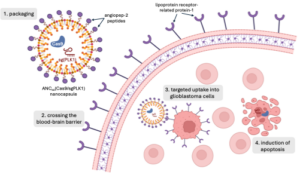
The researchers encapsulated Cas9 and sgPLK1 in a neutrally charged nanoparticle for delivery. The small size of nanoparticles, which are measured in nanometers, allow for easy transport through the bloodstream and uptake by cells. Taking advantage of the high expression of lipoprotein receptor-related protein-1 (LRP-1) on both BBB endothelial cells and glioblastoma tumor cells, they decorated the nanocapsule surface with LRP-1 ligand angiopep-2 peptide to facilitate selective uptake by BBB and glioblastoma cells (Figure 1). Zou and her colleagues bound the nanoparticle together with disulfide bonds as an added layer of selectivity for glioblastoma cell delivery. In the high glutathione environment of a glioblastoma cell, the nanoparticle dissolves, releasing its contents. However, glutathione concentrations are lower in BBB endothelial cells and healthy neurons, reducing the dissolution of the nanoparticles and leaving healthy DNA alone.


Figure 1. Nanoparticles enable permeation of the blood brain barrier (BBB) and selective delivery of the CRISPR/Cas9 system to glioblastoma (GBM) cells. Angiopep-2 peptides, which decorate the nanoparticle’s surface, bind with lipoprotein receptor-related protein-1 (LRP-1) overexpressed on BBB and GBM cells. Following uptake into GBM cells, the nanoparticles dissolve in the high glutathione environment, releasing the Cas9 nuclease and single guide RNA (sgRNA) targeting Polo-like kinase 1 (PLK-1) genes. Zou et al. demonstrated the selective mutation of PLK-1 induced apoptosis in GBM cells with minimal off-target effects.


Through Cas9/sgPLK1 delivery by nanoparticle, Zou et al. demonstrated a 53% reduction in expression of the targeted gene in vitro. PLK1 gene editing was cell selective, with negligible genetic mutation in the healthy surrounding brain tissue. While nanoparticles with and without disulfide cross-linking were capable of gene editing, the disulfide cross-linked nanoparticle induced almost 400% more mutations. Glioblastoma cells treated with disulfide cross-linked nanoparticles were also over 3 times more likely to undergo apoptosis cell death. Mice grafted with patient glioblastoma tumors treated with Cas9/sgPLK1 nanocapsules experienced an almost 3-fold extension in life expectancy, suggesting this treatment as a viable anti-glioblastoma therapy.5

Yet in order to be effectively applied as a cancer therapy, the efficiency of this nanoparticle delivery system must be increased. In mouse glioblastoma models, Zou et al. only achieved a maximum accumulation of 12% and effected a 38% knockdown of PLK1.5 Despite their low magnitude, these values are far greater than similar gene therapy treatments currently studied, suggesting nanoparticles present an innovation in the delivery of CRISPR-Cas9 to difficult to access tumors.

In addition to the improved efficacy over existing systems, the work of Zou et al. opens the door for less invasive administration of treatment. In contrast to previous gene therapies administered directly to the tumor through intercranial injection, the BBB penetration and tumor-specific accumulation of the nanoparticles may permit systemic administration. Zou et al. injected the nanoparticles intravenously, but treatment may even be given as a pill taken orally, eliminating any surgical intervention. Moreover, due to their modular design, the engineered nanoparticles may be adapted as targeted treatments for other tumors. Exchanging the angiopep-2 peptides for another ligand would facilitate uptake by cells expressing the corresponding receptor. The load carried within the nanoparticle could be altered to contain a different sgRNA targeting a new gene or another therapeutic entirely as a complementary treatment. Nanoparticle delivery systems like that studied by Zou et al. contains many layers of selectivity, offering hope for effective delivery of treatment to previously inaccessible tumors.


Works Cited:

  1. Wirsching, H.-G. & Weller, M. Glioblastoma. in Malignant Brain Tumors : State-of-the-Art Treatment (eds. Moliterno Gunel, J., Piepmeier, J. M. & Baehring, J. M.) 265–288 (Springer International Publishing, Cham, 2017). doi:10.1007/978-3-319-49864-5_18.
  2. Delgado-López, P. D. & Corrales-García, E. M. Survival in glioblastoma: a review on the impact of treatment modalities. Clin Transl Oncol 18, 1062–1071 (2016).
  3. Ostrom, Q. T. et al. CBTRUS Statistical Report: Primary Brain and Central Nervous System Tumors Diagnosed in the United States in 2007–2011. Neuro Oncol 16, iv1–iv63 (2014).
  4. Lino, C. A., Harper, J. C., Carney, J. P. & Timlin, J. A. Delivering CRISPR: a review of the challenges and approaches. Drug Delivery 25, 1234–1257 (2018).
  5. Zou, Y. et al. Blood-brain barrier–penetrating single CRISPR-Cas9 nanocapsules for effective and safe glioblastoma gene therapy. Sci Adv 8, eabm8011.
  6. Dotiwala, A. K., McCausland, C. & Samra, N. S. Anatomy, Head and Neck: Blood Brain Barrier. in StatPearls (StatPearls Publishing, Treasure Island (FL), 2024).
  7. Song, R. et al. Selection of rAAV vectors that cross the human blood-brain barrier and target the central nervous system using a transwell model. Molecular Therapy Methods & Clinical Development 27, 73–88 (2022).
  8. Lee, B. et al. Nanoparticle delivery of CRISPR into the brain rescues a mouse model of fragile X syndrome from exaggerated repetitive behaviours. Nat Biomed Eng 2, 497–507 (2018).
  9. Lee, C. et al. Polo-Like Kinase 1 Inhibition Kills Glioblastoma Multiforme Brain Tumor Cells in Part Through Loss of SOX2 and Delays Tumor Progression in Mice. Stem Cells 30, 1064–1075 (2012).

Filed Under: Biology, Chemistry and Biochemistry, Psychology and Neuroscience, Science Tagged With: blood-brain barrier, CRISPR, glioblastoma, nanoparticles

The Solution to Alzheimer’s May Lie in Depression

April 21, 2024 by Nicholas Enbar-Salo '27

Despite being discovered by Alois Alzheimer almost 120 years ago, Alzheimer’s Disease (AD) still remains incurable (Hippius & Neundörfer, 2003). AD causes the brain to break down over time, which is also known as neurodegeneration. AD begins by deterioration of the hippocampus, which is the part of the brain responsible for memory and emotion. It then slowly spreads to other parts of the brain, eventually breaking apart the brain stem, which is responsible for involuntary movements such as breathing and swallowing (Lee et al., 2015). Given that around 39 million people have Alzheimer’s worldwide and that this disease has a 100% fatality rate, scientists across the world have tried to find a cure to this ravaging disease (World Health Organization, 2023). While there is yet to be a cure, recent developments by Stephanie Langella could help with mitigating one of the earliest signs of Alzheimer’s: depressive symptoms.

In this study, Langella and her team studied Presenilin-1 (PSEN1) gene mutations, a major cause of early-onset Alzheimer’s Disease. The PSEN1 gene provides instructions for making the presenilin-1 protein. This protein is an essential part of a protein complex known as gamma-secretase. This complex cleaves toxic proteins such as the amyloid precursor protein (APP) to create nontoxic proteins. When the PSEN1 gene mutates, gamma-secretase struggles to form and break down these toxic proteins, causing APP molecules to join together to create amyloid-beta (also known as amyloid-ꞵ or A-ꞵ), the protein responsible for the neurodegeneration seen in Alzheimer’s Disease (Bagaria et al., 2022). 

A figure of gamma-secretase, APP processing, and generation of Amyloid-β (Aβ). Cleavages of C99 by gamma secretase (ε/ζ/γ) release sAPPβ, a type of APP which is beneficial to cells. When the PSEN1 gene mutates, gamma secretase (γ) produces AICD, a harmful type of APP, into a cell’s liquid (cytosol) and amyloid-β 37-43 into cell organelles. Aβ42 is the form of amyloid-β responsible for neurodegeneration in AD patients. (Steiner et al., 2018).

In particular, they studied its relationship to the neurodegeneration of the hippocampus and depressive symptoms (Langella et al., 2023). They began by creating two groups:  the first group consisted of carriers of the PSEN1 mutation but that had not yet been diagnosed with AD, and the second group consisted of the family members of the respective PSEN1 carriers that did not have the mutation and were not diagnosed with Alzheimer’s. Then, two structural MRIs – a method of neuroimaging which models the brain structures of a patient– with a one-year gap in between the two images were taken of the participants’ hippocampuses to measure the change in the volume of the hippocampus over a year. Participants also took the Geriatric Depression Scale, a 15-item survey that measures depressive symptoms, such as the subjects’ feelings of hopelessness and rating their interest in hobbies, to measure depressive symptoms over one year. 

Once the study was concluded, Langella found that there was no significant difference in the severity of the depressive symptoms between those carrying the PSEN1 mutation and those that did not. However, within the group carrying the PSEN1 mutation, those with smaller hippocampal volumes experienced more depressive symptoms. This association remained even after accounting for the age differences in the participants. This same association was not present in the non-PSEN1 carriers (Langella et al., 2023). Since the volume of the hippocampus did not have any relationship with depressive symptoms with non-PSEN1 carriers, there is likely some relationship between Alzheimer’s and depressive symptoms caused by hippocampal neurodegeneration. 

A).  Structural MRI of the hippocampus from the back of the head (shown in yellow)

C). Top-down structural MRI of the hippocampus (shown in yellow)  (Sato et al., 2021)

There are several important implications of this research. To start, if there is indeed a relationship between the severity of depressive symptoms and the size of the hippocampus in someone with AD, there is a chance that trying to mitigate these depressive symptoms through therapy and antidepressant medication could slow down the deterioration of the hippocampus. By keeping the hippocampus intact for a longer time, people with AD could have better emotional control and memory later in life, which would greatly improve their quality-of-life (Langella et al., 2023). Also, since AD first deteriorates the hippocampus, it is possible that the onset of depressive symptoms in people with the PSEN1 mutation could be used as an indicator to doctors on the severity of the neurodegeneration. For instance, if someone with the PSEN1 gene mutation suddenly begins displaying depressive symptoms, it is possible that AD has just recently started decaying the hippocampus. Doctors can then try to intervene and slow the decay of the hippocampus through administering antidepressants and therapy, but also through encouraging lifestyle changes such as increased exercise. This way, those with Alzheimer’s can live a longer time before their hippocampus fully degrades, letting them keep their memories for a longer time. 

Since this is one of the first studies relating depression and hippocampal decay in people with PSEN1 mutations, there is no theorized mechanism behind why this relationship exists in people with the PSEN1 mutation but not in those without. However, Langella et al. did find a particularly strong association between hippocampal decay in those with the PSEN1 mutation and displaying apathy, one of the measured depressive symptoms in the study (2023). More research should be done on the potential role of certain depressive symptoms on hippocampal decay, along with more research on the neural underpinnings relating the PSEN1 mutation, depression symptoms, and hippocampal decay. There is some evidence linking the formation of amyloid-ꞵ to depression in late-life major depression, but further research into the mechanism underlying this relationship is required (Pomara et al., 2022). 

However, there is a pressing issue with this study; it had a fairly small sample size, with the PSEN1 carrier group having 27 participants and the non-PSEN1 group having 26. Since AD is a disease that affects everyone slightly differently, having such a small sample size makes the results unreliable and hard to generalize to everyone with AD. Regardless of the issues in the study, developments such as the ones created by this study serve to improve the quality of life and life expectancy of people with AD, which promises to improve the lives of almost 39 million people and their families. With every passing discovery into Alzheimer’s, scientists are also getting more information on the mechanisms behind the disease, which could eventually lead humanity to curing the disease altogether. 


Citations

Bagaria, J., Bagyinszky, E., & An, S. S. A. (2022). Genetics, Functions, and Clinical Impact of Presenilin-1 (PSEN1) Gene. International journal of molecular sciences, 23(18), 10970. https://doi.org/10.3390/ijms231810970


Hippius, H., & Neundörfer, G. (2003). The discovery of Alzheimer’s disease. Dialogues in clinical neuroscience, 5(1), 101–108. https://doi.org/10.31887/DCNS.2003.5.1/hhippius


Langella S,  Lopera F,  Baena A, et al.  Depressive symptoms and hippocampal volume in autosomal dominant Alzheimer’s disease. Alzheimer’s Dement.  14 Oct. 2023, 986–994. https://doi.org/10.1002/alz.13501


Lee, J. H., Ryan, J., Andreescu, C., Aizenstein, H., & Lim, H. K. (2015). Brainstem morphological changes in Alzheimer’s disease. Neuroreport, 26(7), 411–415. https://doi.org/10.1097/WNR.0000000000000362


Pomara, N., Bruno, D., Plaska, C.R. et al. Plasma Amyloid-β dynamics in late-life major depression: a longitudinal study. Transl Psychiatry 12, 301 (2022). https://doi.org/10.1038/s41398-022-02077-8


Sato, Jinya, et al. “Lower Hippocampal Volume in Patients with Schizophrenia and Bipolar  Disorder: A Quantitative MRI Study.” Journal of Personalized Medicine, vol. 11, no. 2, 13 Feb. 2021, p. 121, https://doi.org/10.3390/jpm11020121.


Steiner, H., Fukumori, A., Tagami, S., & Okochi, M. (2018, October 28). Making the final cut: Pathogenic amyloid-β peptide generation by γ-secretase. The Journal of Cellular Pathology. https://www.cell-stress.com/researcharticles/making-the-final-cut-pathogenic-amyloid-%ce%b2-peptide-generation-by-%ce%b3-secretase


World Health Organization. “Dementia.” Dementia, 2023, www.who.int/news-room/fact-sheets/detail/dementia.

Filed Under: Psychology and Neuroscience, Science Tagged With: Alzheimer's Disease, Depression, Genes

It’s in the Cards: A Dive into Tarot Card Psychology, Interpretation and Therapeutic Applications

April 21, 2024 by Gabe O'Brien

With a long history dating back nearly 700 years, Tarot cards have maintained a presence in society as a tool that is considered to predict the future and understand one’s inner issues, desires, and motivations. There are many conflicting theories regarding the origin of Tarot cards, with the predominant notion pointing to 14th century northern Italy (Tarot Heritage). Researchers claim that the major arcana of Tarot is based on the Egyptian hieroglyphic book of Thoth (the Egyptian god of wisdom), which is also known as the book of Tarot (Willis 1988). But why do people still use Tarot cards, and what do we get out of using them? The phenomenon surrounding the use and interpretation of Tarot cards can be broken down into two juxtaposing explanations: paranormal and nonparanormal. The paranormal explanation claims that Tarot cards reveal hidden motives, portray opportunities, and offer a reflection of a person’s inner processes, allowing the cards to provide clarity regarding a person’s questions or conflicts. Meanwhile, the nonparanormal explanation claims that the entire phenomenon of Tarot cards can be explained by examining two simple psychological effects: The Barnum effect and “cold reading” (Ivtzan 2007). Additionally, several modern therapeutic approaches have employed the use of Tarot cards as a tool for self-reflection, with Tarot card readings offering clients a sense of order and control in their own lives (Hofer 2009). There are several different reasons for why people use Tarot cards, and the associated applications of the cards can help to improve a person’s mental health when the cards are utilized in a therapeutic context (Hofer 2009).

Many standard Tarot decks follow the same 78-card structure, which is divided into the minor arcana (56 cards), and the major arcana (22 cards). The cards in the major arcana represent the main themes of human life, such as love, death, spirituality, acceptance, etc. The cards in the minor arcana represent subtle mysteries of life, and are considered to be lesser compared to the major arcana (Ivtzan 2007). Additionally, there are several different techniques for choosing the cards in a reading, with the most popular option being for the reader to ask the client to shuffle the cards while focusing on a question, spread the deck, and choose the cards that they feel the most drawn to (Ivtzan 2007). The use of Tarot cards has continued to flourish, even in western societies, and the popularity of Tarot cards is not an indication of reliability or validity, but rather a look into how using the cards can influence our thought processes and mental state. 

Figure 1: The major arcana of Tarot (Medium).

The paranormal explanation surrounding the phenomenon of Tarot cards is the approach that is acclaimed by occultists who believe that the cards reveal information about the quality of a moment for an individual (Ivtzan 2007). They do not believe that the cards predict the future as if it is fixed, but rather reveal information and potential circumstances about changeable events. By creating more awareness about the meaning of a specific moment for a client, this can help to provide the client with important insights, as well as a drive to take control of their own life and make changes that will be beneficial to them in the long run. Comparatively, the nonparanormal explanation examines the use of Tarot cards through the lens of psychological effects, with the Barnum effect being the most emphasized. The Barnum effect is the tendency to believe that vague predictions or general personality descriptions, such as those offered by Tarot or astrology, have specific applications to one’s unique circumstances (American Psychological Association). A Tarot reader may make general, trivial statements that could apply to anyone, and a client, eager to seek guiding information about their life, will accept these statements as truth. The major arcana of Tarot deals with themes that concern every individual’s life, so it is not difficult to come up with general statements about these themes that any person could be susceptible to (Ivtzan 2007). The other psychological effect that the nonparanormal explanation examines is “cold reading,” which is a set of deceptive psychological techniques that give a client the impression that a reader has paranormal abilities. The Barnum effect falls under the umbrella of  “cold reading,” and the techniques behind “cold reading” involve the use of sharp observational skills and a good memory when examining a client. Cues such as a client’s clothing, physical characteristics, and manner of speech can reveal a lot of valuable information to a reader, that a reader can then use to inform the statements that they make to a client regarding the topic of their reading (Ivtzan 2007).

Although there are underlying psychological influences behind the use of Tarot cards, Tarot card readings can still have beneficial effects on a person’s mental health when used in a therapeutic context. A 2009 study investigated how regular users of Tarot cards employed the cards as a tool for self-reflection (rather than for divination). The study involved conducting interviews with several co-researchers who used Tarot cards regularly and in a self-reflective manner, and the interviews from the study were transcribed, with the common themes and qualities that existed between the interviews being extracted (Hofer 2009). Overall, the results of the study found that the co-researchers used Tarot cards as a way to gain insight into their current life situations. The cards were found to be used the most often during difficult times where they could offer a source of comfort. This source of comfort involved providing confirmation that everything was okay and that life had a sense of order. 

On top of this, Tarot cards were also used as a tool for positive reinforcement, where cards were drawn both intentionally and randomly to provide insights about what the co-researchers were seeking in their own lives. With a goal in mind, some of the co-researchers drew a card and then kept it with them until what they were working on or towards had been resolved. They claim that Tarot does not reveal new information to them, but that the use of Tarot cards can help to provide a new perspective on an issue that can influence a plan for a possible course of action (Hofer 2009). 

By examining how therapeutic techniques involving Tarot have been successful for co-researchers who have consistently employed these techniques in their own lives, this study outlines how Tarot has the potential to be used as an effective therapeutic tool. Despite the foundational psychological effects behind the mainstream use of Tarot, Tarot cards can still have beneficial impacts on a person’s mental health and inner psychological processes. Further research surrounding the beneficial impacts of Tarot in a therapeutic setting would involve examining a greater number of participants from a wider variety of backgrounds, so that this research could be generalized to a larger audience. Regardless of the reasoning behind why a person may use Tarot cards, there is no doubt that Tarot cards have maintained a strong presence in society, and these cards have the potential to do more than just “predict the future.”

Literature Cited 

  1. APA Dictionary of Psychology. “APA Dictionary of Psychology.” Apa.org, 2014, dictionary.apa.org/barnum-effect.
  2. “History.” Tarot Heritage, 24 July 2011, tarot-heritage.com/history-4/. Accessed 13 Apr. 2024.
  3. Hofer, Gigi Michelle. “Tarot cards: an investigation of their benefit as a tool for self reflection.” University of Victoria PhD diss (2009).
  4. Ivtzan, Itai. “Tarot cards: a literature review and evaluation of psychic versus psychological explanations.” Journal of Parapsychology 71 (2007).
  5. Macsparrow, Mark. “Many Major Arcana Cards in a Reading Means Many Changes Ahead.” Medium, 12 May 2021, tarotreadings.medium.com/many-major-arcana-cards-in-a-reading-means-many-changes-ahead-516becf2faf5. Accessed 13 Apr. 2024.
  6. Willis, T. Magick and the tarot. Wellingborough, UK: Aquarian (1988).

Filed Under: Psychology and Neuroscience, Science Tagged With: barnum effect, mental health, tarot cards, therapy

Expanding Brain Cells: Improvements in Molecular Expansion Technology a Possible Milestone in Neuropathology Diagnosis

April 21, 2024 by Vincent Chen '27

The immune system identifies cells in the body by examining molecular stages on the surface of cells, which are known as antigens. Both the human body and mind utilize this information to decide if cells within our body pose a threat to our well-being. Nevertheless, science today continues to struggle with differentiating harmless and harmful cells. For example, cancerous cells may be so similar in antigen identities that they can’t be told apart. However, with a deeper look, researchers may be able to uncover smaller antigens that allows humanity to successfully distinguish between such cells.

Scientists have been using technology that expands cells in uniform across three dimensions to identify such smaller antigens that have previously been unaccounted for. The most recent advancement involved dExPath (decrowding expansion pathology), an improvement with four times greater expansions and stronger resolutions compared to its predecessor, ExPath. dExPath solved many problems by offering multi-round immunostaining, maximized protein epitope (another name for antigens) preservation, and elimination of fluorescent overlaps. Immunostaining normal brain tissue and brain tumor tissue has helped identify numerous improvements and advantages to dExPath. (Valdes et al., 2021)

dExPath is a form of microscopy technology that facilitates the immunostaining of cells, so far only applicable to the brain and brain stem cells. Immunostaining utilizes antibodies and conjugated enzymes to generate fluorescence as an indicator of certain antigens and markers on cells of interest. At its core, dExPath is a form of expansion microscopy, a preparation and imaging technique used to visualize biological nanostructures. Wassie et aldescribed the process as synthesizing a dense and even network of swellable polyelectrolyte hydrogel to react with specific nanostructures and physically expand them prior to imaging. Once expanded, immunostaining agents were able to label antigens of interest on cells, all of which were visible from an ordinary laboratory microscope.

Experimental uses of dExPath have allowed researchers to detect distinguishing epitopes and their combinations on glial cell tumors, offering a possibility for the technology’s use in detecting brain cancers. dExPath also facilitated multi-round immunostaining, where multiple forms of staining were implemented to highlight different structures in one image, eliminating the need to run multiple images for each layer of staining.

dExPath has solved many issues that persisted in ExPath. The predecessor failed to preserve some binding sites on protein epitopes, leaving a number of antigens unaccounted for before the use of dExPath. During imaging, the new technology also addressed the prior issue of fluorescent overlaps, particularly due to the presence of lipofuscin, a common waste product among cells. Lipofuscin is often mistaken for a different cellular marker of interest, but decrowding allowed for improved access of antibodies to epitopes, allowing the imaging technology to more clearly recognize its intended targets and ignore the lipofuscin autofluorescence. dExPath’s capacity outperformed Sudan Black B, a stain that reduced the autofluorescence of lipofuscin but weakened other relevant fluorescent signals.

Valdes et al. (2021) used dExPath to expand and investigate human and animal brain cell samples. Samples of normal, low-grade (slow-growth) tumors and high-grade (faster-growth) tumors were examined before and after the expansion process to detect differences in marker visibilities. The samples were immunostained for markers vimetin, Iba1, and GFAP. The presence and frequency of particular markers helped convey new indicators for stages of malignant growth in brain tissues. For example, GFAP and vimentin-labeled low-grade cells were interpreted as an indication of potential future malignancy. Additionally, cells labeled with Iba1 and GFAP indicated a possible increase in tumors with phagocytic (ingestive) properties that made them more invasive.

Figure 2, Immunostaining examples of vimetin, Iba1, and GFAP staining via. dExPath. Row 1 exhibits pre-expansion staining. Rows 2 & 3 exhibit pre-decrowding staining, and rows 4 & 5 exhibit post post-decrowding staining. Pre and post-expansion staining alternate starting from row 2. (Valdes et al., adapted from Figure 7)

Fluorescence imaged by dExPath is of significantly higher quality and consistency. dExPath revealed that there are significantly more cells of interest, with many corresponding to more different classes of cells, such as macrophages and glioma cells, that are deemed important to glioma pathology than previously expected.

dExPath is already demonstrating tremendous potential in brain disease analysis and diagnosis, as well as accuracy and reliability to an unprecedented degree. The technology’s compatibility with low-cost, conventional microscopes and more commercially available reagents and instruments in basic laboratories makes the technology tremendously accessible and convenient. Its advantages uncovered features after molecular expansion that uncovered cell demographics previously unaccounted for and redefined the aggressive nature of diseased cells. However, there are many more tests that dExPath must undergo, such as assessing its visualization of other neurological diseases or cancers. Continued research concerning dExPath may allow the world of science and medicine to uncover more about mysteries in neurobiology, and possibly introduce dExPath to clinical settings as a new assessment tool for neuropathology diagnosis.

Sources

Valdes, Pablo A., et al. Decrowding Expansion Pathology: Unmasking Previously Invisible Nanostructures and Cells in Intact Human Brain Pathology Specimens. 7 Dec. 2021,

Wassie, Asmamaw T., et al. “Expansion Microscopy: Principles and Uses in Biological Research.” Nature Methods, vol. 16, no. 1, 2019, pp. 33–41, https://doi.org/10.1038/s41592-018-0219-4.

Filed Under: Psychology and Neuroscience

  • Page 1
  • Page 2
  • Page 3
  • Go to Next Page »

Primary Sidebar

CATEGORY CLOUD

Biology Chemistry and Biochemistry Computer Science and Tech Environmental Science and EOS Honors Projects Math and Physics Psychology and Neuroscience Science

RECENT POSTS

  • Biological ChatGPT: Rewriting Life With Evo 2 May 4, 2025
  • Unsupervised Thematic Clustering for Genre Classification in Literary Texts May 4, 2025
  • Motor Brain-Computer Interface Reanimates Paralyzed Hand May 4, 2025

FOLLOW US

  • Facebook
  • Twitter

Footer

TAGS

AI AI ethics Alzheimer's Disease antibiotics artificial intelligence bacteria Bathymetry Beavers Biology brain Cancer Biology Cell Biology Chemistry and Biochemistry Chlorofluorocarbons climate change Computer Science and Tech CRISPR Cytoskeleton Depression dreams epigenetics Ethics Genes honors Luis Vidali Marine Biology Marine Mammals Marine noise Medicine memory Montreal Protocol Moss neurobiology neuroscience Nutrients Ozone hole Plants Psychology and Neuroscience REM seabirds sleep student superintelligence Technology therapy

Copyright © 2025 · students.bowdoin.edu