• Skip to primary navigation
  • Skip to main content
  • Skip to primary sidebar
  • Skip to footer

Bowdoin Science Journal

  • Home
  • About
    • Our Mission
    • Our Staff
  • Sections
    • Biology
    • Chemistry and Biochemistry
    • Math and Physics
    • Computer Science and Technology
    • Environmental Science and EOS
    • Honors Projects
    • Psychology and Neuroscience
  • Contact Us
  • Fun Links
  • Subscribe

Lily Warmuth

The Association Between Tooth Loss and Cognitive Decline

May 4, 2026 by Lily Warmuth

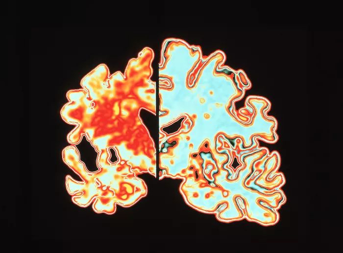
Imaging of a vertical (coronal) slice through the brain of an Alzheimer patient (left) compared with a normal brain ( right).
“Could Magnetic Brain Stimulation Help People with Alzheimer’s? | Scientific American.” n.d. Accessed May 4, 2026. https://www.scientificamerican.com/article/could-magnetic-brain-stimulation-help-people-with-alzheimer-rsquo-s/.

Cognitive decline with age is a major concern in medicine and public health. In 2021, the World Health Organization reported 57 million people were affected by dementia worldwide (World Health Organization, 2023). Well-established risk factors include alcohol intake, lower education level, physical inactivity, obesity, and diabetes, and preventive strategies have developed steadily. However, one potential contributor is often overlooked in major dementia research: tooth loss. Galindo-Moreno et al. (2022) examined this relationship through a large-scale analysis of over 100,000 US Americans, making a case for oral health as an underrecognized factor in cognitive decline. 

Edentulism refers to the partial or complete loss of permanent teeth. Edentulism can be caused by a multitude of factors, including biological processes such as caries (tooth decay) and periodontal disease (infection or inflammation of gums and bone), pulpal pathologies (damage to nerves, tissue, and blood vessels in the center of a tooth), trauma, or oral cancer. In addition to biological causes, edentulism can result from factors affecting dental care: patient preference, access to care, treatment options, and health insurance (Felton 2009). A study found 37% of edentulism cases were due to extraction from caries, 29% from periodontal diseases, and 12% due to trauma (Al-Rafee 2020).  

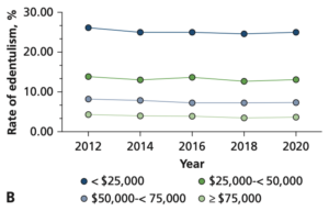
Although oral health care has developed significantly in the last few decades, edentulism remains a prevalent and irreversible condition (Al-Rafee 2020). It can occur at all ages, but the highest incidence occurs between the ages of 75-79 [Figure 1] (Chen et al. 2025). Those most affected by tooth loss typically have a lower socioeconomic standing, which makes health care less affordable and accessible [Figure 2] (Vemulapalli et al. 2024)  
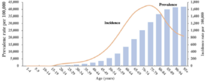
Graph of global incidence and prevalence of edentulism per 100,000 across all ages. Highest incidence rate at ages 75-79. Prevalence per 100,000 gradually increases as age increases.
Figure 1: Global prevalence and incidence rates of edentulism in 2021. Chen, Hui Min, Kuo Shen, Ling Ji, Colman McGrath, and Hui Chen. 2025. “Global and Regional Patterns in Edentulism (1990-2021) With Predictions to 2040.” International Dental Journal 75 (2): 735–43. https://doi.org/10.1016/j.identj.2024.11.022. December 31, 2024: 738


Prevalence rate of complete edentulism in US adults 65 years and older across different socio-economic status'. As income increases, the rate of complete edentulism decreases.
Figure 2: Prevalence rate of complete edentulism in US adults 65 years and older according to demographic characteristics: Behavioral Risk Factor Surveillance System 2012-2020. Income level. Vemulapalli, Abhilash, Surendra Reddy Mandapati, Anusha Kotha, Hemanth Rudraraju, and Subhash Aryal. 2024. “Prevalence of Complete Edentulism among US Adults 65 Years and Older.” The Journal of the American Dental Association 155 (5): 399–408. https://doi.org/10.1016/j.adaj.2024.02.002. May 6, 2024: 407

Galindo-Moreno et al. proposed multiple pathways by which tooth loss can lead to cognitive decline. Two that play directly into known factors are the “diet and nutrition mechanism” and the masticatory mechanism. The number of teeth and which teeth are present affect what we can eat and how we eat. Mastication — chewing of food (Xu et al. 2008) — is directly influenced by edentulism due to the reduced bite force one can exert with missing teeth or dentures (Galindo-Moreno et al. 2022; Weijenberg et al. 2011). Changes to mastication may impact cognition by decreasing sensory input, which would reduce cell growth and development, impairing the cholinergic neurotransmitter system responsible for regulating memory, muscles, and attention, and reducing the generation of new neurons triggered by exercise (Weijenberg et al. 2011). Mastication additionally restricts our diet and therefore directly plays into the diet and nutrition mechanism. Often, with altered dentition, chewing can be an immense hurdle, for which the solution is a softer yet less nutritious diet.Nutrients such as omega-3 fatty acids, B vitamins, and antioxidants have important neuroprotective properties that help preserve the blood brain barrier, an essential layer that prevents toxins from entering the brain,additionally reducing inflammation, lowering the risk of cognitive decline (Power et al. 2019). Both the masticatory and diet and nutrition mechanisms are intertwined with diabetes and obesity, which are known risk factors for cognitive decline (Galindo-Moreno et al. 2022). 

Another pathway this study mentions is the inflammation/infection mechanism. A leading cause of edentulism is periodontitis, a severe gum infection often driven by the bacterium Porphyromonas gingivalis. This bacterium induces the local release of cytokines, proinflammatory proteins (Galindo-Moreno et al. 2022). Once in the bloodstream, cytokines promote the production of amyloid-β, a peptide whose accumulation is associated with Alzheimer’s disease (Leira et al. 2020). Simultaneously, Porphyromonas gingivalis increases the permeability of the blood-brain barrier (Lei et al. 2023). The heightened permeability of the BBB causes accumulation of overproduced amyloid-β in the brain tissue [Figure 3] (Galindo-Moreno et al. 2022; Leira et al. 2020)  
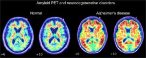
Amyloid PET scan of patient with Alzheimer's Disease (right), and patient without Alzheimer's (left). Patient with Alzheimer's Disease shows higher detection of Amyloid plaques.
Figure 3: Amyloid PET scan comparison of healthy brain and Alzheimer’s disease. Chapleau, Marianne, Leonardo Iaccarino, David Soleimani-Meigooni, and Gil D. Rabinovici. 2022. “The Role of Amyloid PET in Imaging Neurodegenerative Disorders: A Review.” Clinical Investigation. Journal of Nuclear Medicine 63 (Supplement 1): 13S-19S. https://doi.org/10.2967/jnumed.121.263195.

To investigate the relationship between tooth loss and cognitive decline, the researchers analyzed data from over 100,000 Americans drawn from two large national health surveys, NHIS (2014-2017) and NHANES (2005-2018). The NHIS survey was particularly well-suited for assessing cognitive state, as it included four questions on concentration and memory. However, the survey included only one binary dental question asking whether the participants had a complete dentition or had lost ≥1 teeth. The NHANES survey complemented this with a thorough section on dental records. The exact number and location of lost teeth were documented. However, it assessed cognitive state with only one question on memory and confusion (Galindo-Moreno et al. 2022).   

Their primary statistical tool was multinomial logistic regression, a method used when an outcome has more than two categories. In this case, the categories were cognitive difficulty, ranging from “none” to “some” to “a lot.” By using this model, the researchers simultaneously accounted for other factors known to affect cognitive health, including age, income, education level, depression, anxiety, cardiovascular health, and lifestyle habits such as smoking and exercise, which were included in the health surveys. By modeling these variables together, the researchers could estimate the independent contribution of tooth loss to cognitive decline.  

The results were expressed as odds ratios (ORs), which indicate how much more likely a given outcome is in one group than in a reference group. Here, the reference was a fully toothed person reporting no cognitive difficulties. An OR above 1.0 indicated higher odds of cognitive problems among people with missing teeth. This held true even after the other variables were statistically accounted for. The researchers also used a technique called ROC curve analysis on the NHANES data that included exact tooth counts, allowing them to identify a meaningful threshold below which cognitive risk measurably increased (Galindo-Moreno et al. 2022).  

The researchers found that, overall, the presence of teeth was statistically associated with a better cognitive state. The NHIS data showed that people with edentulism (partial or complete) had an OR > 1 across all cognitive categories, especially memory, even after accounting for other risk factors. This trend was also observed across categories of gender, socio-economic status (SES), education, and cardiovascular risk — all of which negatively impact cognition. Notably, socioeconomic status emerged as one of the strongest predictors, alongside edentulism, reflecting how directly financial circumstances shape access to dental care and, through it, long-term cognitive health. 

Using ROC curve analysis of the NHANES data, they determined the threshold for cognitive risk to be 20.5 teeth, indicating that a person with fewer than 21 teeth has an increased risk of cognitive decline compared to a fully dentulous person (Galindo-Moreno et al. 2022). Importantly, the study analyzed the NHANES survey and found a gradient effect: the fewer teeth a person had, the worse their cognitive outcomes tended to be, which strengthens the case that the association is meaningful rather than coincidental. Furthermore, a threshold could be determined for each individual tooth category: 5.5, 5.5, 3.5, 4.5, respectively, for molars, premolars, canines, and incisors. The multinomial regression of the NHANES data determined molars had the highest OR. The researchers linked this to the masseter, an important masticatory muscle supported by molars, which may, through its activity, stimulate the release of neurotrophic factors that support brain health. 

The link between edentulism and cognitive decline is still scarcely researched. As of March 2026, there are only 66 results on PubMed, 142 on ScienceDirect, and 148 on Wiley on the correlation between edentulism and cognitive decline. To put this into perspective, there are 2,277 results on PubMed, 18,967 on ScienceDirect, and 10,546 on Wiley on the relationship between diet and cognitive decline. The discussed research article combines two USA national health surveys with diverse samples, NHIS and NHANES, making it one of the largest in scope to date on tooth loss and cognitive decline. Although Galindo-Moreno and his team compellingly demonstrate the correlation, they recognize that their findings cannot answer whether edentulism leads to poorer cognition or rather poor cognition leads to edentulism (Galindo-Moreno et al. 2022, 3498). Some of the issues the researchers faced were the binary assessment of dentition in the NHIS survey, the single question on cognitive condition in the NHANES survey, and the overall lack of records on the reasons for tooth loss (Galindo-Moreno et al. 2022).   

Nevertheless, this study is a step in the right direction. Galindo-Moreno et al. showed that edentulism is correlated with cognition, thereby providing meaningful epidemiological evidence for a relatively young field. Consequently, this study and further research could have great clinical implications for cognitive health, not only in cost-effective treatment and prevention, but also in an important personal impact for those struggling with cognitive impairments and dental hygiene. 


Al-Rafee, Mohammed A. 2020. “The Epidemiology of Edentulism and the Associated Factors: A Literature Review.” Journal of Family Medicine and Primary Care 9 (4): 1841–43. https://doi.org/10.4103/jfmpc.jfmpc_1181_19.  

Chapleau, Marianne, Leonardo Iaccarino, David Soleimani-Meigooni, and Gil D. Rabinovici. 2022. “The Role of Amyloid PET in Imaging Neurodegenerative Disorders: A Review.” Clinical Investigation. Journal of Nuclear Medicine63 (Supplement 1): 13S-19S. https://doi.org/10.2967/jnumed.121.263195.  

Chen, Hui Min, Kuo Shen, Ling Ji, Colman McGrath, and Hui Chen. 2025. “Global and Regional Patterns in Edentulism (1990-2021) With Predictions to 2040.” International Dental Journal 75 (2): 735–43. https://doi.org/10.1016/j.identj.2024.11.022.  

“Dementia.” n.d. Accessed March 27, 2026. https://www.who.int/news-room/fact-sheets/detail/dementia.  

Felton, David A. 2009. “Edentulism and Comorbid Factors.” Journal of Prosthodontics 18 (2): 88–96. https://doi.org/10.1111/j.1532-849X.2009.00437.x.  

Galindo-Moreno, Pablo, Lucia Lopez-Chaichio, Miguel Padial-Molina, et al. 2022. “The Impact of Tooth Loss on Cognitive Function.” Clinical Oral Investigations 26 (4): 3493–500. https://doi.org/10.1007/s00784-021-04318-4.  

Lei, Shuang, Jian Li, Jingjun Yu, et al. 2023. “Porphyromonas Gingivalis Bacteremia Increases the Permeability of the Blood-Brain Barrier via the Mfsd2a/Caveolin-1 Mediated Transcytosis Pathway.” International Journal of Oral Science15 (January): 3. https://doi.org/10.1038/s41368-022-00215-y.  

Leira, Yago, Álvaro Carballo, Marco Orlandi, et al. 2020. “Periodontitis and Systemic Markers of Neurodegeneration: A Case–Control Study.” Journal of Clinical Periodontology 47 (5): 561–71. https://doi.org/10.1111/jcpe.13267.  

Power, Rebecca, Alfonso Prado-Cabrero, Ríona Mulcahy, Alan Howard, and John M. Nolan. 2019. “The Role of Nutrition for the Aging Population: Implications for Cognition and Alzheimer’s Disease.” Annual Review of Food Science and Technology 10 (1): 619–39. https://doi.org/10.1146/annurev-food-030216-030125. 

Vemulapalli, Abhilash, Surendra Reddy Mandapati, Anusha Kotha, Hemanth Rudraraju, and Subhash Aryal. 2024. “Prevalence of Complete Edentulism among US Adults 65 Years and Older.” The Journal of the American Dental Association 155 (5): 399–408. https://doi.org/10.1016/j.adaj.2024.02.002.  

Weijenberg, R. A. F., E. J. A. Scherder, and F. Lobbezoo. 2011. “Mastication for the Mind—The Relationship between Mastication and Cognition in Ageing and Dementia.” Neuroscience & Biobehavioral Reviews 35 (3): 483–97. https://doi.org/10.1016/j.neubiorev.2010.06.002.  

World Health Organization. 2023. “Dementia” Fact Sheets. https://www.who.int/news-room/fact-sheets/detail/dementia 

Xu, W. L., J. E. Bronlund, J. Potgieter, et al. 2008. “Review of the Human Masticatory System and Masticatory Robotics.” Mechanism and Machine Theory 43 (11): 1353–75. https://doi.org/10.1016/j.mechmachtheory.2008.06.003. 
Filed Under: Psychology and Neuroscience, Science Tagged With: Alzheimer's Disease, brain, cognitive, Dentistry, Edentulism, neurobiology, Psychology and Neuroscience, Tooth loss

Effect of Dental Malocclusions on Posture in Children

December 12, 2025 by Lily Warmuth

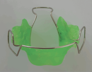
Photograph of a Binator device, an orthodontic appliance made of acrylic resin and wire that resembles a traditional retainer.

It is estimated that over six million patients seek orthodontic treatment every year to improve their malocclusion, or misalignment of teeth (Hung et al. 2023). Seeing as many people value this treatment, it is not surprising to learn that the way our teeth fit into one another affects the way we eat, talk, breathe, and even our posture. Musculoskeletal (shoulders, spine, muscles) and stomatognathic (teeth, jaws, chewing muscles, tongue, lips) are separate systems of our bodies that interact in intricate ways. For example, a misalignment of teeth alters the muscle-use patterns in our cheeks to compensate for this disparity, which in turn affects the neck muscles which are connected to our face muscles. Through a slight discrepancy in teeth-alignment, the whole head can shift into a different position, impacting one’s health (Bardellini et al. 2022). Unfortunately, the intersection of posture and dental malocclusions is a scarcely researched field. Seeing how impactful dental alignment is to the rest of the body, it is important to research and understand the factors that influence it.    

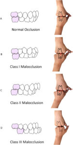
One study published in 2022 by a group of Italian researchers (Bardellini et al.) examined how these systems work together, and the effects of correcting dental malocclusions through orthodontic treatment on the posture of children. While there are many different classifications and types of dental malocclusions, this article specifically analyzes patients using Angle’s classification. Angle’s classification shows three types of malocclusions: class I, II, or III (Fig. 1). Each is described by the position of the lower (mandible) and upper (maxillary) molars. Class I is defined as the molars fitting together in a standard way, however, malocclusions are still present in other teeth besides the molars. In Angle’s class II, the lower molar is farther back (distal) than the upper molar. Lastly, class III shows the lower molar too far in front of the upper molar (Campbell and Goldstein 2021).  

Angle's classification of occlusion illustrated with dental diagrams and hand analogies: normal occlusion, Class I, Class II, and Class III malocclusions.
Figure 1: Simulate Angle’s classification of malocclusion by hands. Xie, Zhiwei, Fuying Yang, Sujuan Liu, and Min Zong. 2023. “The ‘Hand as Foot’ Teaching Method in Angle’s Classification of Malocclusion.” Asian Journal of Surgery 46 (2): 1063

The patients that participated in the study were assessed by two clinicians who evaluated their dental occlusions according to Angle’s classification. While deciding which patients to include in the study, the type of dental-skeletal malocclusion within Angle’s classification did not play a role. Most patients observed in this study exhibited a class II malocclusion, followed by class I and III. Patients that had scoliosis, required physical therapy, chronic diseases affecting balance, macro trauma, cleft lip or palate were excluded to ensure that the improvement in posture depended only on malocclusions and orthodontic treatment. Since this study aimed to find a connection between misalignment of teeth and posture in children, the patients belonged to the age group of 9-12 (Bardellini et al. 2022).    

Bardellini and her team investigated the postures and weight distribution of patients before and after the treatment using multiple methods, such as vertical laser line (VLL) and stabilo-baropodometric analysis.   

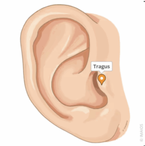
To examine the posture through VLL, the patients were positioned in a standardized position (relaxed posture and arms at side) in front of a white wall. A singular vertical laser line (VLL) was projected onto the patients (Bardellini et al. 2022). The posture was then examined for two factors, the position of the head in relation to the VLL and an excess of extension or flexion. A standard position means the head is centered so that it crosses the tragus—the pointy piece of cartilage close to the cheek (Fig. 2).

Anatomical illustration of human ear with labeled pin above the tragus.
Figure 2: Tragus – anatomical structures. Source: IMAIOS, “Tragus – Anatomical Structures,” accessed November 14, 2025.

If the cartilage did not cross the VLL, the patients’ head was either in a forward or backwards position. Extension and flexion were examined by asking the patients to open their mouths as wide as possible. If the head moved away from the VLL line, it indicated either excess of extension—head bent backwards—or of flexion—the head bent forwards (Fig. 3, Bardellini et al. 2022).

Orthodontic treatment outcomes displayed as paired lateral profile photographs of six patients labeled a through f. Each pair shows pre-treatment (left) and post-treatment (right) views with a vertical line for reference. Arrows on some cases indicate anterior or posterior shifts in facial profile. The images demonstrate improvements in head posture following treatment.
Figure 3: Improvement of the head position (evidenced with the “open mouth test”) in six patients (a-b-c-d-e-f). Bardellini, Elena, Maria Gabriella Gulino, Stefania Fontana, Francesca Amadori, Massimo Febbrari, and Alessandra Majorana. 2022. “Can the Treatment of Dental Malocclusions Affect the Posture in Children?” May 1, 2022: 245

The VLL test indicated that 16 out of 60 patients had a backwards position of the head, 29 a forward position, 10 showed excess of extension while opening their mouths, and 31 an excess of flexion. Only seven patients already had a correct position, meaning that in 75% of patients, dental misalignment influenced head position in relation to VLL line, and 68.33% either flexion or extension.  

After determining the posture of the head, the researchers then examined the weight distribution of the participants using a stabilo-paropodometric platform. The patients were asked to stand on a carpet under which a stabilo-paropodometric platform (40x40cm) was placed. The platform measured the typology of the foot and weight distribution across the two feet. The typology of feet can be divided into three kinds: normal, cavus (extreme arch), or flat (underdeveloped arch). Typology can differ between feet, with either both feet showing the same type or different types. The ideal distribution of body weight between feet should be symmetrical at about 50% on each foot (Bardellini et al. 2022).  

Through measurements obtained with the stabilo-baropodometric platform, the study found 45 cases (both or one side) with cavus feet, and 6 with flat feet (both sides). Hence, 85% of patients had a typology that incorrectly supported their body. Additionally, about 70% of patients had an unequal weight distribution between their two feet, exacerbating bad posture. An incorrect spread of body weight can be identical on both feet—either too much pressure on the ball of the foot or heel—or it can vary between feet (i.e. one foot shows increased pressure at heel, and the other at the ball of the foot) (Bardellini et al. 2022).  

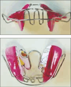
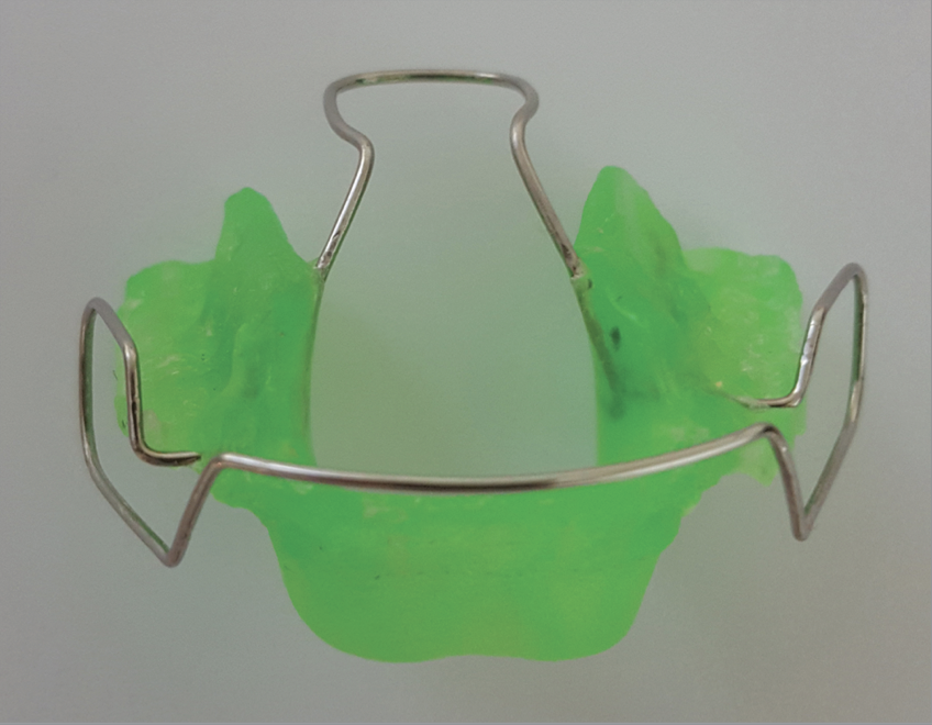
After the classification of malocclusion was identified and the posture (VLL) and weight distributions (Stabilo-baropodometric platform) were measured, the patients were treated with an individually prepared Mouth Slow Balance (Fig. 4), which works by repositioning the tongue, widening the maxilla (upper jaw), and keeping the mandible’s (lower jaw) relation to the maxilla (Bardellini et al. 2019, Bardellini et al. 2022). They describe the MSB device as a “evolution of the Binator”, a retainer like appliance adjusting the bite (Fig. 5, Bardellini et al. 2019 p. 243).  

Photograph of a Mouth Slow Balance (MSB) device, an orthodontic appliance made of acrylic resin and wire that resembles a traditional retainer.
Figure 4: The MBS (mouth slow balance) Class III device Bardellini, E., M. G. Gulino, S. Fontana, J. Merlo, M. Febbrari, and A. Majorana. 2019. “Long-term evaluation of the efficacy on the podalic support and postural control of a new elastic functional orthopaedic device for the correction of Class III malocclusion.” European Journal of Paediatric Dentistry, no. 3: 200.
Photograph of a Binator device, an orthodontic appliance made of acrylic resin and wire that resembles a traditional retainer.
Figure 5: The Binator appliance. Pakshir, Hamidreza, Ali Mokhtar, Alireza Darnahal, Zinat Kamali, Mohammad Hadi Behesti, and Abdolreza Jamilian. 2017. “Effect of Bionator and Farmand Appliance on the Treatment of Mandibular Deficiency in Prepubertal Stage.” Turkish Journal of Orthodontics 30 (1): 16

The patients were observed during their treatments for four years (2014-2018), and by the end, 51 out of 60 patients exhibited a correction of malocclusions, either fully aligned or class I (Bardellini et al. 2022). Other patients either dropped out of the study (3 patients) or reached a correction after the observed time frame (6 patients). 

Of the 53 patients, 23 obtained the ideal position and 19 saw an improvement but did not complete correction of head-position. In 10 cases, patients were found to have been overcorrected.  In the beginning of the four-year observation period, 15 patients had a correct position regarding VLL posture assessment. After treatment, 7 kept their correct position, while 8 now developed a forward position. Additionally, two patients that showed a backwards position before treatment developed a forward position by the end (Bardellini et al. 2022).  

Bardellini et al. (2022) also found significant improvements of the posture in VLL open mouth exams. 53.3% now kept their tragus on the laser line while opening their mouths, when they used to hyper-extend or –flex.  

53 participants (88%) improved their foot typology, of which 17 achieved a complete correction. Before treatment, only 15% of participants had a “normal” typology, which increased to 28% after treatment. However, weight distribution that varied between feet significantly increased from 18 to 37, of which seven patients developed a weight distribution imbalance they previously didn’t show. Overall, cases also exhibited an improvement without complete correction which decreased the median of support discrepancies over the course of the treatment (Bardellini et al. 2022).  

These findings provide evidence for Bardellini et al.’s hypothesis that posture is in fact altered by dental malocclusions. They explain that through a complex chain of muscles across different systems, muscles alter their patterns which disturb the posture, specifically in the position of the head and support of feet.  Muscles around our cheeks (masticatory) and neck (cervical) were already discovered to have a connection in previous research (Bardellini et al. 2022). Furthermore, trunk muscles (abdomen, chest, back) are also connected to these muscles. Since the misalignment of teeth affects the so-called mandibular elevator muscles that are a part of our cheek muscles, this change flows over into other muscle systems (cervical and trunk) acting on our posture. Our strategies for balancing are primarily spread across the trunk, head, and pelvis, which means that the misposition of the head leads our body to try and balance it using other methods (trunk and pelvis) (Bardellini et al. 2022). So, the wrong posture shifts the center of gravity. 

Although Bardellini et al. have found significant evidence that there is a correlation between dental malocclusions and posture, they acknowledge that they are one of few studies that focus on this specific alteration in posture, hence emphasizing that more research needs to be done.  

Furthermore, the results may have been skewed because the team did not consider that the natural changes occurring in growing children may also influence their posture, weight distribution, and more. However, for this specific study it would have been unethical to have a control group of untreated children to compare the effects of treatment vs no treatment (Bardellini et al. 2022).   

Bardellini and her team are one of the few trailblazing research articles that examine the impact of malocclusions on posture, specifically targeting the head and feet. As mentioned before, not much research has been done in this field that examines this topic especially, yet it can prove to be vital for child development. Correcting posture early on can improve a person’s life-quality for the rest of their lives, impacting everyday tasks. Hopefully, in the future more researchers will recognize the importance of this subject and contribute new findings.


References:

Bardellini, E., M. G. Gulino, S. Fontana, J. Merlo, M. Febbrari, and A. Majorana. 2019. “Long-term evaluation of the efficacy on the podalic support and postural control of a new elastic functional orthopaedic device for the correction of Class III malocclusion.” European Journal of Paediatric Dentistry, no. 3: 199–203. https://doi.org/10.23804/ejpd.2019.20.03.06. 

Bardellini, Elena, Maria Gabriella Gulino, Stefania Fontana, Francesca Amadori, Massimo Febbrari, and Alessandra Majorana. 2022. “Can the Treatment of Dental Malocclusions Affect the Posture in Children?” May 1. DOI: 10.17796/1053-4625-46.3.11 

Campbell, Stephen, and Gary Goldstein. 2021. “Angle’s Classification–A Prosthodontic Consideration: Best Evidence Consensus Statement.” Journal of Prosthodontics (United States) 30 (S1): 67–71. https://doi.org/10.1111/jopr.13307. 

Hung, Man, Golnoush Zakeri, Sharon Su, and Amir Mohajeri. 2023. “Profile of Orthodontic Use across Demographics.” Dentistry Journal 11 (12): 291. https://doi.org/10.3390/dj11120291. 

IMAIOS. “Tragus.” e-Anatomy, accessed November 20, 2025. https://www.imaios.com/en/e-anatomy/anatomical-structures/tragus-1536888748. 

Pakshir, Hamidreza, Ali Mokhtar, Alireza Darnahal, Zinat Kamali, Mohammad Hadi Behesti, and Abdolreza Jamilian. 2017. “Effect of Bionator and Farmand Appliance on the Treatment of Mandibular Deficiency in Prepubertal Stage.” Turkish Journal of Orthodontics 30 (1): 15–20. https://doi.org/10.5152/TurkJOrthod.2017.1604. 

Xie, Zhiwei, Fuying Yang, Sujuan Liu, and Min Zong. 2023. “The ‘Hand as Foot’ Teaching Method in Angle’s Classification of Malocclusion.” Asian Journal of Surgery 46 (2): 1062–64. https://doi.org/10.1016/j.asjsur.2022.07.130. 

Filed Under: Biology, Science Tagged With: Dentistry, Orthodontics, Posture, Treatment Outcomes

Primary Sidebar

CATEGORY CLOUD

Biology Chemistry and Biochemistry Computer Science and Tech Environmental Science and EOS Honors Projects Math and Physics Psychology and Neuroscience Science

RECENT POSTS

  • Carcinization: Is it Happening to Everybody? May 13, 2026
  • Efficacy of Neoantigen Vaccines against Triple Negative Breast Cancer May 12, 2026
  • New developments in understanding plankton transport May 12, 2026

FOLLOW US

  • Facebook
  • Twitter

Footer

TAGS

AI AI ethics Alzheimer's Disease antibiotics artificial intelligence bacteria Bathymetry Biology brain Cancer Biology Cell Biology Chemistry and Biochemistry Chlorofluorocarbons climate change cognitive Computer Science and Tech CRISPR Dentistry Depression Dermatology dreams emergency medicine epigenetics Ethics Genes Gut microbiota honors Marine Biology Marine Mammals Marine noise Medicine memory Montreal Protocol neurobiology neuron neuroscience Nutrients Ozone hole Psychology and Neuroscience seabirds sleep student Technology therapy Women's health

Copyright © 2026 · students.bowdoin.edu